Infrared, ultraviolet, and nuclear magnetic resonance spectroscopies differ from mass spectrometry in that they are nondestructive and involve the interaction of molecules with electromagnetic energy rather than with an ionizing source. Before beginning a study of these techniques, however, let’s briefly review the nature of radiant energy and the electromagnetic spectrum.
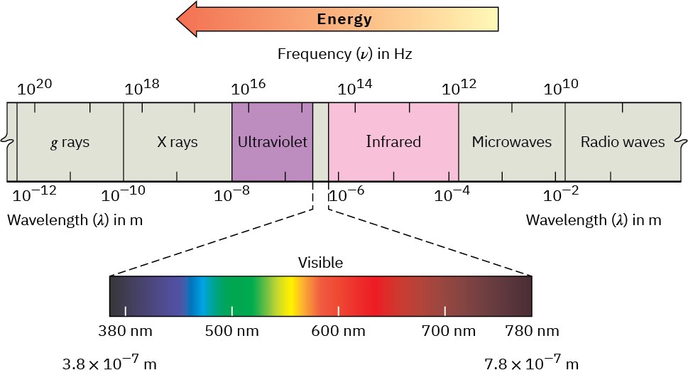
Visible light, X rays, microwaves, radio waves, and so forth are all different kinds of electromagnetic radiation. Collectively, they make up the electromagnetic spectrum, shown in Figure 12.17. The electromagnetic spectrum is arbitrarily divided into regions, with the familiar visible region accounting for only a small portion, from 3.8 × 10⁻⁷ m to 7.8 × 10⁻⁷ m in wavelength. The visible region is flanked by the infrared and ultraviolet regions.

Figure 12.17 The electromagnetic spectrum covers a continuous range of wavelengths and frequencies, from radio waves at the low-frequency end to gamma (γ) rays at the high-frequency end.
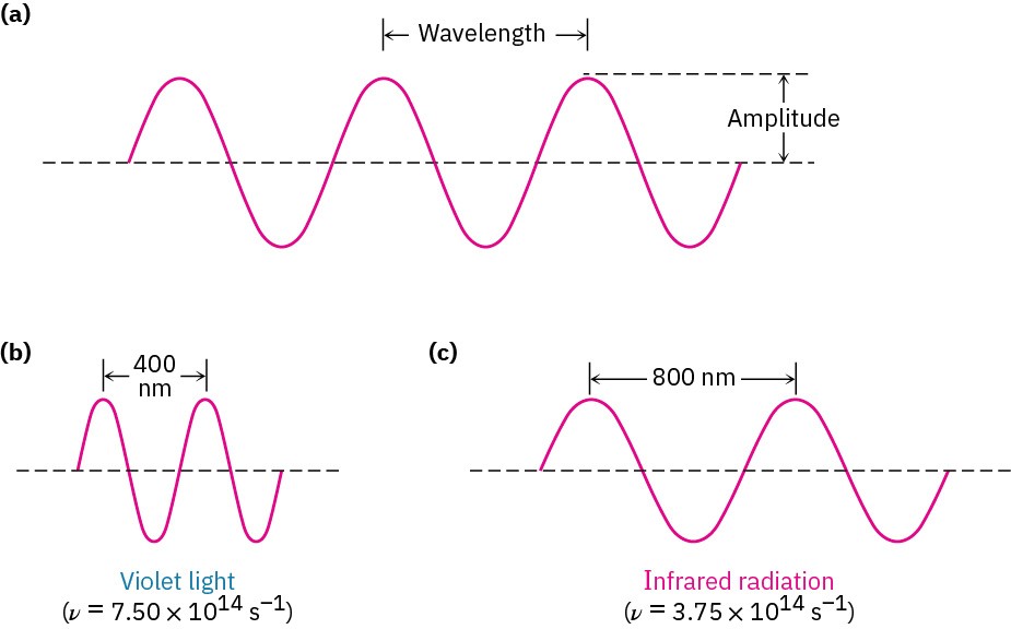
The familiar visible region accounts for only a small portion near the middle of the spectrum. Electromagnetic radiation is often said to have dual behavior. In some respects, it has the properties of a particle, called a photon, yet in other respects it behaves as an energy wave. Like all waves, electromagnetic radiation is characterized by a wavelength, a frequency, and an amplitude (Figure 12.18). The wavelength, λ (Greek lambda), is the distance from one wave maximum to the next. The frequency, ν (Greek nu), is the number of waves that pass by a fixed point per unit time, usually given in reciprocal seconds (s–1), or hertz, Hz (1 Hz = 1 s–1). The amplitude is the height of a wave, measured from midpoint to peak. The intensity of radiant energy, whether a feeble glow or a blinding glare, is proportional to the square of the wave’s amplitude.
Figure 12.18 Electromagnetic waves are characterized by a wavelength, a frequency, and an amplitude. (a) Wavelength (λ) is the distance between two successive wave maxima. Amplitude is the height of the wave measured from the center. (b)–(c) What we perceive as different kinds of electromagnetic radiation are simply waves with different wavelengths and frequencies.
Multiplying the wavelength of a wave in meters (m) by its frequency in reciprocal seconds (s–1) gives the speed of the wave in meters per second (m/s). The rate of travel of all electromagnetic radiation in a vacuum is a constant value, commonly called the “speed of light” and abbreviated c. Its numerical value is usually rounded off to 3.00 × 10⁸ m/s.
Wavelength × Frequency = Speed
𝜆 (m) × 𝜈 (s-1) = 𝑐 (m/s)
𝜆 = 𝑐/𝑣 or 𝑣 = 𝑐/𝜆
Just as matter comes only in discrete units called atoms, electromagnetic energy is transmitted only in discrete amounts called quanta. The amount of energy ε corresponding to 1 quantum of energy (1 photon) of a given frequency ν is expressed by the Planck equation
𝜀 = ℎ𝑣 = ℎ𝑐/𝜆
where h = Planck’s constant (6.62 × 10⁻³⁴ J ‧ s = 1.58 × 10⁻³⁴ cal ‧ s).
The Planck equation says that the energy of a given photon varies directly with its frequency ν but inversely with its wavelength λ. High frequencies and short wavelengths correspond to high-energy radiation such as gamma rays; low frequencies and long wavelengths correspond to low-energy radiation such as radio waves. Multiplying ε by Avogadro’s number NA gives the same equation in more familiar units, where E represents the energy of Avogadro’s number (one “mole”) of photons of wavelength λ:
𝐸 = 𝑁Aℎ𝑐/𝜆
When an organic compound is exposed to a beam of electromagnetic radiation, it absorbs energy of some wavelengths but passes, or transmits, energy of other wavelengths. If we irradiate the sample with energy of many different wavelengths and determine which are absorbed and which are transmitted, we can measure the absorption spectrum of the compound.
An example of an absorption spectrum—that of ethanol exposed to infrared radiation—is shown in Figure 12.19. The horizontal axis records the wavelength, and the vertical axis records the intensity of the various energy absorptions in percent transmittance. The baseline corresponding to 0% absorption (or 100% transmittance) runs along the top of the chart, so a downward spike means that energy absorption has occurred at that wavelength.

Figure 12.19 An infrared absorption spectrum for ethanol, CH3CH2OH. A transmittance of 100% means that all the energy is passing through the sample, whereas a lower transmittance means that some energy is being absorbed. Thus, each downward spike corresponds to an energy absorption.
The energy a molecule gains when it absorbs radiation must be distributed over the molecule in some way. With infrared radiation, the absorbed energy causes bonds to stretch and bend more vigorously. With ultraviolet radiation, the energy causes an electron to jump from a lower-energy orbital to a higher-energy one. Different radiation frequencies affect molecules in different ways, but each provides structural information when the results are interpreted.
There are many kinds of spectroscopies, which differ according to the region of the electromagnetic spectrum used. We’ll look at three: infrared spectroscopy, ultraviolet spectroscopy, and nuclear magnetic resonance spectroscopy. Let’s begin by seeing what happens when an organic sample absorbs infrared energy.
Worked Example 12.3 Correlating Energy and Frequency of Radiation
Which is higher in energy, FM radio waves with a frequency of 1.015 × 10⁸ Hz (101.5 MHz) or visible green light with a frequency of 5 × 10¹⁴ Hz?
Strategy – Remember the equations ε = hν and ε = hc/λ, which say that energy increases as frequency increases and as wavelength decreases.
Solution – Since visible light has a higher frequency than radio waves, it is higher in energy.
Problem 12-5
Which has higher energy, infrared radiation with λ = 1.0 × 10⁻⁶ m or an X ray with λ = 3.0 × 10⁻⁹ m?
Radiation with ν = 4.0 × 10⁹ Hz or with λ = 9.0 × 10⁻⁶ m?
Problem 12-6
It’s useful to develop a feeling for the amounts of energy that correspond to different parts of the electromagnetic spectrum. Calculate the energies in kJ/mol of each of the following kinds of radiation:
(a) A gamma ray with λ = 5.0 × 10⁻¹¹ m
(b) An X ray with λ = 3.0 × 10⁻⁹ m
(c) Ultraviolet light with ν = 6.0 × 10¹⁵ Hz
(d) Visible light with ν = 7.0 × 10¹⁴ Hz
(e) Infrared radiation with λ = 2.0 × 10⁻⁵ m
(f) Microwave radiation with ν = 1.0 × 10¹¹ Hz

