
58 Characterization Techniques
Brigitte Gallagher; Jenna Diemel; and Rachel Falkowski
Fluorescence microscopy is used to sorts cells by size and complexity based on how light reflects off the cells. this is used to determine the size of the nanoparticles that are being made and some of the nanoparticle structural topography.
Video about is Fluorescence microscopy [7].
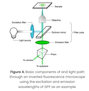
Fluorescence microscopy can give 3D data including measurements, topographical images, and time lapses. Some limitations are that contracts are needed, the wavelength must be the same as the membrane that you are trying to see and it could take time to get the right wavelength.
Fluorescence helps to identify the proteins and outer layer of the nanoparticle and can record how the particle reacts to stimuli over time to give us a better understanding of how it reacts within the body.
Media Attributions
- A scope
