
Learning Objectives
- Define and identify external features and regions of the common integument with an emphasis on clinically relevant structures.
- Perform left sided skin reflection of carnivore cadavers
- Demonstrate recognition of, and appropriate use of, dissection instruments.
The purpose of this lab is to examine certain structures and features of the common integument and then to reflect the skin off the left side of the cadaver as directed. Team members are to rotate dissection responsibilities for this lab, to allow all students a chance to use instruments and to reflect skin.
Prelab reading of this introduction is recommended and portions of it are highlighted in the common integument lecture.
Introduction to the common integument
(adapted from Hudson and Hamilton, Atlas of Feline Anatomy for Veterinarians and from König and Liebich, Veterinary Anatomy of Domestic Mammals)
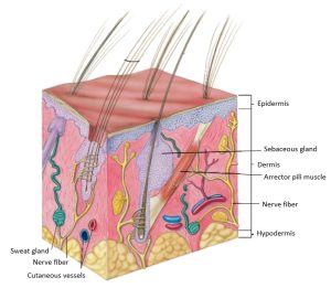
The common integument ([L] in-tegere – in, to cover, integumentum – a covering) is the vital outer covering and barrier of the animal, interfacing with the environment. The common integument encompasses the skin and its appendageal structures (e.g. hair, arrector pili muscles, glands, pads, claws, hooves, horns), the subcutaneous tissue, and the vascular and nervous supply. It is the largest organ of mammals.
The common integument fulfills many functions, to include:
- A protective barrier to external mechanical, chemical, physical and biological factors encountered in the environment, and for preventing unregulated loss of endogenous body substances.
- A reservoir and/or excretory organ for electrolytes, water, vitamins, fat, and other elements and molecules.
- A primary sense organ for touch, pressure, pain, itch, heat, and cold.
- Thermoregulation
- Immunological defense
- Communication
The common integument has unique properties with regards to coat color, skin pigmentation, inflammatory response, and specialized integumentary structures.
Clinical relevance:
The ‘skin’
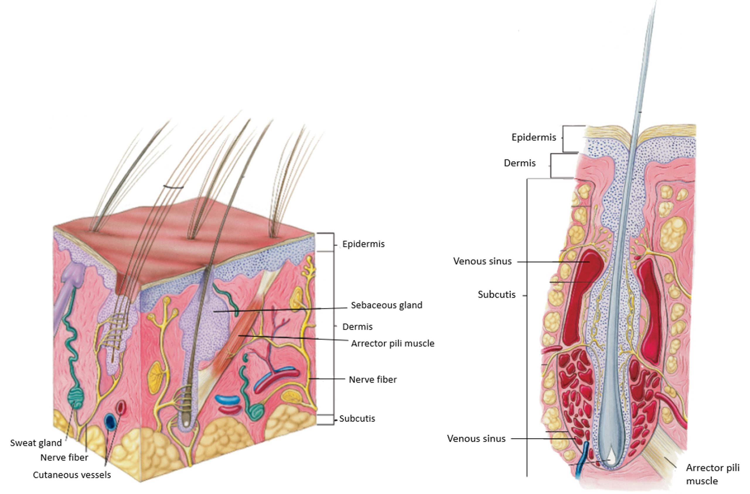
‘Integument’ is commonly used (somewhat inaccurately) when referring to the skin. Cutis is Latin for skin. Dérma is Greek for skin, and derm– is the root combining word for skin-related terms (e.g. dermatitis, pachyderm, taxidermy). The skin includes the epidermis ([L] epi – upon, above) and the dermis. The hypodermis ([L] hypo – below, under, less, and also known as subcutis, subcutaneous tissue, tela subcutanea) is the layer deep to the dermis. In clinical terms, reference to the ‘skin’ implies all three layers, ie epidermis, dermis and hypodermis. The epidermis of skin is an epithelial tissue (one of the four major tissue types in the body) and specifically is classified as stratified squamous epithelium (one of eight subtypes of epithelial tissue). The dermis is a form of connective tissue (another of the major tissue types in the body). The hypodermis (subcutis or subcutaneous layer) is a loose connective tissue between the dermis and the superficial fascia. At the mucocutaneous junction, skin is continuous with the mucous membranes at the various openings of the body’s digestive, respiratory, urinary and genital tracts. The microscopic structure and function of the skin will be considered in physiology and pathology content. Below are a few more details regarding these layers for gross anatomy context.
Epidermis
The epidermis consists of four to five distinct layers: the stratum corneum (the most superficial layer we see), stratum lucidum (the regionally present 5th layer), stratum granulosum, stratum spinosum, and stratum basale. The thickest epidermis of the body of carnivores is found on the pads and nasal plane (discussed later). Stratum corneum is Latin for horn layer. The substance within cornified cells (aka keratinocytes, from [Gk] keras = horn) of the stratum corneum is called ‘horn’. The basal cell layer of the epidermis is referred to as the stratum germinativum or germinal epithelium, because it is the site of epidermal mitotic cell division (i.e. the germination of new cells). The basal cells lie on the basement membrane, a critical structure that separates the epidermis from the dermis. Basal cells actively divide and push their progeny upward to replenish the more superficial layers of the epidermis. Additional cell types found in the basal layer include melanocytes, and macrophages (Langerhans cells).
Clinical relevance:
-
- Schematic illustration of tactile hair. 4

-
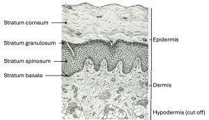
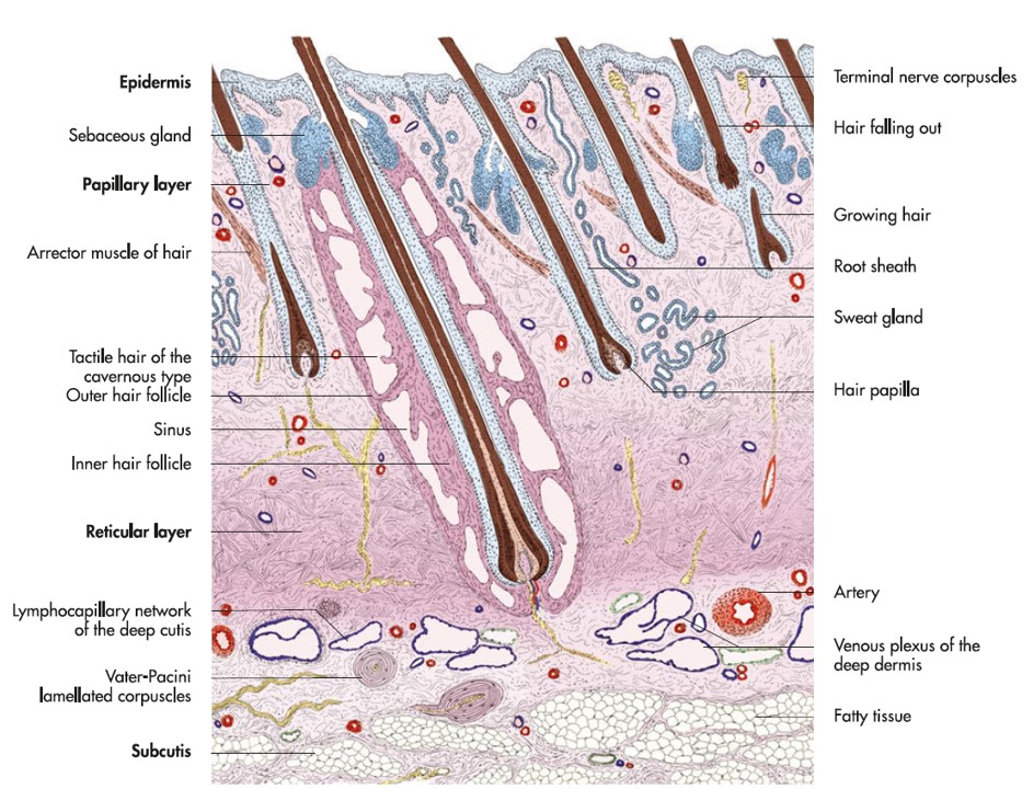
- Layers of skin.

Dermis (or Corium, from [L] corium = skin)
The dermis is a connective tissue composed of collagen, elastic and reticular fibers, and interstitial ground substance. Nervous tissue, blood vessels, lymphatics, glands, hair follicles, arrector pili muscles, and various cellular elements are present throughout. The dermis provides the nutritional needs of the non-vascularized epidermis. The dermis contributes most to the thickness of skin and forms the familiar leather used to make products. In our domestic animals, cattle generally have the thickest dermis (which makes IV catheterisation of the external jugular vein challenging!) and sheep and cats the thinnest. The dermis decreases in thickness from dorsal to ventral on the abdomen and proximal to distal on the limbs.
Hypodermis (subcutis or subcutaneous tissue)
The hypodermis is composed of bands of collagen fibers and abundant, smaller elastic fibers that interweave and enclose fatty adipose tissue. Compared to the dermis, hypodermis is relatively loosely arranged with tissues separated by (microscopically) small spaces. Hence the term areolar tissue ([L] areola = small space) is used in reference to the hypodermis. The dog, cat and sheep have abundant hypodermis. The hypodermis of the horse, ox and goat is thinner and more tightly adhered to the underlying trunk. Where skin movement is minimal in general, the hypodermis is very thin or absent e.g. lips, eyelids, ears. Modified hypodermis forms specialized padding in the feet of animals e.g. the digital cushion of the horse. Closely related on the deep side of the hypodermis is fascia, a stronger connective tissue surrounding the body.
-
- Schematic illustration of tactile hair. 4
-
- Detailed schematic illustration of tactile hair. 4
Clinical relevance:
Subcutaneous fluids are deposited in the loose connective tissue of the hypodermis, particularly in carnivores, where it exists in greater amounts e.g. the interscapular region. Pigs have substantial fat accumulation in the hypodermis (backfat measurements of pigs determine carcass quality and reproductive soundness), and a cresty neck in the horse is due to regional fat accumulation in the hypodermis (a sign of Equine Metabolic Syndrome). Inflammation of the subcutaneous fat is termed pansteatitis or yellow fat disease and causes painful subcutaneous nodules. Inflammation of the subcutaneous tissues in general is called panniculitis, with various causes.
Clinical relevance: the ‘skin pinch’.
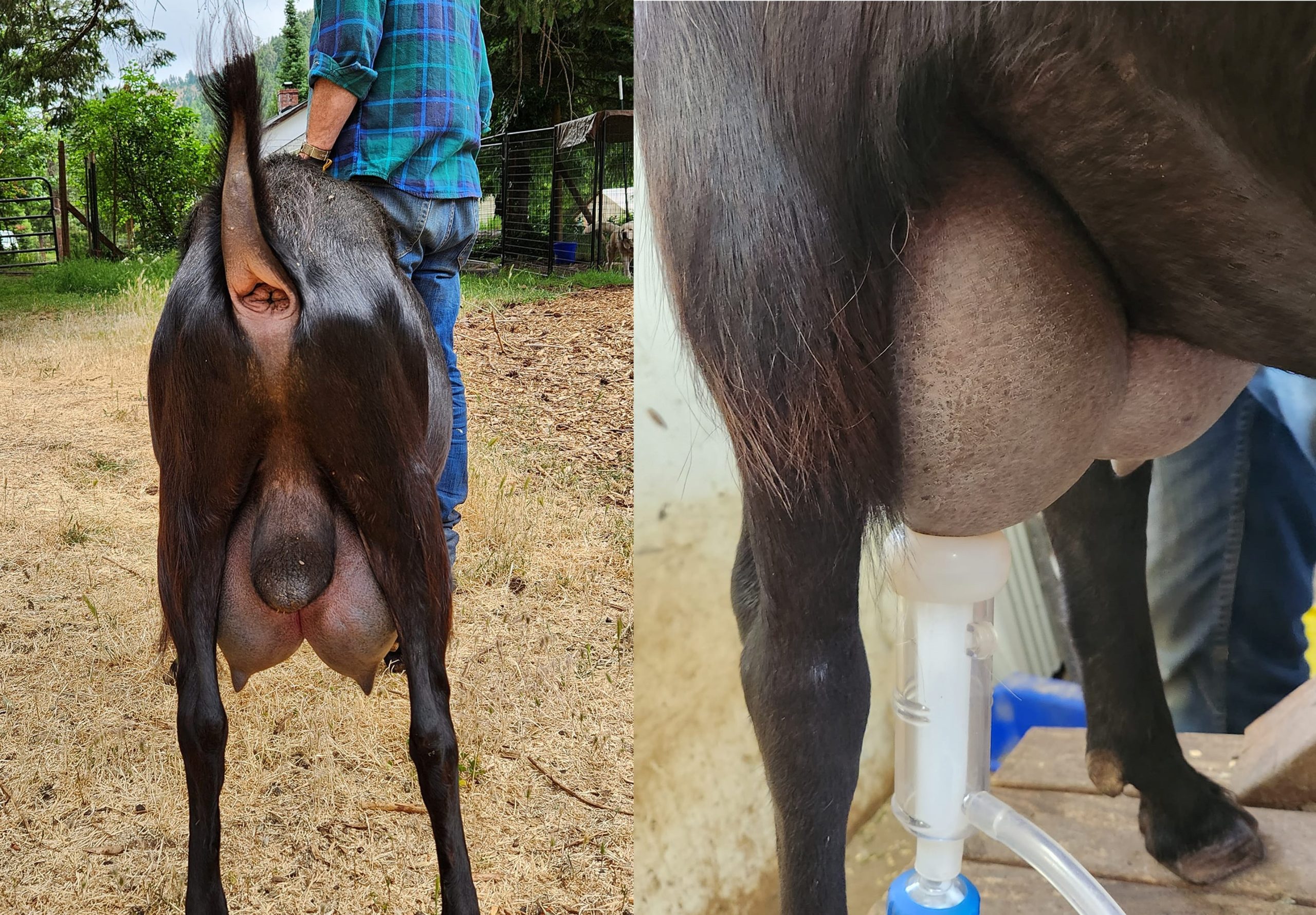
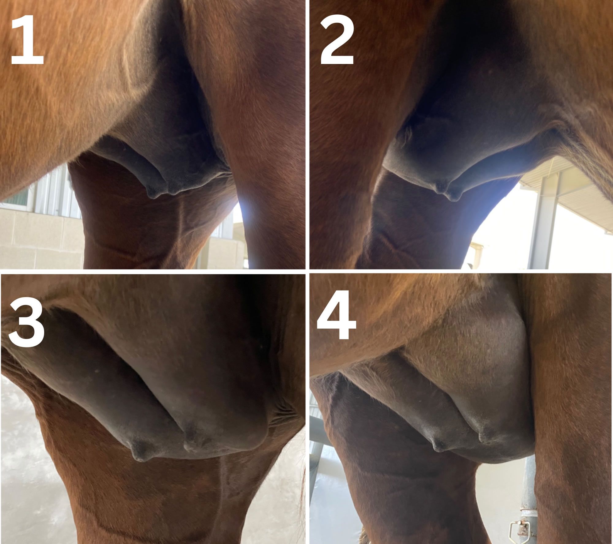
As mentioned, skin thickness varies on different parts of the body, mostly depending on the extent of the dermis. Normal skin is quite pliable due to its elasticity, with variation between species and regions on the body as to how this changes. The ‘skin pinch’ used clinically to assess hydration status is an observation of the change in the skin’s turgor or elasticity. In botany, turgor (from Latin, turgere ‘to swell’), refers to the normal state of turgidity or rigidity of plant cells, due to pressure exerted by fluid within the cells. Plants that lose turgor, due to loss of water from cells, are observed to wilt. With the skin pinch we are observing how the skin returns to its normal position after a change in shape produced by the tenting or pinching of the skin. Reduced skin turgor is seen as a delay in skin returning to its normal contour, and this may be a sign of systemic dehydration.
Compare the skin pinch on a well hydrated horse to the dehydrated horse:
Appendageal structures
Hairs ([L] pili)
Hair is a modified epidermis. Long, thin columns of elastic horn tissue are produced at the root of the hair in the hair follicle. Hair follicles are invaginations of the epidermis, extending well into the dermis and sometimes hypodermis. Sebaceous and apocrine sweat glands discharge into the space of the hair follicle, around the shaft of the hair. These secretions are distributed over the skin and coat the hair as it grows, providing the sheen of a healthy coat. Tiny smooth muscles, arrector pili muscles, acting involuntarily, straighten hairs to increase thermal properties and to give an animal a threatening appearance (raising its hackles). They are present in all haired skin areas, being most highly developed in the dorsal lumbar, sacral and tail regions
Relatively stiff guard hairs (capilli) provide the outer coat of hair in domestic animals (except sheep) and the undercoat consists of fine, wavy wool hairs (pili lanei). The fleece of sheep is of entirely wool hair. Guard hairs show body region, and species, and breed variation. Horses have a mane and forelock, goats have a beard (barba), ungulates have long tail hairs. Modified guard hairs are present at the nostril (vibrissae), at the entrance of the ear canal (tragi), and form eyelashes (cilia).
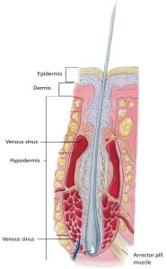
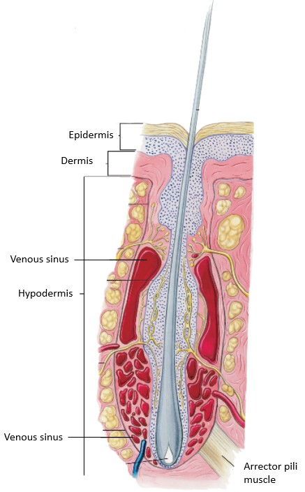
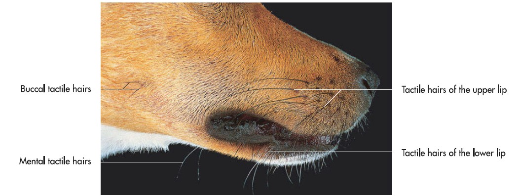
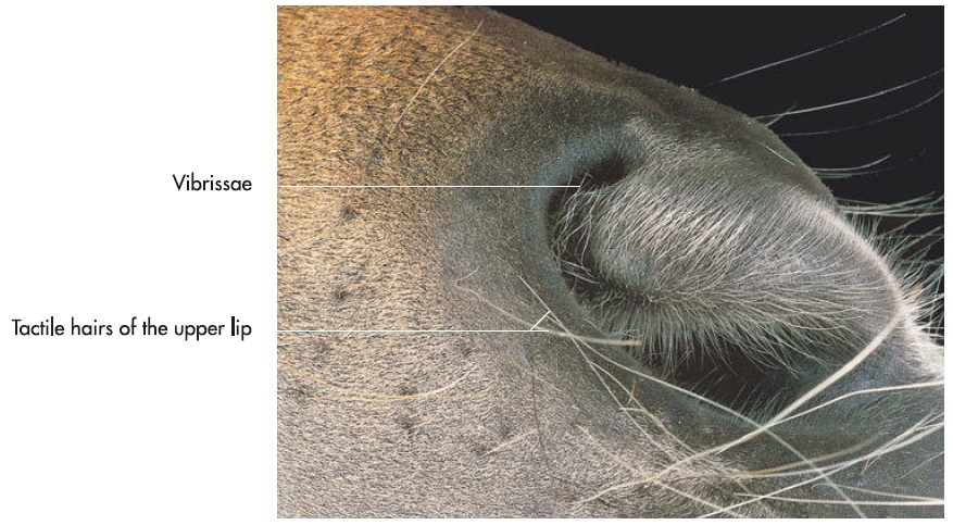
Tactile hairs are a particularly specialized and modified guard hair. They are substantially thicker and longer than normal guard hairs. The tactile hair follicle is surrounded by a venous (blood) sinus. Therefore, yet another name for these specialized hairs is sinus hairs. In the walls of the venous sinus are nerve endings responsive to movement of the tactile hair, which is amplified by wave action of the surrounding blood. Most tactile hairs are found on the face, principally on the upper lip and around the eyes, though others are scattered on the lower lip, chin, and elsewhere on the head. The slightest movement of tactile hairs, even by air currents, stimulates the nerve endings and provides information on the animal’s immediate surroundings. The attached arrector pili muscles move the tactile hairs and the hair can be “put on the alert” when required.
-
- Schematic illustration of tactile hair. 4
-
- Detailed schematic illustration of tactile hair. 4
-

- Skin with sinus hair. 7
Clinical relevance:
Sebaceous Glands
Sebaceous glands are holocrine glands producing sebum and are mostly associated with hair follicles, discharging into the follicular space. Sebum is an oily secretion that (mixed with the secretion of apocrine sweat glands) keeps the skin soft, pliable and waterproof, and gives the hair shafts their glossy sheen. The sebaceous glands of the lips and face are numerous and larger than in other regions. The cat has an increased number of these glands on the chin region, referred to as the circumoral glands (or submental organ). The dorsal aspect of the tail (caudal glands) is another concentrated site. Contraction of the arrector pili muscles helps express the sebaceous glands. The oil glands of the eyelids (Meibomian glands) are specialized sebaceous glands. The cerumin glands of the external ear canal are a combination of sebaceous and modified tubular glands, secreting the characteristic waxy substance (ear wax) called cerumen. Pigs have sparse and rudimentary sebaceous glands.
Clinical relevance:
Sweat Glands (tubular glands)
Apocrine sweat glands are coiled or saccular, and discharge their protein-rich (albuminous) sweat into hair follicle sacs. Their secretion provides the individual odor of an animal. They are distributed throughout all haired skin. Apocrine glands are relatively numerous in the horse, with additional openings, and they are responsible for the frothy, ‘lathering up’ of a working horse, due to the rich protein content of the sweat. Apocrine glands are not present in pads or the nasal plane. They are largest and most numerous near mucocutaneous junctions, in interdigital spaces, and over the dorsal neck, and rump. Large apocrine glands may be found on the dorsal tail.
Clinical relevance:
Hyperfunction of these glands on the dorsal tail along with the associated large sebaceous glands may result in the buildup of waxy, greasy debris (“stud tail”).
Eccrine sweat glands are small, tightly coiled merocrine (secretions are discharged directly on the skin surface) glands which are found in the footpads of carnivores and the frog of the horse foot. The excretory duct opens directly to the pad surface. The secretion is watery. In contrast, these glands predominate throughout the body of primates.
Clinical relevance:
-

- Schematic illustration of tactile hair. 4
-
- Skin with sinus hair. 7
Mammary gland
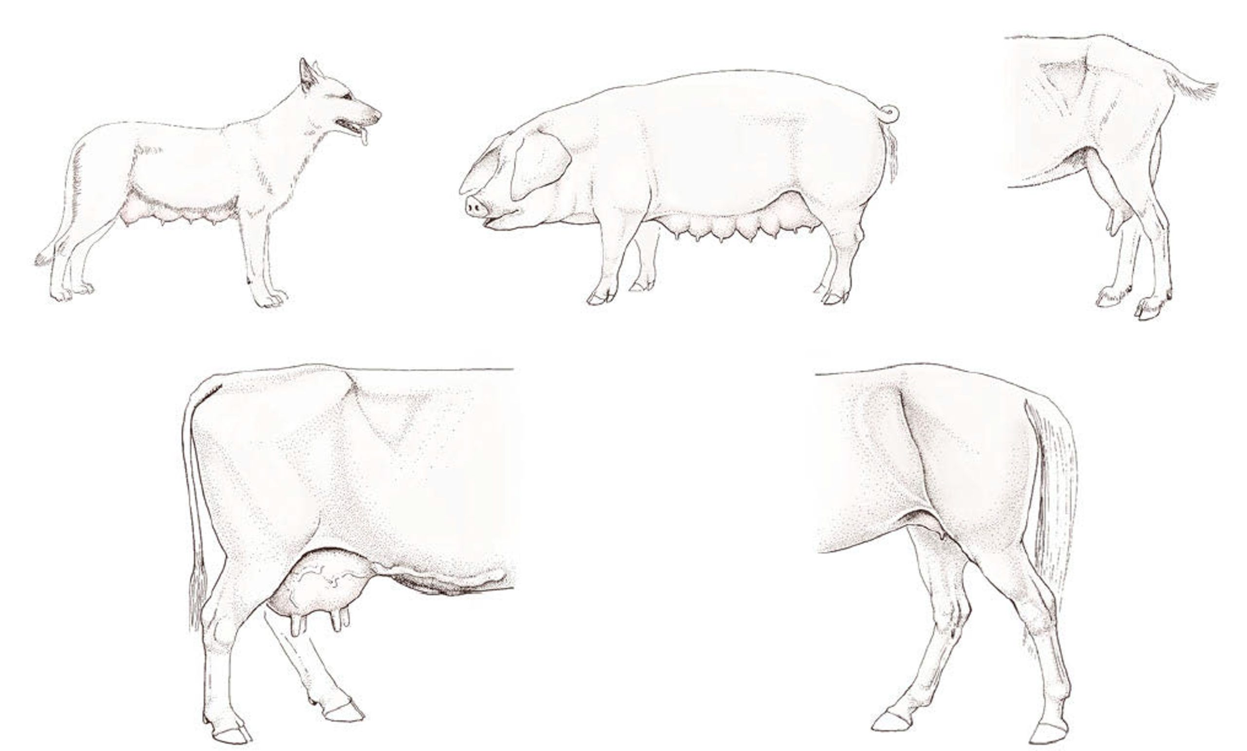
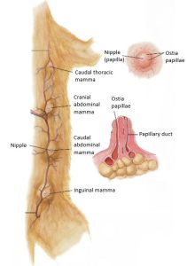
The mammary gland is a series of highly modified sweat glands, occupying the hypodermis, and they are not fused to the body wall (which makes removal of the gland, a mastectomy, less difficult to perform). A mammae is the complex of tissue associated with one teat. This complex consists of skin, glandular tissue and associated connective tissue. Clinically the terms mammary glands and mammae are often used interchangeably. Mammary gland is the collective term for the mammae of mammals with thoracic, abdominal and inguinal mammae, i.e. the sow, bitch, queen. In ruminants and the horse, the mammary gland is restricted to the inguinal region and the collective term in these species is udder. A left and right set of mammary glands is present. Supernumerary teats are additional teats that are typically nonfunctional. Mammary glands receive extensive blood supply, ramped up during lactation. Lymphatic drainage is cranial and caudal to regional lymphocenters.
Male mammals typically have rudimentary or vestigial mammae, with teats visible (but not always! Stallions/geldings don’t normally have teats, but jacks frequently do, bucks and bulls do, and bucks may develop functional glands).
Detailed internal anatomy of the mammary gland will be considered in the reproductive system unit.
Clinical relevance:
Milk production. Mastitis (inflammation of the mammae, usually due to a bacterial infection) and tumors are two clinical conditions of note for the mammary gland.
-

- Mammae of the bitch and sow. Udders of the doe, cow and mare. 7
-
- Mammary gland of nonlactating (top) and lactating (bottom) queen cat with magnification of nipple and sagittal section of nipple. 4

-

- Lactating lamancha buck, Forrest-Pride Blak Falcon. Cathlin Williams
Cutaneous muscles
Very thin muscles are located in the superficial fascia of the head, neck and trunk, immediately deep to the hypodermis. These muscles directly act on the skin, causing it to twitch and tense. Cutaneous muscles of the head contribute to facial expressions. Examples of cutaneous muscles include m. cutaneus faciei, m. platysma, m. cutaneus colli, and m. cutaneus trunci. A few cutaneous muscles will be highlighted as they are encountered during the dissection (in the musculoskeletal unit). During skin reflection it is not uncommon for cutaneous muscles to be elevated with the skin, because they are so closely associated.
Lab dissection instructions
Carnivore common integument and additional external body features
Study the cadaver before skin reflection is commenced and consider the following structures, features and terms. Should you have access to a live companion animal, they are now officially ‘supplementary anatomy resources’ and generally tolerate the curious veterinary student intent on studying anatomy, especially if you combine with a good grooming:)
Observe: Pick up a fold of skin in different locations on the cadaver. Notice that the skin is thickest in the neck region, thinner over the sternum, and thinnest on the ventral surface of the abdomen; also notice that the skin of the dorsum of the neck and thorax is relatively loosely attached.
Starting at the head, observe the locations of tactile hairs (pili tactiles). Those around the mouth are commonly referred to as whiskers.
-
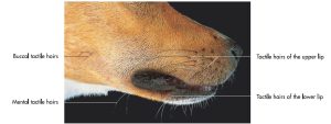
- Tactile hairs on the head of a dog. 7
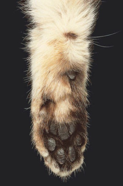
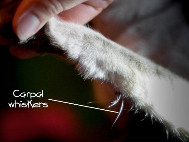
In the cat, tactile hairs are also found on the caudomedial distal antebrachium, close to the carpus.
Observe: Observe the carpal tactile hairs in the feline cadaver.
-
- Schematic illustration of tactile hair. 4
-
- Detailed schematic illustration of tactile hair. 4
-
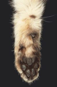
- Tactile hairs at the carpus of a cat. 7
-

- Carpal tactile hairs Logan Forbes
Observe: While focused on the head, examine the nose.
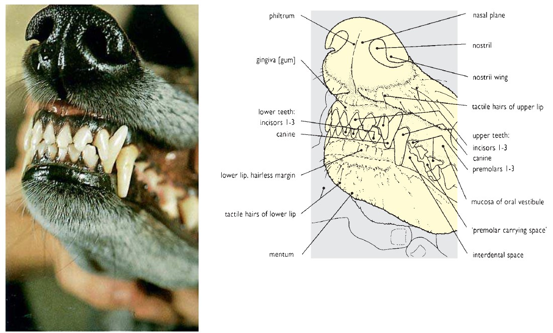
The nasal plane (planum nasale) is the hairless, non-glandular portion of the apex of the nose (a moist nose is due to secretions from nasal glands elsewhere, and tear drainage). A groove (philtrum) divides the nasal plane vertically. The nasal plane skin is composed of thickened and highly cornified epidermis that is elevated into ridges, creating patterns unique to the individual. In dogs, nose prints may be used similar to fingerprints as a form of identification.
-

- Surface features of the dog muzzle. 25
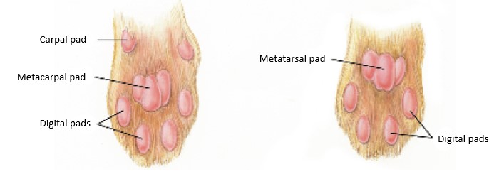
Observe: Now direct your attention to the distal limbs and paws. Identify pads and claws.
Pad ([L] torus)
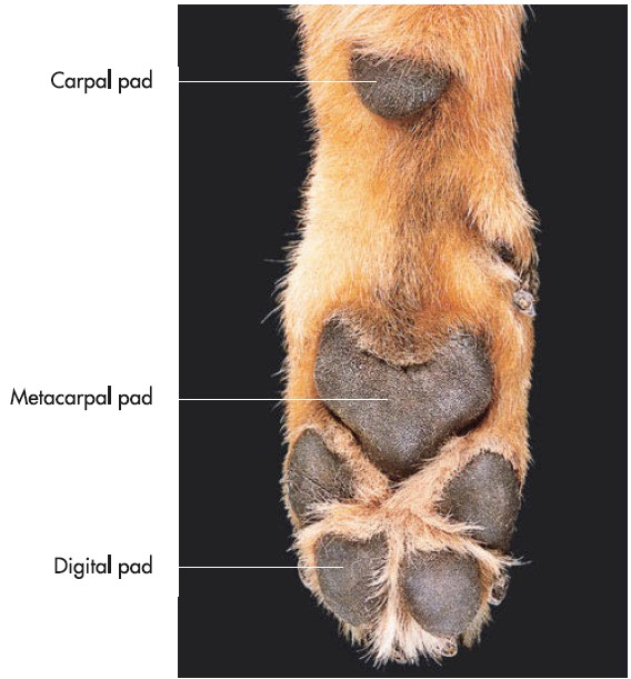
The pads (tori) of carnivores are thickened, highly cornified and pigmented, hairless portions of the skin. The dog and cat weight bear on the digital, metacarpal, and metatarsal pads. Carnivores also have a carpal pad on the palmar carpus (which does not normally contact the ground) but no corresponding tarsal pad. The epidermis of pads is the thickest of the body. The dermis of the pad shows prominent dermal papillae interdigitating with epidermal pegs. Very pronounced subcutaneous cushions of adipose tissue are present in the pads that serve as shock absorbers. Sweat glands (eccrine glands) are present.
-
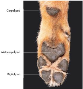
- Footpads of a dog. 7
-

- Digital pad of the dog. 26
-
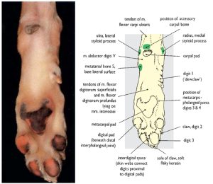
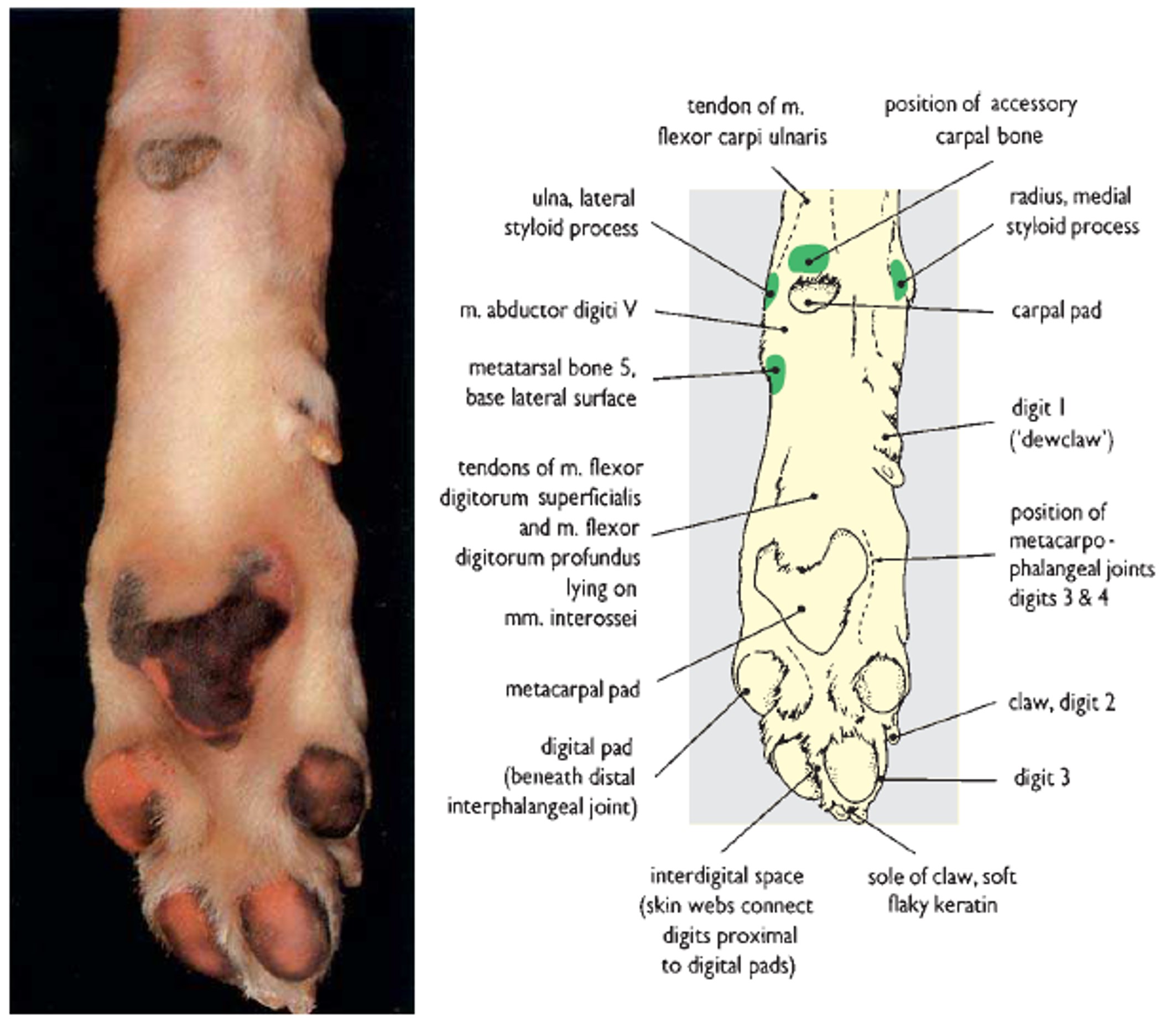
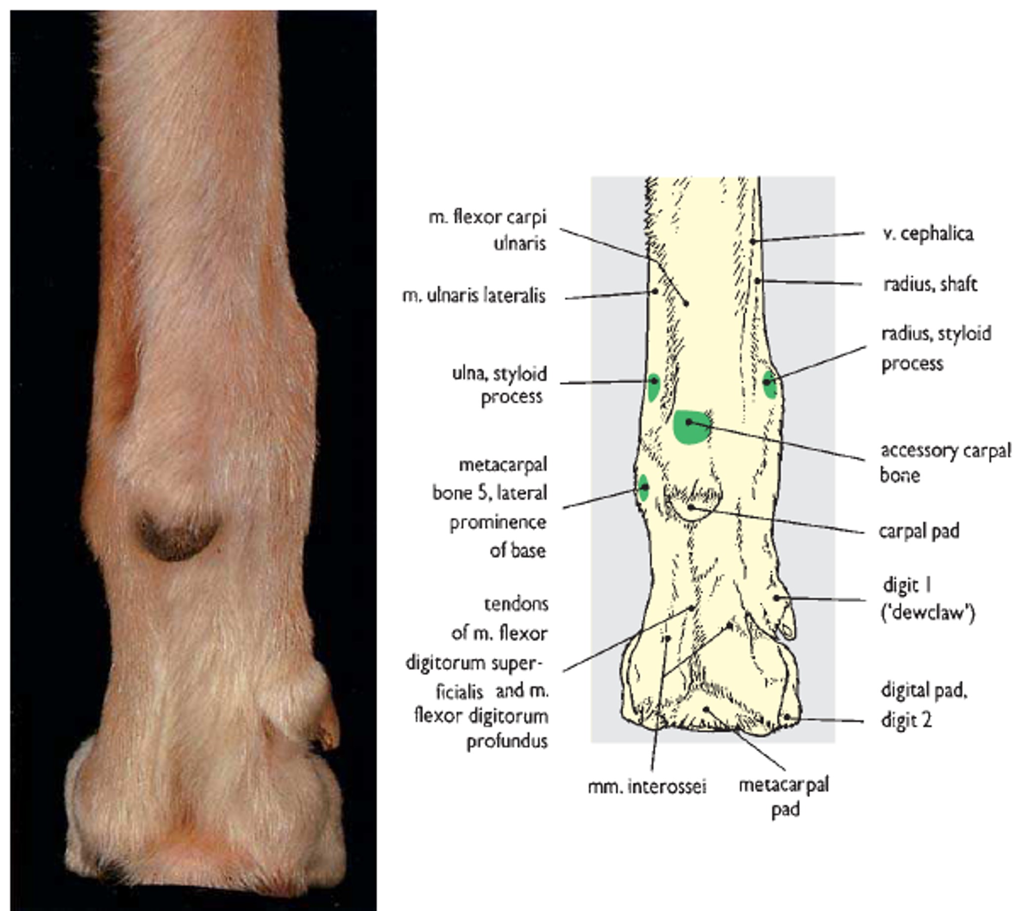
- Surface features of the left carpus and manus of the dog. 26
-
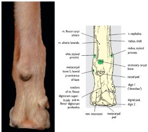
- Surface features of the left carpus and manus of the dog. 26
-
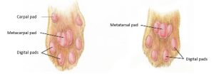
- Tori (pads) of thoracic limb (left) and pelvic limb of the cat. 4
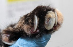
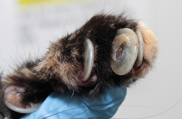
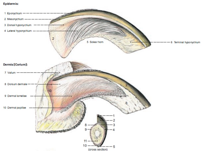
Claw ([L] Unguicula)
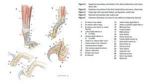
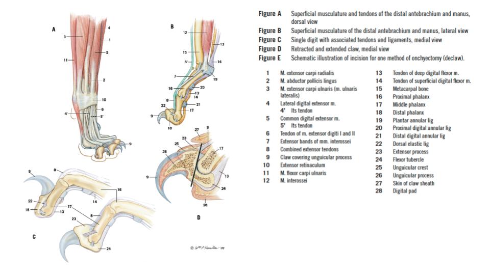
The claw is a specialized, curved, cutaneous structure that is a direct continuation of the dermis and epidermis. The most prominent part is the superficial layers of the epidermis, modified to form the continuously growing, hard, horny claw (or ‘nail’). Grossly, the claw consists of a central dorsal ridge of thicker, horny material, and two thinner walls and a sole. At the base (or root) of the claw, extending from the distal phalanx, the dermis (corium) is a deeper, vascular, innervated, connective tissue that nourishes the epidermis. Trimming a claw too short breaches the dermis, resulting in hemorrhage and discomfort. Previously stated, the stratum basale of the epidermis is the source of the proliferating epidermal cells that constantly produce new growth of the horny claw. A fold of skin covers the base of the claw dorsally, and palmarly or plantarly the digital pad does the same. One obvious difference in the claws of dogs and cats is that cats retract (ensheath) their claws due to the passive pull of the elastic dorsal ligaments of the distal interphalangeal joint, whereas dog claws are consistently extended.
Clinical relevance:
-
- Ingrown/overgrown cat claw Mercedes Lackey
-
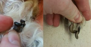
- Overgrown dog nails nhvpethealth.com

-

- Claw of the dog. 26
-
- Cat claws 4
Umbilicus
Observe: Next, examine the ventral abdomen of the cadaver and find the umbilicus. It can be surprisingly difficult to find on a cadaver, and is more easily found on a live animal.
The umbilicis (belly button) is represented by a scar that may be either flat or slightly raised and is located on the midventral line, one third to one fourth of the distance from the xiphoid cartilage of the sternum to the scrotum or vulva. It is often hidden by hair. The umbilicus is irregularly oval and may be from a few millimeters to a centimeter in length. The umbilicus serves as a landmark in abdominal surgery and it is where the umbilical cord extending from the placenta was attached to the developing fetus in utero.
Mammary Gland
Observe: While on the ventral abdomen and thorax identify the external mammary gland anatomy.
In the dog, the mammary gland consists of typically five pairs of mammae (i.e. total of 10, with a range of 8 to 12). They are situated in two rows, usually opposite each other. The number is usually reduced in the smaller breeds. When ten glands are present, the cranial four are the thoracic mammae, the following four are the abdominal mammae, and the caudal two are the inguinal mammae. When the abdominal and inguinal mammae are maximally developed, the glandular tissue in each row appears to form a continuous mass. Each mamma has a teat, that is partly hairless and contains about 12 teat canal openings, but these vary and are difficult to see if the animal is not lactating.
The mammary gland of the cat consists of generally four pairs of mammae (i.e. eight total). In some male cats only a pair of inguinal teats are obvious. Four to seven teat canal openings can be identified on each teat.
Clinical relevance:
Mammary tumors are more common in the dog, and ~45% are malignant. In the cat, although less common to occur, most mammary tumors (~85%) are malignant adenocarcinomas. These tumors show profoundly aggressive behavior and are associated with a poor prognosis. Spaying animals before their first heat cycle dramatically reduces the risk of developing mammary tumors.
-
- Mammary gland of nonlactating (top) and lactating (bottom) queen cat with magnification of nipple and sagittal section of nipple. 4
Observe: Now view the perineum of the cadaver.
Anal Sacs ([L] sinus paranalis)
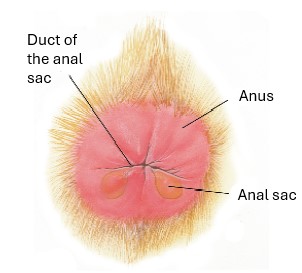
The anal sacs (sinus paranalis) of the carnivore are ovoid cutaneous pouches located in a lateral position between the internal (smooth muscle) and external (skeletal muscle) anal sphincters. Anal sacs are lined by a smooth, keratinized, stratified squamous epithelium that is perforated by the efferent ducts of sebaceous and apocrine tubular glands (the paranal sinus glands). The efferent ducts of the anal sacs are narrow and terminate at the transition between the anus and skin. The anal sacs expel their serofatty, unpleasant smelling secretion during defecation or by voluntary contraction of the external anal sphincter m. The scent is utilized for marking.
Clinical relevance:
Observe: Examine the anus and look for the openings of the anal sacs at the 4 and 8 o’clock positions. A blunt probe can assist with locating these often discrete openings.
-
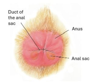
- Location of anal sacs in relation to anus of the cat, caudal view. 4
-
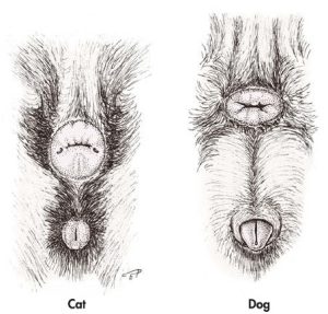
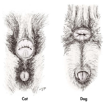
- Anus and external female genital organs. 7
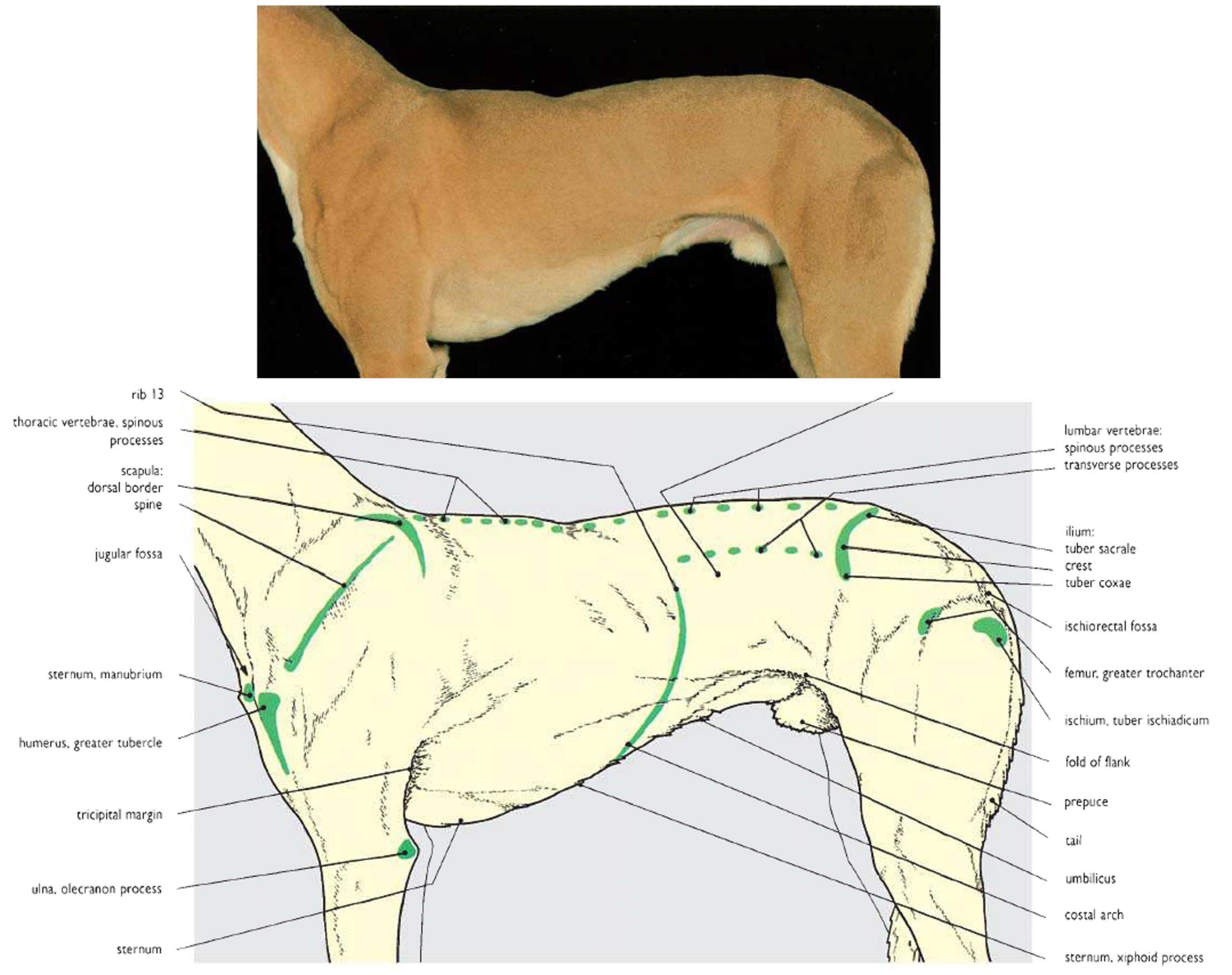
Costal arch
A rib consists of a bony head, neck, and shaft with its distal (ventral) end changing to a cartilaginous structure, called the costal cartilage. The costal cartilages of the tenth, eleventh, and twelfth ribs in the carnivore unite with each other to form the costal arch.
Observe: Palpate the curved, caudal border of the ‘rib cage’ – this is the costal arch. Try to palpate the caudal border and free end of the last or thirteenth rib. This rib does not attach to other ribs and is therefore called a “floating rib”.
-

- Costal arch of the dog. 26
Before skin incisions are made through the epidermis and dermis, let’s reconsider what to anticipate in the subcutaneous (hypodermis) layer.
Subcutaneous tissue and fascias
When skin is incised and reflected we are typically cutting through epidermis and dermis and as we peel back the skin we view the subcutaneous tissue. As previously described the subcutaneous tissue is composed of areolar tissue, a layer of loose, irregularly arranged connective tissue that often contains fat. The areolar tissue is often distended with embalming fluid (and subcutaneous injections are made into this tissue – e.g. subcutaneous fluids provided to a cat with chronic renal disease). Dissection through the areolar tissue plane is quick and easy based on the nature of the tissue being incised. Fascia, on the other hand, is a denser, regularly arranged thin layer of connective tissue. It is more fibrous, and it envelops the body beneath the skin and encloses individual muscles or groups of muscles. Superficial fascia is directly under and associated with the areolar tissue and covers the entire body. Cutaneous muscles are located in the superficial fascia. Some superficial fascia (and cutaneous muscles) may be reflected with the areolar tissue. Superficial fascia blends with the deep fascia, which is more firmly attached to the muscles that it encloses. The superficial and deep fascias are not always easily distinguished from each other. As a point of reference, the external jugular vein is surrounded by superficial fascia. Fascia may receive a more specific name based on its location in the body e.g. the thoracolumbar fascia. When the fatty areolar tissue and fascia with vessels and nerves are removed from a muscle, the muscle is said to be “cleaned.”
Skinning instructions
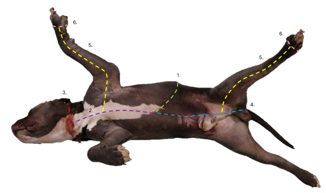
Dissect: Alright, time to incise and reflect skin! The objective is to remove the skin from the left side of the cadaver (except for the head). Examine the images and follow the instructions below. Ensure the cadaver is lying in right lateral recumbency (i.e. the left side of the animal is facing up towards you). We wish to reflect the skin from the left side of the body. Once reflected the skin will be removed and discarded.
Review of directional terminology.
- Palpate the left costal arch, the dorsal midline and the ventral midline. Make a skin incision from dorsal midline to ventral midline that follows the line of the costal arch.
- From ventral midline extend the skin incision cranially onto the ventral neck, to a point level with just behind the palpable jaw line.
- Then extend a transverse incision from the ventral cranial neck around to the dorsal midline, passing just caudal to the ear.
- Back to the ventral midline starting point, extend the skin incision caudally, skirting around external genitalia, and around the left margin of the anus, to end at the butt of the tail on its dorsal surface.
- Roll the cadaver into dorsal recumbency. Abduct the left limbs. From a point on the ventral incision directly opposite the left thoracic limb and left pelvic limb, extend an incision onto the medial surface of the limb and incise distally to the paw.
- Make a complete circular incision through the skin at the paws, just above the level of the digits (additional skin can be removed later as needed).
- Lastly, extend an incision from cranial to caudal (back of head to butt of tail), along dorsal midline, to complete the incision of the skin borders.
- Now, with incisions made, (and depth increased as necessary), carefully reflect the skin off the left side of the body. More incisions can be made to create smaller sections of skin for removal, as preferred.
- The skin will be intimately ‘fused’ with the thin underlying cutaneous muscles over the neck, thorax, and abdomen. These muscles may be left on the specimen as is practical, otherwise they may be reflected with the skin.
-

- Skin incisions
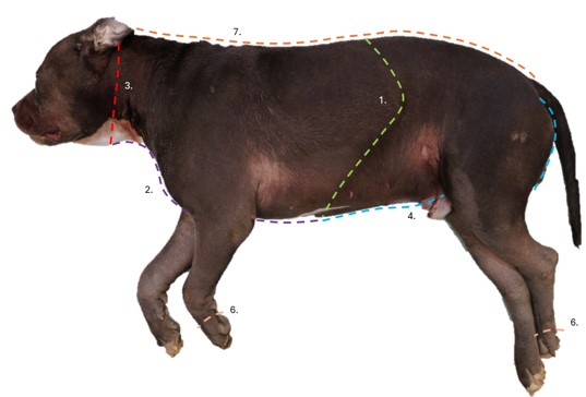
-
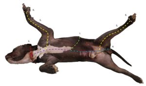
- Skin incisions

Ungulate common integument: comparative and unique features
Observe: Examine available ungulate (horse, goat, ox calf, pig) cadavers to consider the following integument features. The skin should be present and intact on the right side of the cadaver for evaluation. The left side of the cadaver has had the skin reflected (but it may still be attached at certain points)
Equine
Observe: Examine the head region. Note tactile hairs, nasal hairs, mane and forelock (likely clipped in the cadavers).
Tactile hairs are present on the upper and lower lips and around the eyes. The hairless nasal plane feature of other animals is not present in the horse.
-

- Guard hairs (vibrissae) at the vestibule of the nares of a horse. 7
Observe: Examine the side of the throat region. Palpate the borders and identify Viborg’s triangle.
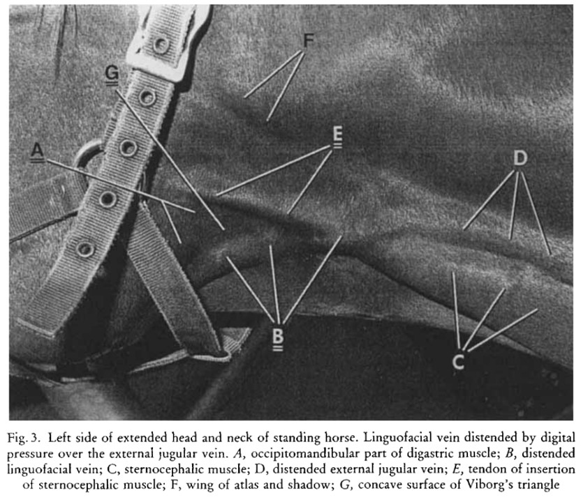
Viborg’s triangle (trigonum viborgi) – this external feature sits over the pharyngeal region and was described by the Danish veterinarian Viborg, in 1795, as a surgical approach to the guttural pouch of the horse (a unique air-filled cavity related to respiratory tract). Although the surgical approach is less commonly used today, the region and term remain clinically significant. The triangle is demarcated by the caudal margin of the ramus of the mandible, the tendon of insertion of the sternocephalicus muscle and the linguofacial vein. This anatomy will be considered again in later units.
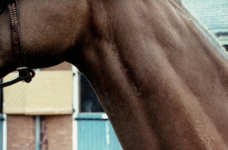
Jugular groove – This is a linear depression extending down the lateral neck, defined by sternocephalicus and cleidocephalicus muscles. The external jugular vein (an important venous access vessel in ruminants and the horse) passes in this visible and palpable groove. This anatomy will be considered again in later units.
Observe: Examine the neck region, observe and palpate the jugular groove.
-
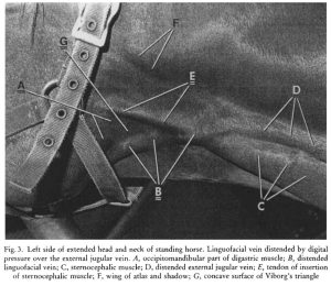
- Viborg’s triangle. 27
-

- Horse jugular groove Royal Veterinary College
Chestnut and ergot
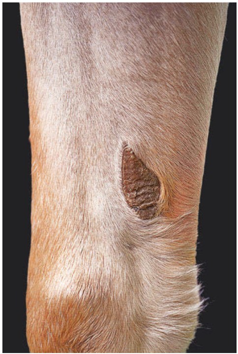
Observe: Examine the thoracic and pelvic limbs on their medial aspect close to the carpus and tarsus. Identify the raised, hairless, somewhat irregular, epidermal structures. These are the chestnuts, analogous to the carpal pad in the carnivore, and the equivalent of a tarsal pad, that, recall, carnivores do not have.
Chestnuts are unique in structure and can be used as a form of identification, as has been done for certain breed registries. Donkeys and zebras normally don’t have pelvic limb chestnuts and a few horse breeds don’t either e.g. the Icelandic horse. Chestnuts on horses should not be confused with the Horse Chestnut tree……..
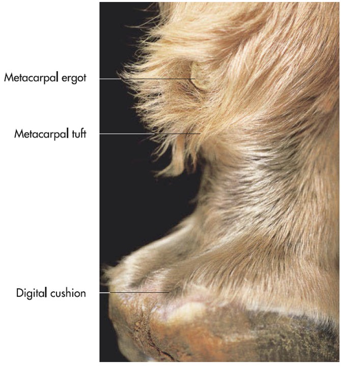
Observe: Palpate the palmar/plantar aspect of the metacarpo- and metatarsophalangeal joint (fetlock or ankle in the horse) to find a raised, hairless, epidermal structure. This is the ergot, analogous to the metacarpal and metatarsal pad of carnivores.
-

- Chestnut of the horse proximomedial to the carpus of a horse as a rudimentary carpal pad. 7
-

- Horse ergots as rudimentary metacarpal pads and heel bulbs. 7
Observe: View the udder.
Mammary gland
Mares have an udder, the collective term for the mammae of mammals with inguinal mammae, i.e. cow, ewe, doe, mare. The mare has two inguinal mammae (like the doe and ewe). A mammae is the complex of tissue associated with one teat. The skin of the teat of the mare contains sweat and sebaceous glands. Sebaceous secretions occur just prior to parturition that give the teat a characteristic appearance; this is called ‘waxing’. A gland complex is the single gland and associated duct system within a mammae. On each mammae, the number of gland complexes coincides with the number of teat orifices. Mares have two gland complexes per mammae, therefore two teat orifices per teat are visible. Internal mammary gland anatomy will be considered in the reproductive system unit.
-

- Weekly progression of the mammary gland as foaling approaches. Mad Barn
Observe: View the hooves.
Hoof ([L] ungula) – horses bear weight through a single digit per limb, i.e. digit 3. The critical structure of the horse hoof will be studied in detail in the musculoskeletal unit. For now, recognize it is a complex, specialized modification of the common integument.
-
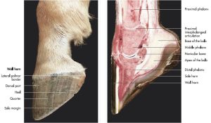
- Lateral aspect and sagittal section of the foot of a horse. 7

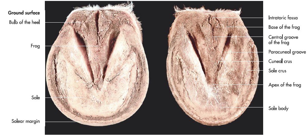
-
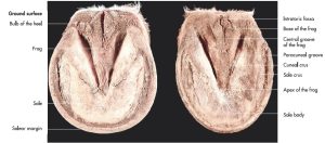
- Round ground surface of a front foot (left) and oval gound surface of a hind foot (right) of the horse. 7
Ruminant
Observe: Examine the head region
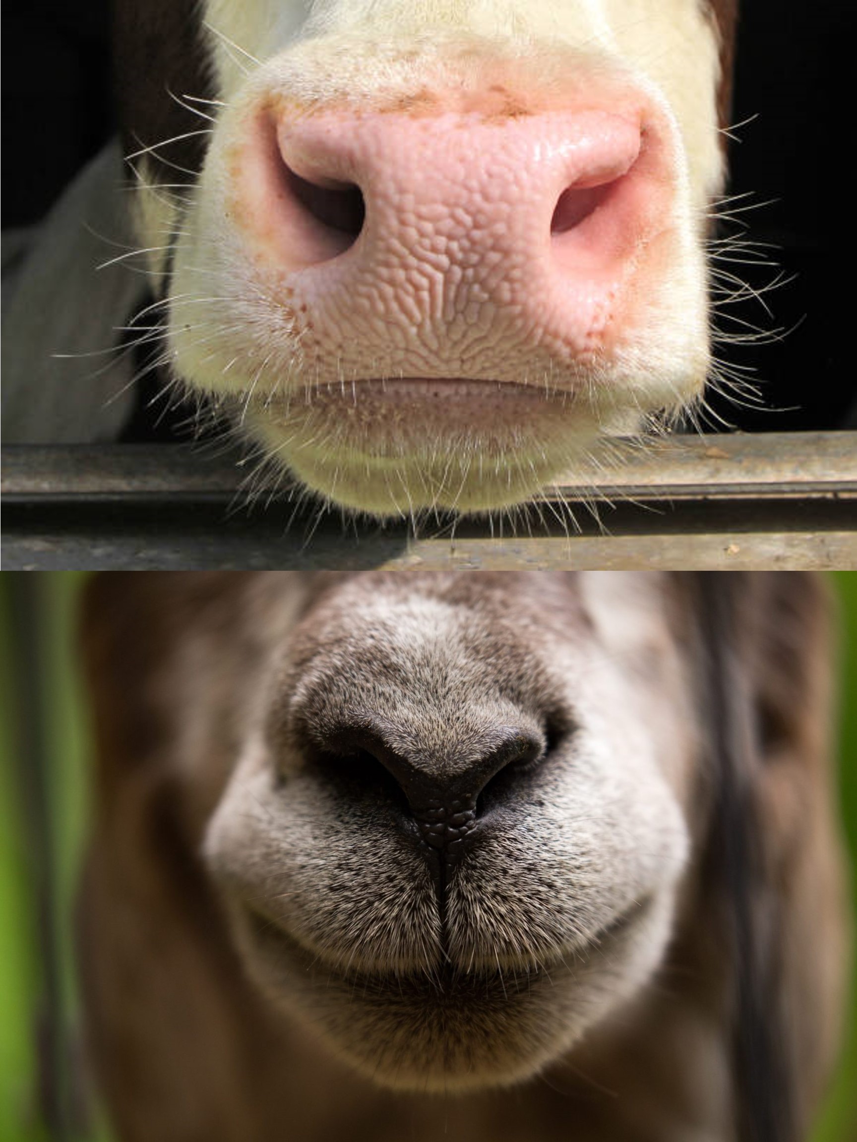
The ox has a hairless nasolabial plane (planum nasolabialis) incorporating the nose and upper lip. The small ruminant has a nasal plane, like the carnivore.
The skin covers the body forming characteristic folds and grooves that are prominent in various species and breeds.
The dewlap is the pendulous midline fold of skin in the distal neck of the ox, very prominent in certain male breeds of cattle. Merino sheep have large skin folds on their neck. Appendages of skin, containing cartilage, hang from the ventral cranial neck, commonly in goats, and are called wattles.
-

- Cow nasolabial plane vs goat nasal plane iStock
-
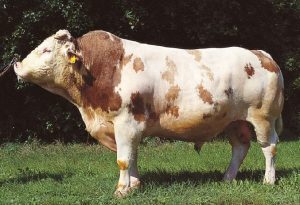
- Pedigree bull with characteristic median skin fold at the caudal end of the neck (dewlap). 7

-
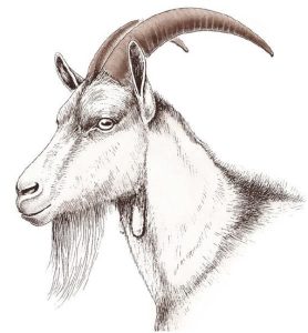
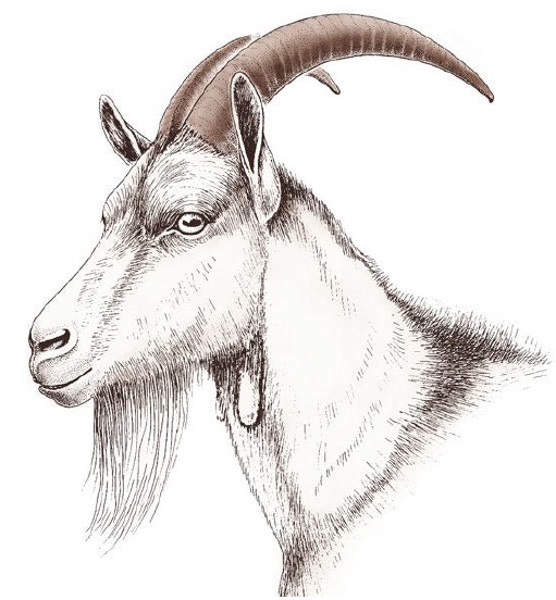
- Head of a buck goat with species-characteristic modifications of the common integument: wattles, long beard hairs and horns. 7
Observe: Examine the skin between the lateral abdomen and the thigh.
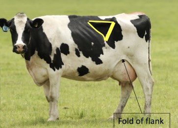
The flank fold connects the ventrolateral abdomen caudally to the medial thigh region. It is well developed in ungulates due to the cutaneus trunci m. contained within, and can be grasped to help position a small ruminant in lateral recumbency (‘flank a calf down’).
-
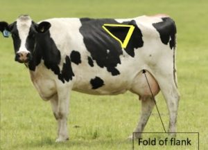
- Fold of the flank. M. Gerard
Observe: Jugular groove – observe/palpate this feature in the ruminant.
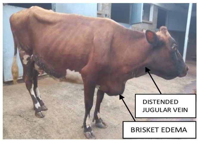
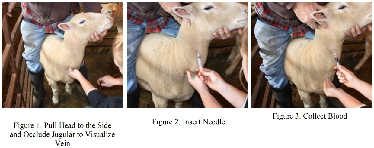
Jugular groove – This is a linear depression extending down the lateral neck, defined by sternocephalicus and cleidocephalicus muscles. The external jugular vein (an important venous access vessel in ruminants and the horse) passes in this visible and palpable groove. In cattle, the dermis of the neck is particularly thick, making intravenous catheterization in the adult ox difficult. A ‘cut down’ procedure may be pursued whereby the skin is incised through most of its dermal thickness to facilitate catheter insertion. Sheep lack a part of the sternocephalicus m. and therefore have a less defined groove, which is also difficult to see when the wool coat is long!
-
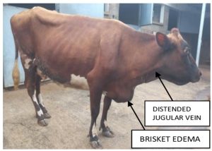
- Complications of traumatic reticulopericarditis, showing the location of the jugular vein.E. Serem
-

- Blood draw in sheep. Virginia Tech
-
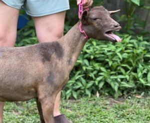
- Goats have a well defined jugular groove, especially when vocalizing. L. Cobb

Skin glands of note
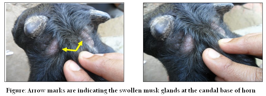
Goats have cornual (horn) glands, one at the caudomedial base of each horn. These are sebaceous glands, and are typically difficult to see grossly.
Sheep have numerous specialized skin pouches or sinuses, similar to the anal sacs of dogs and cats. These sinuses are lined with sebaceous and sweat glands. The resulting secretion is considered a scent marker. Location and number in the sheep include: Infraorbital (aka lacrimal) sinus (2), interdigital sinus (4), inguinal sinus (2).
-
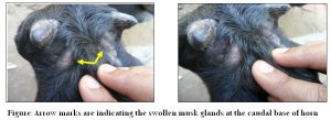
- Musk Gland Swelling in an Anemic Black Bengal Goat; Case Study. M. Ahaduzzaman
-
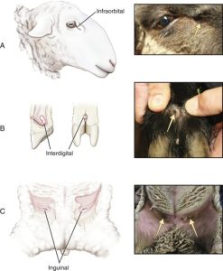
- Sinuses of sheep. A. Crane

Horns
Observe: Observe horns, as present, in the goat.
Find the epidermal bud, or location of by identifying the hair whorl, in the ox calf.
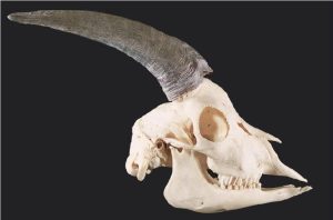
The horns (cornu) of domestic ruminants consist of a bony process (cornual process, extending from the frontal bone) encapsulated in a heavily cornified epidermal tissue, i.e. horn tissue. Horns are hairless and glandless. Particularly in wild ruminants, horns are used as weapons during breeding season and to establish and maintain hierarchies. Horns are present in both sexes in domestic ruminants, typically larger in males. Polled breeds, by definition, do not have horns.
Horns are permanent, continuously growing structures, following appearance post-natally, with shape and size strongly characteristic of the species, breed, age, and sex. As an aside, antlers are not horns. The mature antler is a construct of bone, having developed from a cartilaginous model covered in finely haired tissue, known as velvet. The antlers of the deer family are grown (rapidly!) and shed on an annual basis, under hormonal influence.
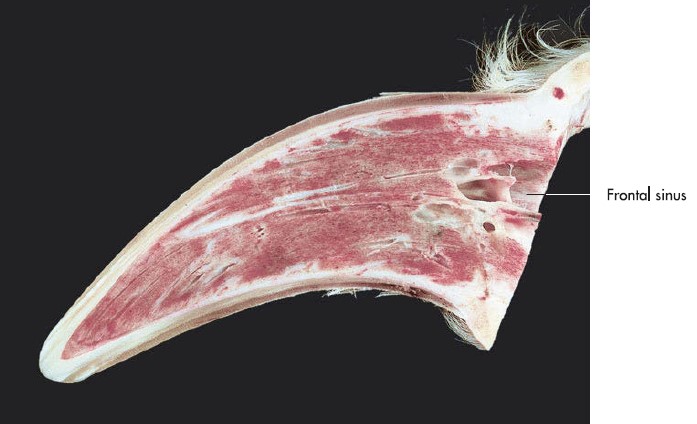
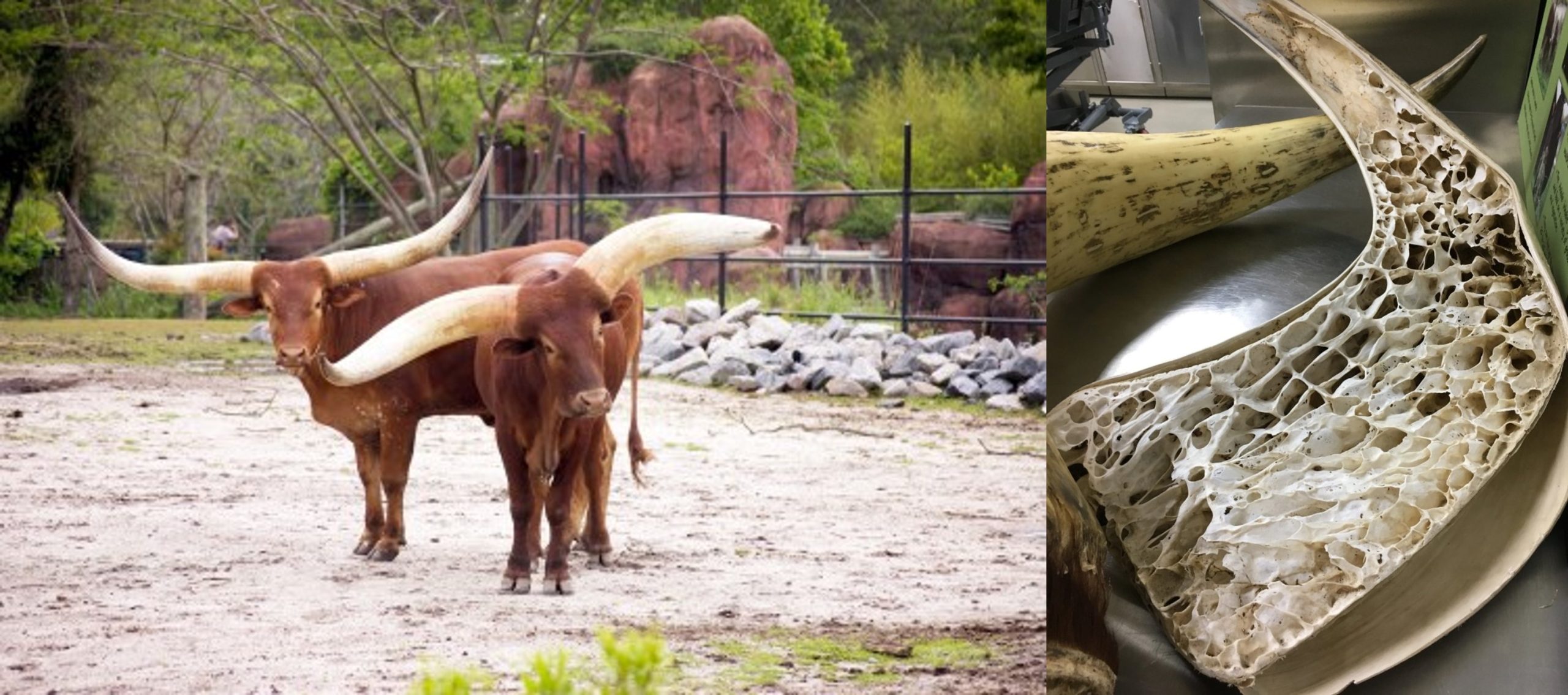
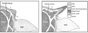
Horns have a base, body and apex. The cornual process becomes pneumatized over time, until the base and body are primarily hollow, while the apex remains solid. This air-filled space is called the cornual diverticulum of the frontal sinus. Dehorning practices or fracturing of the horn can lead to open communication of the frontal sinus and an infection (sinusitis) as a consequence.
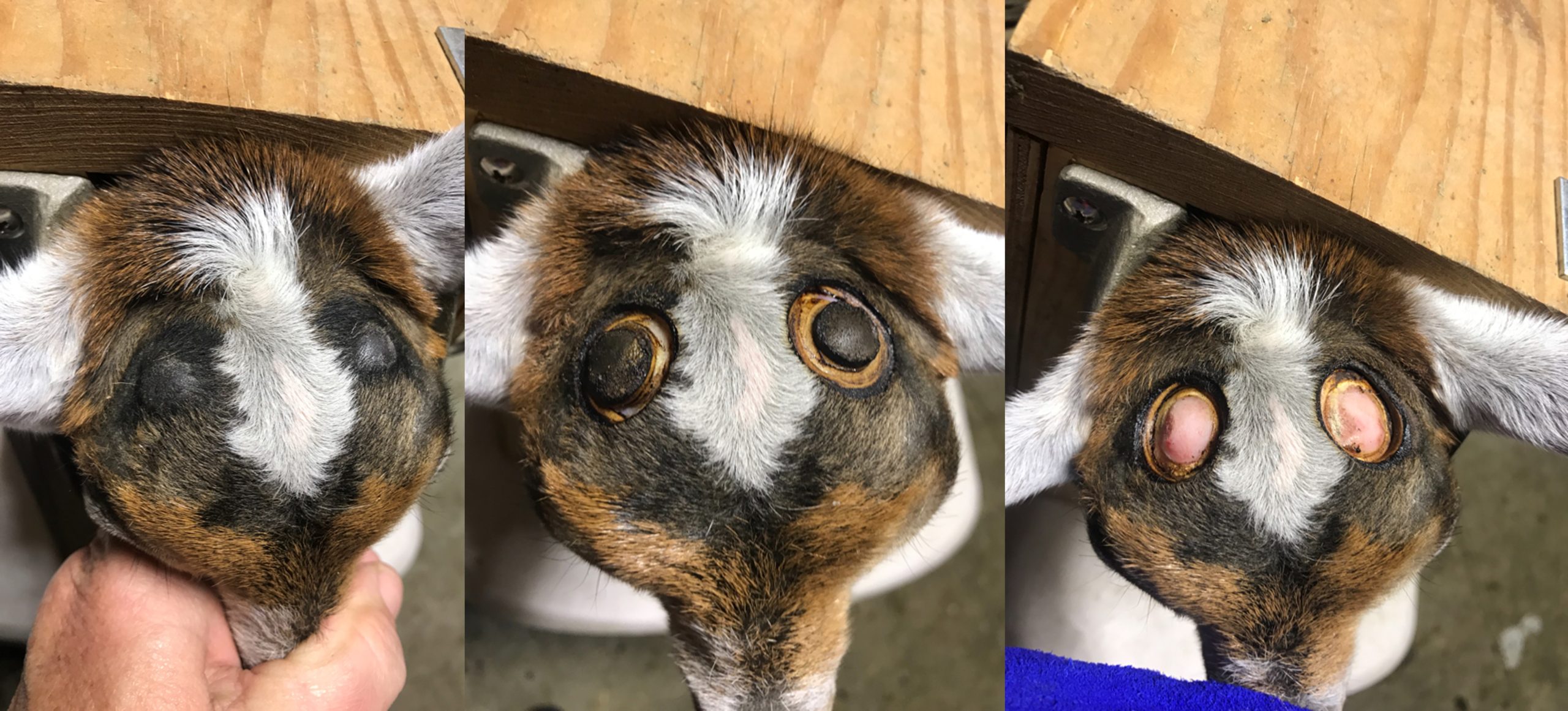
There is no hypodermis present in the horn. Horn dermis directly adheres to the underlying bone and projects papillae into the overlying epidermis. In the neonate, an epidermal bud (hillock) can be identified as the horn development site, usually indicated by a hair whorl at birth. Effective debudding at this early age destroys the germinal epidermis, preventing horn and cornual process growth.
Horn growth is primarily from the base, with newer horn pushing older horn layers towards the tip, elongating the structure. The epidermis, layered over the scaffold of dermal papillae, forms tubular horn (akin to tubular horn in equine hooves). Horn growth rate is tied directly to nutritional status of the animal, and energy demands elsewhere (e.g. lactation, pregnancy).
Horn dermis receives a significant blood supply (cornual a.) to nourish the highly active germinal epidermis and is innervated by the cornual branch (sensory nerve that ties back to the Trigeminal n. [CNV]. The nerve can be blocked before dehorning.
Small ruminant horns:
- Have the same basic anatomy as the ox
- Arise much closer to the orbit
- Sheep horns grow a helical form, goats are more straight and directed caudally
- Growth is more intermittent resulting in a corrugated surface
- Blood supply and innervation are more direct based on the horn location.
- The goat horn has additional innervation that should be blocked before dehorning.
- Goats have a sebaceous cornual gland at the caudomedial base of each horn
-

- Longitudinal section of the horn of a 1.5-year-old bull with beginning pneumatisation of the cornual process. 7
-

- Skull of a sheep with helical horns. 7

-
-
Skull of a goat with caudally growing
horns. 7

-

- Watusi horn, pneumatized inside. Virginia Zoo
-
- Calf horn bud before 8 weeks old (left) and around 6 months old (right) DairyNZ

-

- Cautery iron goat disbudding at 1 week old. Left – horn buds. Middle – skin around bud cauterized. Right – horn bud removed until skull visualized. Prancing Pony Farm
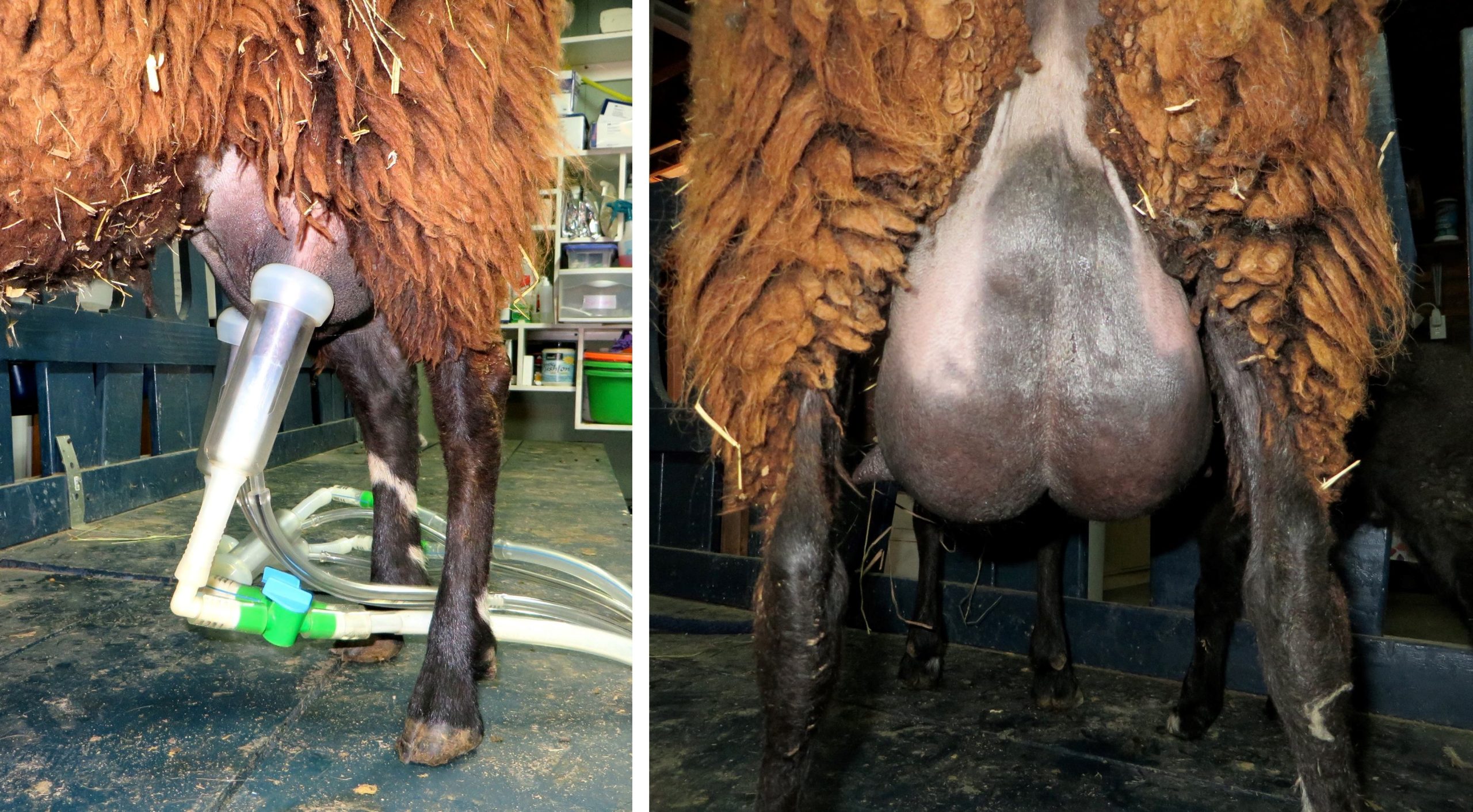
Observe: View the udder.
Mammary gland
Ruminants have an udder, the collective term for the mammae of mammals with inguinal mammae, i.e. cow, ewe, doe, mare. The cow has four inguinal mammae, more commonly referred to as quarters. The doe and ewe have two inguinal mammae (like the mare). A mammae is the complex of tissue associated with one teat. A gland complex is the single gland and associated duct system within a mammae. On each mammae, the number of gland complexes coincides with the number of teat orifices. Ruminants have one gland complex per mammae, therefore one teat orifice per teat is visible. Previously mentioned, the ewe has cutaneous inguinal sinuses on the dorsolateral aspect of her udder that contain secretions thought to scent mark her lambs. Internal mammary gland anatomy will be considered in the reproductive system unit.
-
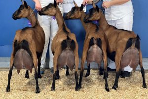
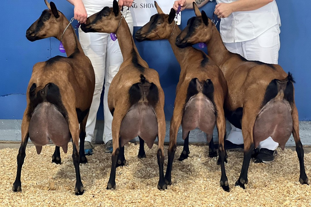
- Oberhasli dairy goats L. Cobb
-

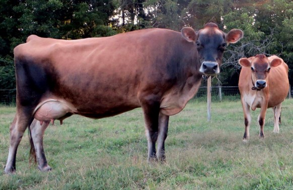
- Jersey dairy cow L. Cobb
-
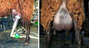
- East Friesian dairy sheep L. Cobb
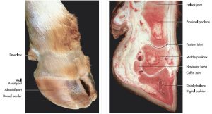
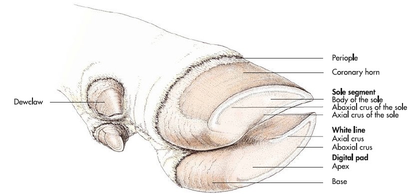
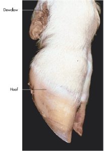
Observe: View the hooves.
Hoof ([L] ungula) – the ruminant bears weight on digits 3 and 4, per limb, with non-weight bearing, vestigial digits 2 and 5 persisting as dewclaws. The critical structure of the ruminant hoof will be studied in detail in the musculoskeletal unit. For now, recognize it is a complex, specialized modification of the common integument.
-
-
Hoof of the cow. 7

-

- Ground surface of the principal hooves and dewclaws of the ox, showing the white line. 7
-
- Thoracic foot of a sheep. 7
Swine
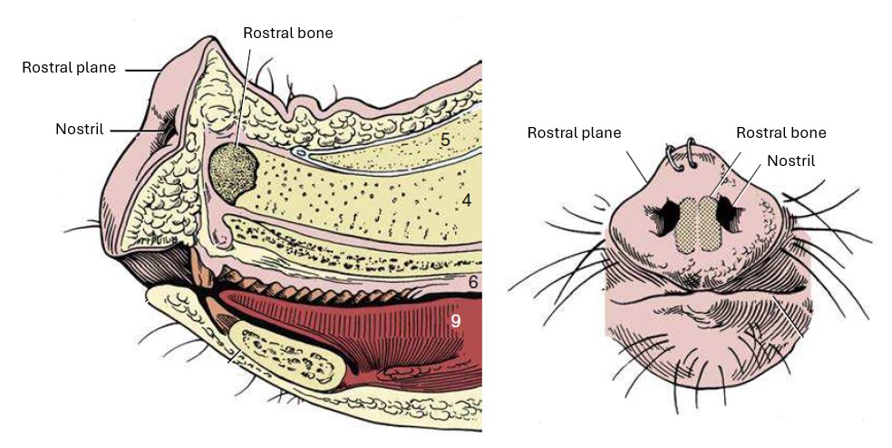
Observe: Examine the head region.
The pig has a rostral plane (planum rostralis) that is the thickened disk-shaped rostral margin of their snout, supporting rooting behavior. The rostral plane has a high density of sweat glands, responsible for the moist snout.
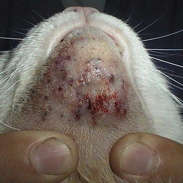
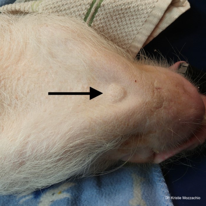
On the ventral mandible, rostrally, the mental gland of the pig is identified as a slightly raised circular mound, with a few elongated mental hairs. Sebaceous and sweat glands secrete a scent marker.
-

- The pig snout from the front and in median section. 8
-
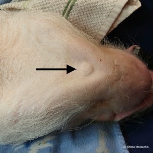
- Pig mental gland K. Mozzachio
Hair coat
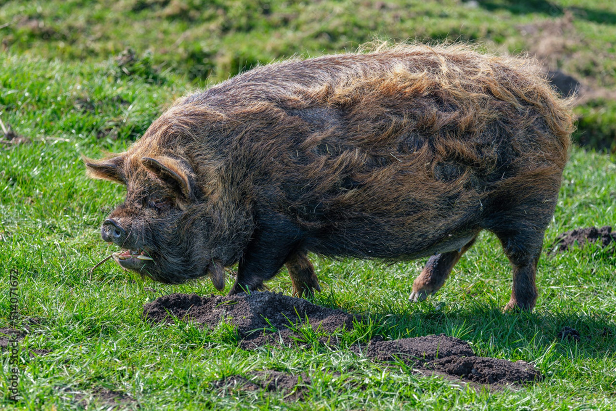
The course, stiff, sparsely scattered guard hairs of the pig are a species-specific modification. These hairs are referred to as bristles ([L] setae).
Observe: Examine the neck region.
The pig lacks a distinct jugular groove along its neck. Instead, a jugular fossa is observed distally in the ventral neck as a depression adjacent the point of the sternum.
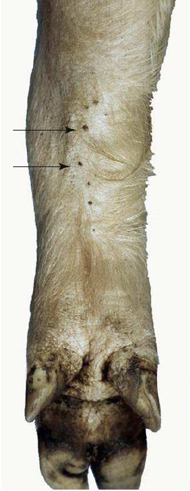
Observe: Examine the thoracic limbs at the carpal level.
Carpal glands (the ‘carpal organ’) are located in a linear array on the palmaromedial carpus/distal antebrachium. These glands secrete a seromucous substance, likely a sexual marker. They may appear as a series of dark dots on the skin.
-

- Kune Kune pig hair coat Adobe Stock
-
- Pig blood collection from jugular vein. Virginia Tech

-
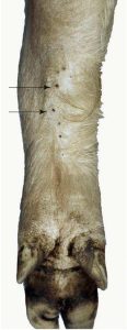
- Carpal glands (arrows) of a pig, palmar view. 8
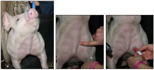
Observe: View the mammary gland.
Mammary gland
Sows typically have 14 mammae, i.e. 7 pairs, a row each side of ventral midline, similar to carnivores. Thoracic, abdominal and inguinal mammae are present. A mammae is the complex of tissue associated with one teat. A gland complex is the single gland and associated duct system within a mammae. On each mammae, the number of gland complexes coincides with the number of teat orifices. Sows, like mares, have two gland complexes per mammae, therefore two teat orifices per teat are visible. Internal mammary gland anatomy will be considered in the reproductive system unit.
-
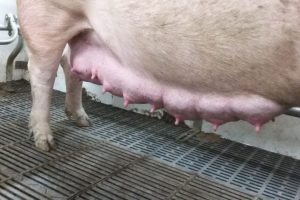
- Pig mammary glands V. Moustsen

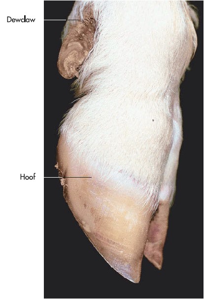
Observe: View the hooves.
Hoof ([L] ungula) – the ‘trotters’ of pigs represent digits 3 and 4, per limb, along with the non-weight bearing, but complete, paradigits 2 and 5. The paradigits are also known as dewclaws. The critical structure of the pig hoof will be studied in the musculoskeletal system unit. For now, recognize it is a complex, specialized modification of the common integument.
-
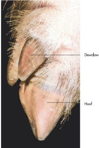
- Thoracic foot of a pig. 7

-

- Bulbs of the principal and accessory hooves of a pig. 7

Terms List
Print optimized PDF terms list
| Term | Species |
| Epidermis | |
| Dermis | |
| Hypodermis | |
| Tactile or sinus hairs | |
| Nasal plane | Carnivore, goat |
| Nasolabial plane | Ox |
| Rostral plane | Pig |
| Digital pad | Carnivore |
| Metacarpal pad | Carnivore |
| Metatarsal pad | Carnivore |
| Carpal pad | Carnivore |
| Ergot | Horse |
| Chestnut | Horse |
| Costal arch | Carnivore |
| Umbilicus | Carnivore |
| Anal sacs | Carnivore |
| Carpal gland | Pig |
| Mental gland | Pig |
| Cornual gland | Goat – know the location, not identified |
| Horn | Ruminant |
| Epidermal bud | May view in ox calf |
| Mammary gland | Carnivore, pig |
| Udder | Cow, mare, doe, ewe |
| Teat | All |
| Jugular groove | Horse, ruminant |
| Jugular fossa | Pig |
| Flank fold | Ruminant |
| Viborg’s triangle | Horse |
| Claw | Carnivore |
| Hoof | Horse, ruminant, pig |
| Dewclaw | Ruminant, pig |
Toward or relatively near the topside/backline (as opposed to the underside or supporting surface) of the head, body, and tail.
Toward or relatively near the underside, the 'belly' (or supporting) surface and the corresponding surface of the head, neck, thorax, and tail.
Toward or relatively near the head.
Cuts across the body at a right angle to its longitudinal axis.
Toward or relatively near the tail.
To lie on the back, with abdomen facing upwards. Dorsal means toward or relatively near the upper surface or back line of the body.
The movement of a part away from the median plane.
Toward or relatively near the median plane.
Down the limb, ie more distant from the attachment to the body; away from the main mass or origin.