MSK LAB 1A – Thoracic Limb Osteology and Arthrology
Learning Objectives
- Identify the regions of the thoracic limb, the bones and key bone features in each region, the joints and joint structures.
- Start to recognize and state sites of attachments for muscles, ligaments and tendons.
- Identify flexor and extensor sides of joints.
Lab instructions:
Part A: An instructor will provide an overview of this lab and highlight a few examples of comparative bone features across the species being studied.
Part B: Use ‘bone boxes’ (containing carnivore skeletons), bone models, articulated skeletons and images, to study the regions, bones and joints of the thoracic limb. Refer to the terms tables for focused studying. Be able to identify thoracic limb regions, the bones and their features, and joints and structures related to joints, in any species (i.e. dog, cat, horse, ruminant and pig), as applicable to that species. It is understood that not all material will be covered in lab and it is anticipated that continued learning and review of this content will continue as self-study effort.
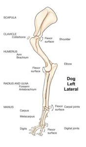
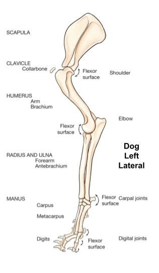
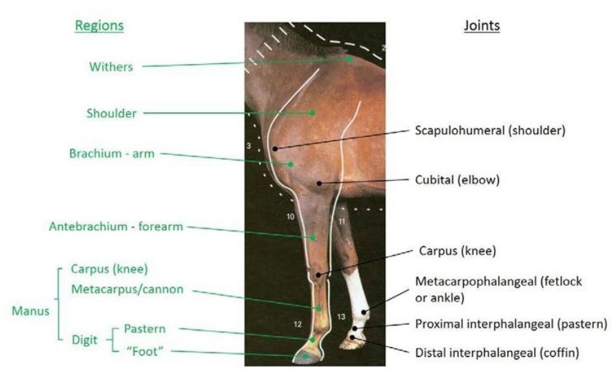
Thoracic Limb Regions
| Regions of the thoracic limb (proximal to distal) | Bones of the region |
Common Name | Species |
| Withers | Highest point of spinous processes of thoracic vertebrae | Horse | |
| Shoulder | Scapula | All | |
| Brachium | Humerus | Arm | All |
| Antebrachium | Radius, ulna | Forearm | All |
| Manus | Bones of carpus, metacarpus, digits | All | |
| Carpus | Carpal bones | ‘Knee’ – particularly in horse | All |
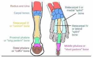
| Metacarpus | Metacarpal bones | ‘Cannon’ – ungulate | All |
| Digit | Phalanges (proximal, middle, distal) | In ungulates, includes pastern and “foot” | All |
| Pastern | Proximal and middle phalanges | Ungulates | |
| Foot | Distal phalanx | Hoof | Ungulates |
| Joints of the thoracic limb (proximal to distal) | Common Name |
| Scapulohumeral or glenohumeral joint | Shoulder |
| Cubital joint | Elbow |
| Carpus | “Knee” in horse |
| antebrachiocarpal joint | |
| middle carpal joint | |
| carpometacarpal joint | |
| Metacarpophalangeal joint | Fetlock, Ankle (ungulates) |
| Proximal interphalangeal joint | Pastern joint – ungulates |
| Distal interphalangeal joint | Coffin joint – ungulates |
-
- Dog TL regions 1
-

- Regions and joints (anatomical and lay terms) of the equine thoracic limb 17
-

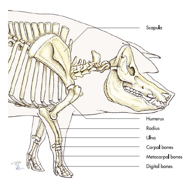
- Pig Thoracic Limb Bones 7
Clinical Application: Q0
You receive a phone call from a distraught owner who miniature horse “Viserion” has a laceration, as described by the owner, between Viserion’s knee and ankle, in her right front limb. What region is injured?
Interactive review content:
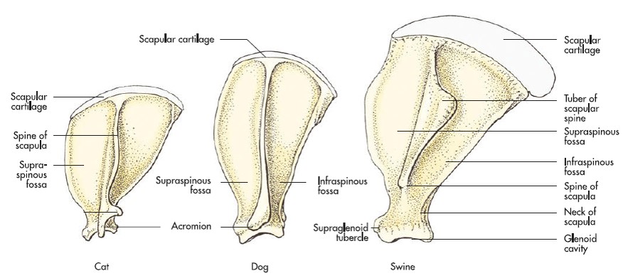
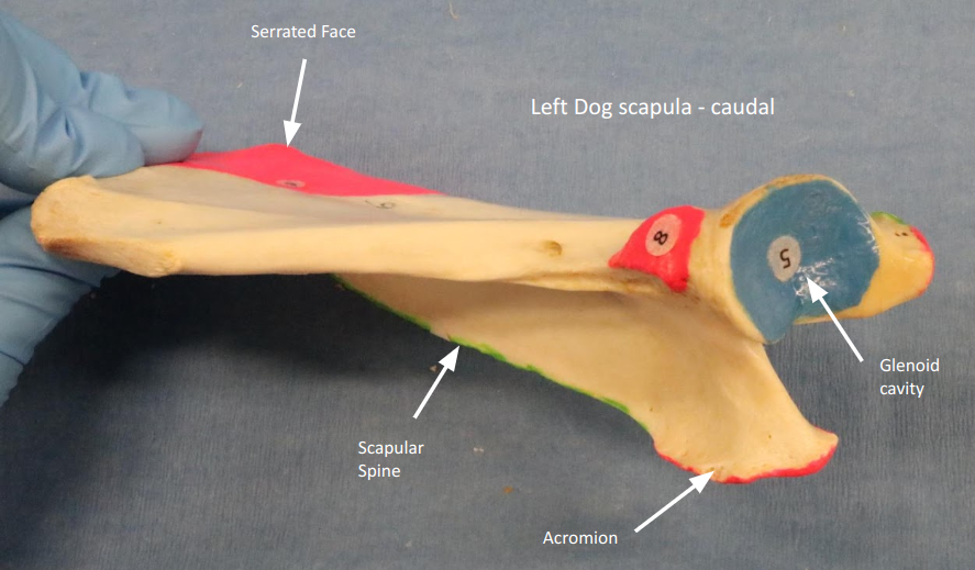
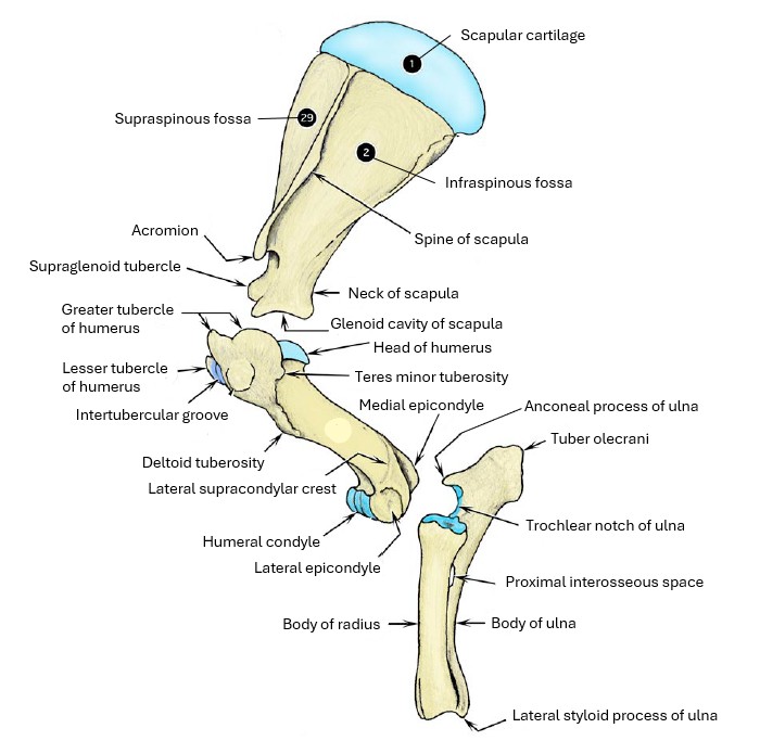
Scapula
| Osteology | Features | Species differences/comments |
| Scapula | Scapular spine | |
| Acromion | Two parts in cat; acromion not present in horse and pig | |
| Supraspinous fossa | ||
| Infraspinous fossa | ||
| Subscapular fossa | ||
| Supraglenoid tubercle | ||
| Serrated face | ||
| Glenoid cavity | ||
| Neck of scapula | ||
| Scapular cartilage (attached at dorsal margin of scapula) | Ungulate |
-
- Dog Scapula 1

-
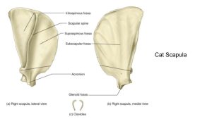
- Cat Scapula 5

-
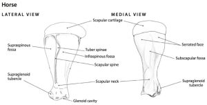
- Horse Scapula 6
-
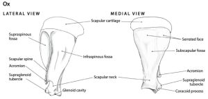
- Ox Scapula 6
-
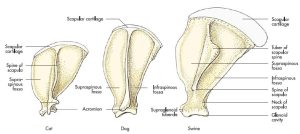
- Cat, dog, & pig scapula 7
-
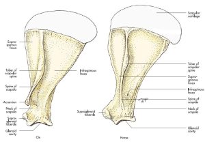
- Ox and horse scapula 7

-
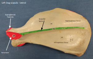
- Dog scapula – lateral

-
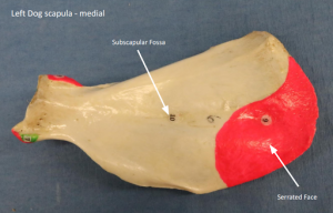
- Dog scapula – – medial

-

- Dog scapula – distal
-
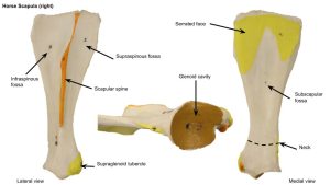
- Horse scapula

-
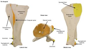
- Ox scapula
Interactive review content:
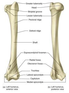
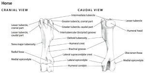
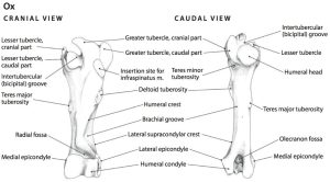
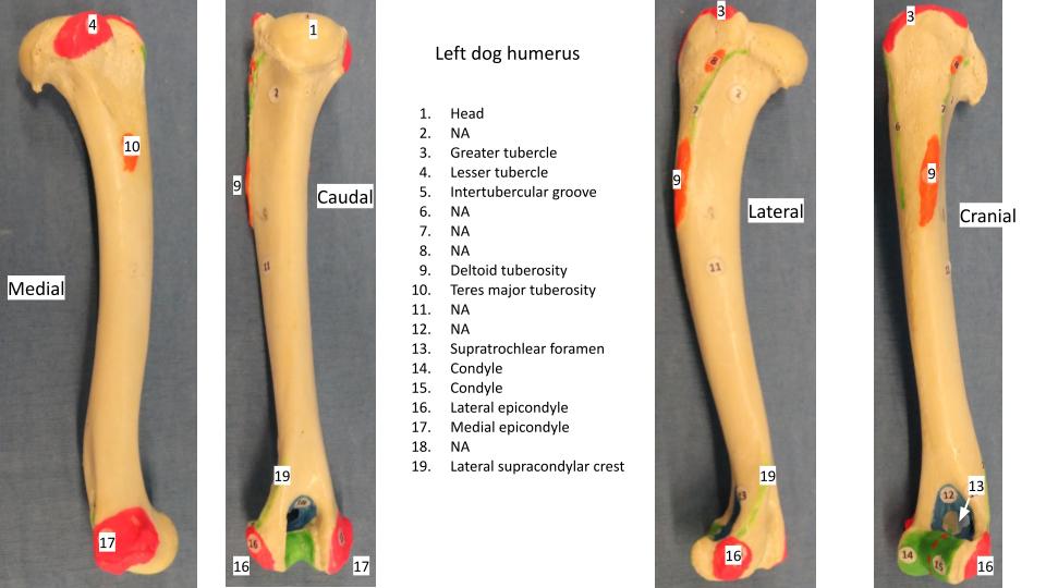
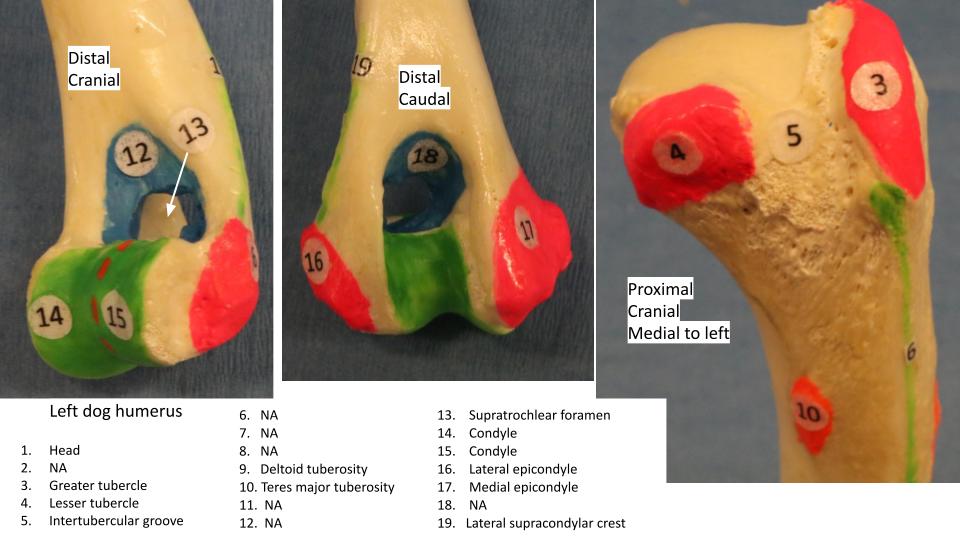
Humerus
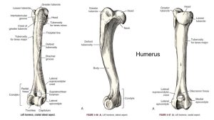
| Osteology | Features | Species differences/comments |
| Humerus | Head of humerus | |
| Greater tubercle | Horse – cranial and caudal parts | |
| Lesser tubercle | ||
| Intermediate tubercle | Horse | |
| Intertubercular (bicipital) groove | ||
| Neck of humerus | ||
| Shaft or body of humerus | ||
| Deltoid tuberosity | ||
| Teres major tuberosity | ||
| Teres minor tuberosity | ||
| Lateral supracondylar crest | ||
| Lateral/medial epicondyles | ||
| Humeral condyle | ||
| Supratrochlear foramen | Dog | |
| Supracondylar foramen | Cat |
-
- Dog humerus. 1
-
- Cat humerus 5
-
-
Skeleton of left shoulder, arm, and forearm of ox,
lateral view. 2
-
- Horse humerus 6
-
- Ox humerus 6
-
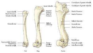
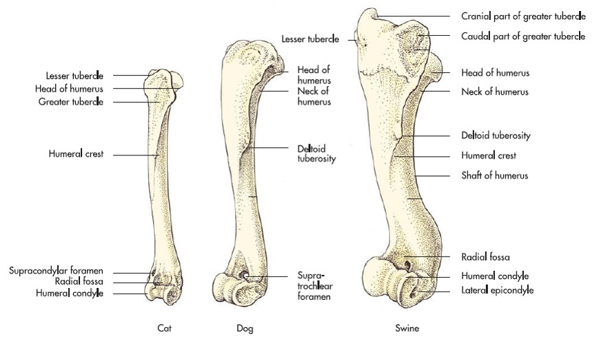
- Cat, dog & pig humerus 7
-
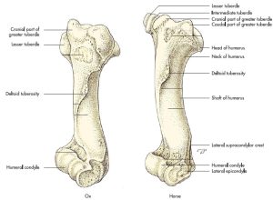
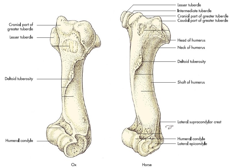
- Ox and horse humerus 7
-
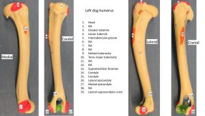
- Dog humerus
-
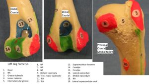
- Dog humerus
-
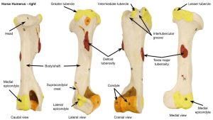
- Horse humerus
-
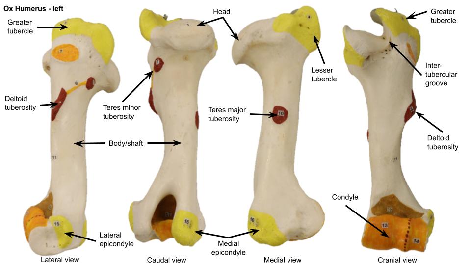
- Ox humerus
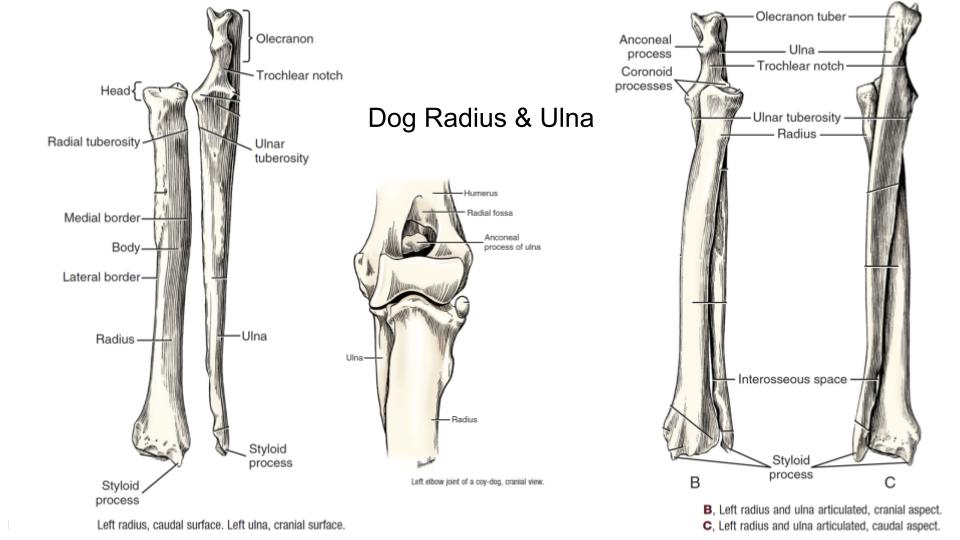
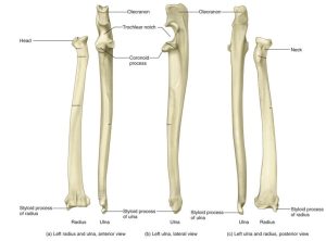
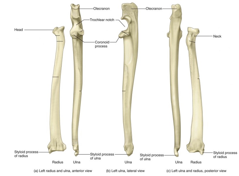
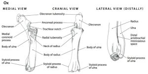
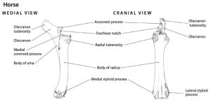
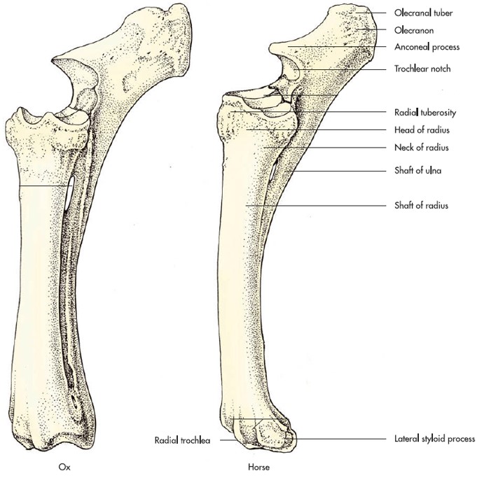
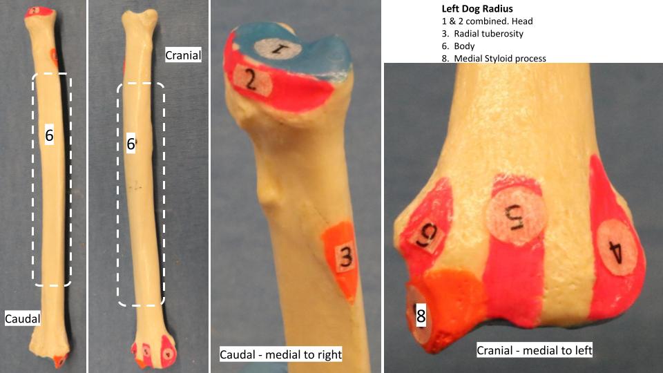
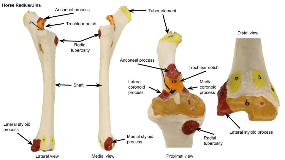
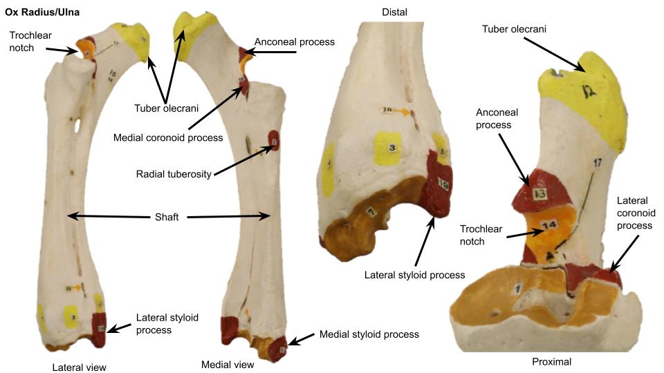
Radius & Ulna
| Osteology | Features | Species differences/comments |
| Radius | Head of radius | |
| Radial tuberosity | ||
| Shaft or body of radius | ||
| Medial styloid process | ||
| Ulna | Olecranon of ulna/tuber olecrani/olecranon tuberosity | |
| Trochlear notch | ||
| Anconeal process | ||
| Medial and lateral coronoid process | ||
| Ulnar tuberosity | ||
| Lateral styloid process | Fused to radius in horse |
-
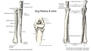
- Dog radius and ulna 1
-
- Cat radius and ulna5
-
- Horse radius and ulna 6
-
- Ox radius and ulna 6
-
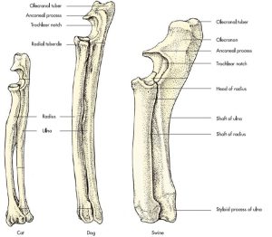
- Cat, dog, and pig radius and ulna 7
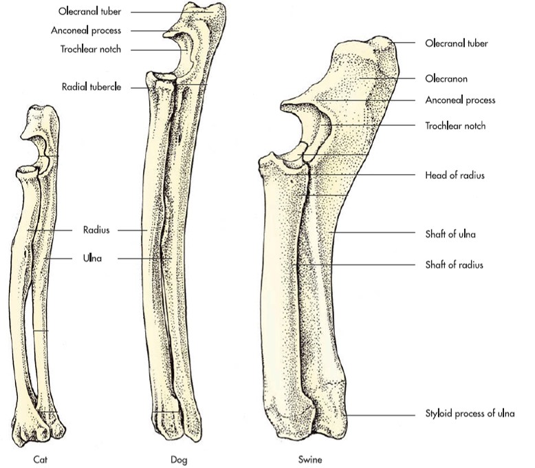
-
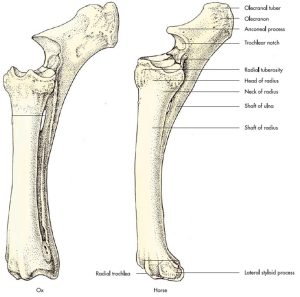
- Ox and horse radius and ulna 7
-
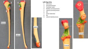
- Dog ulna

-

- Dog radius
-

- Horse radius/ulna
-

- Ox radius/ulna
Interactive review content:
Clinical Application: Q1
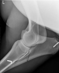
Review the equine radiograph and describe the fracture.
Your radiographic description should include the bone, the location of the fracture on the bone, the direction of the fracture line (eg transverse, oblique), is it comminuted or not (may be very hard to tell from one view), and does the fracture extend into a joint or not (articular or non-articular). What else is present in the view?
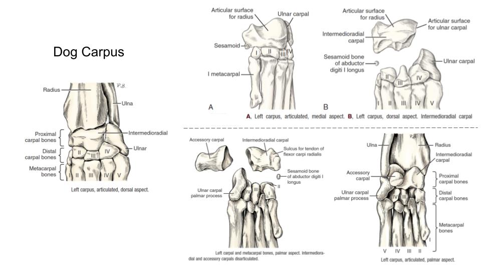
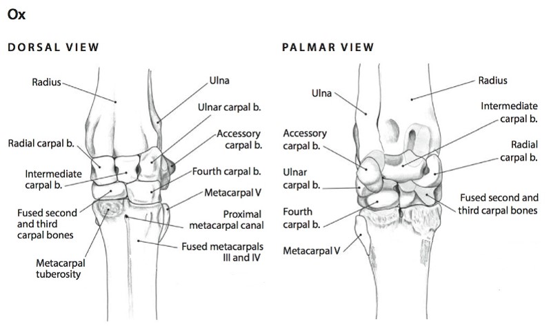
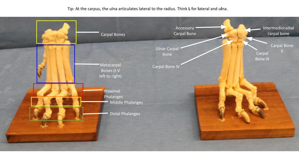
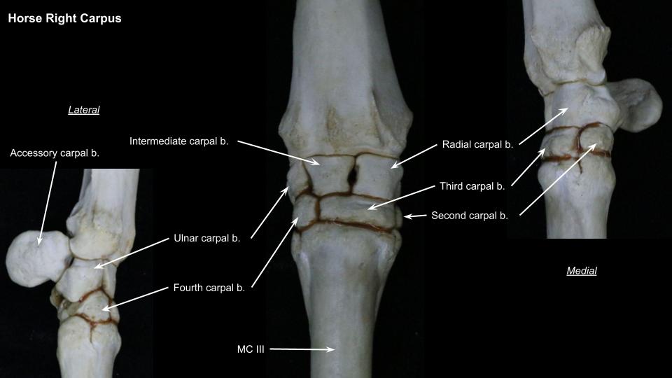
Carpus
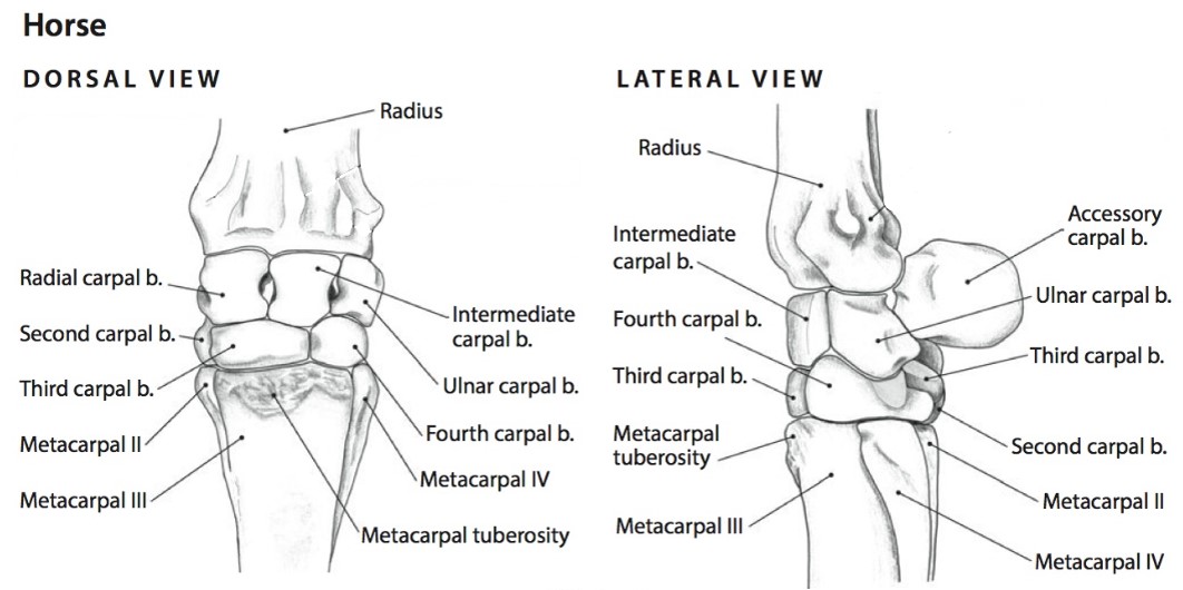
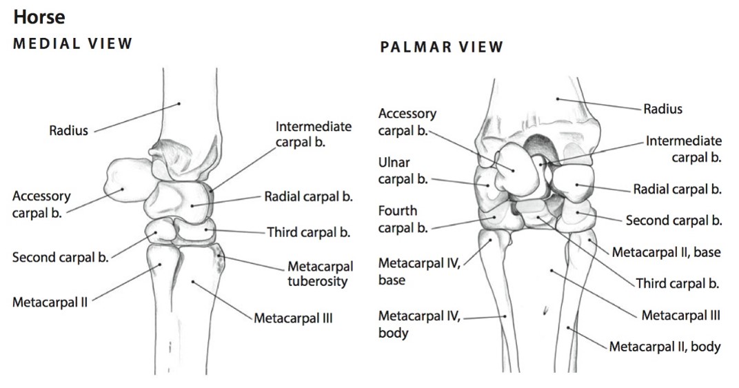
| Osteology | Species differences/comments |
| Intermedioradial carpal bone | Carnivore only (= fused intermediate and radial carpal bones) |
| Radial carpal bone | Ungulate only |
| Intermediate carpal bone | Ungulate only |
| Ulnar carpal bone | |
| Accessory carpal bone | projects palmarly as very palpable prominence |
| Carpal bones I, II, III, IV | Carnivore – 1, 2, 3, 4
Pig – 1, 2, 3, 4 Ruminant – Fused 2/3, 4 Horse – [inconsistent 1], 2, 3, 4 |
| Sesamoid in abductor pollicis longus | Carnivore |
-
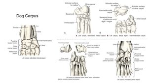
- Dog carpus 1
-
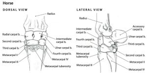
- Horse carpus 6
-
- Horse carpus 6
-
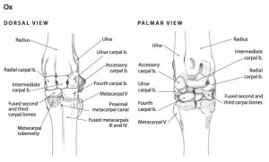
- Ox carpus 6
-
- Comparative carpi7

-

- Dog carpus
-

- Horse carpus
-
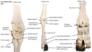
- Ox carpus

Metacarpals & Phalanges
| Osteology | Features | Species differences/comments |
| Metacarpal bones | Base (proximal end); Head (distal end) | |
| Metacarpal bone I | Carnivore only | |
| Metacarpal bone II | Horse – “button” of splint bone at distal end | Horse – MC2 is the medial splint bone |
| Metacarpal bone III
aka Cannon bone of ungulates |
Sagittal ridge of MC3 | Horse |
| Metacarpal tuberosity | Ungulate – Insertion of ECR m.
Ruminant – MC3+MC4 fused |
|
| Metacarpal bone IV | Horse – “button” of splint bone at distal end | Horse – MC4 is the lateral splint bone
Ruminant – MC3+MC4 fused |
| Metacarpal bone V | Carnivore present, and ruminant – vestigially present | |
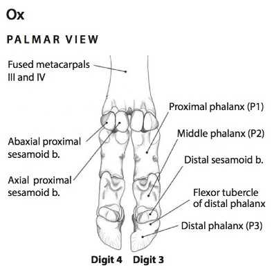
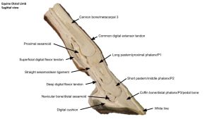
| Proximal sesamoid bones | ‘Base’, ‘apex’, axial and abaxial surfaces | Ungulate only; at level of palmar side of metacarpophalangeal joint |
| Dorsal sesamoid bones | Carnivore only | |
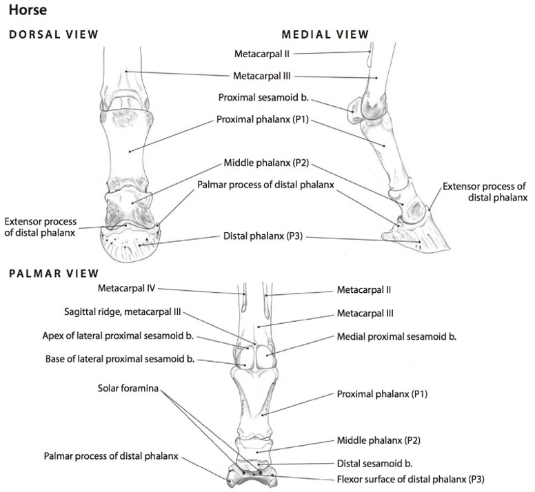
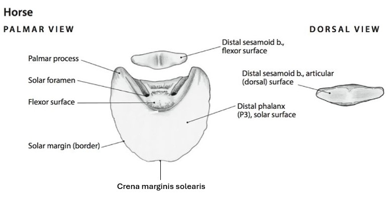
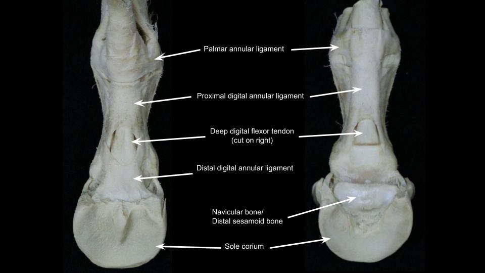
| Distal sesamoid/Navicular bone | proximal border, distal border, flexor and articular surfaces, sagittal ridge of navicular bone | Ungulate at level of palmar distal interphalangeal joint. |
| Digits I-V – species variation | Consist of proximal, middle and distal phalanges; axial and abaxial side to each digit (or medial and lateral sides in horse since only one digit) | Carnivores – 5 digits (2-5 are weight bearing)
Ruminant and Pig – 4 digits (3 and 4 are weight bearing; 2 and 5 = dewclaws) Horse – 1 digit (3rd digit, bears all weight of limb!) |
| Proximal phalanx | Base (proximal end); Head (distal end) | Ungulate = Pp, P1, long pastern bone |
| Middle phalanx | Base (proximal end); Head (distal end) | Ungulate = Pm, P2, short pastern bone
Carnivore – Pm is absent in digit 1. |
| Distal phalanx | Ungulate = Pd, P3, coffin or pedal bone | |
| Extensor process | All (attachment of CDE) | |
| Flexor tubercle of Pd | Carnivore, Ruminant | |
| Solar border (margin) | Horse | |
| Crena marginis solearis | Horse | |
| Medial and lateral palmar processes (‘wings’) of Pd | Horse | |
| Solar surface | Horse (DDFT attaches to solar surface) |
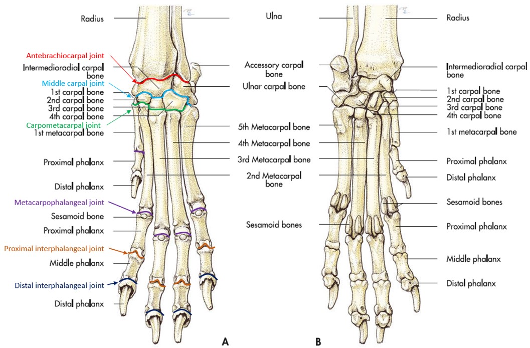
-
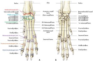
- Skeleton and joints of the left forepaw of the dog: (A) dorsal and (B) palmar aspect. 7
-
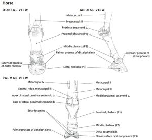
- Left Digit of the Horse, Dorsal, Medial, and Palmar Views. 6
-
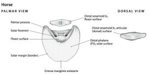
- Distal Phalanx and Distal Sesamoid (Navicular) bone of the Horse – Palmar and Dorsal View.6
-
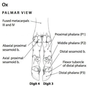
- Left Digits of the Ox, Palmar View. 6
-
- Horse vs human bone comparison. Bones in gray have been lost in horses. David Hankin

-
- Horse distal limb

Clinical Application: Metacarpal Fractures in Carnivores
Fractures of the metacarpal (MC) and metatarsal (MT) bones are common and can occur in single or multiple bones. Treatment can be either conservative or surgical, with the former being necessary most commonly. Two or more intact bones provide a natural splint that is further augmented by soft tissue swelling around the fracture. Conservative management should be considered in these cases if the fractures feel relatively stable and the digits are aligned. Surgical treatment may be used for fractures of three or four bones, if a compound fracture exists (the bone has gone through the skin), or for grossly unstable fractures of one or two bones. Another consideration is whether or not the primary weight-bearing bones, metacarpal/metatarsal III and IV, are affected. Treatment recommendations are often based on the clinician’s judgement. The clinician’s experience with surgical correction, the availability of proper surgical tools and implants, the patient’s signalment, and the client’s ability to care for the patient, financially or otherwise, all need to be considered when making a recommendation for treatment.
Clinical Application: Q2
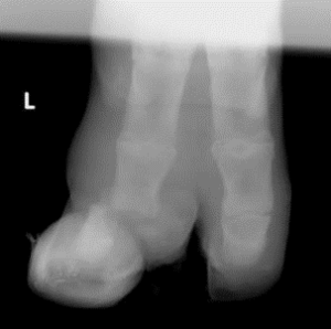
Review the radiograph.
Perissodactyl or artiodactyl?
What bone is fractured? (be specific).
What joint is affected?
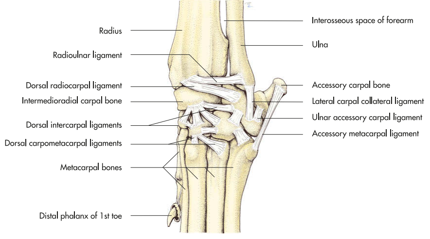
Joints & Ligaments
The ungulate specific ligaments will be covered in depth during lab 7; you may wish to focus on those terms in that lab.
| Landmarks | Species differences/comments |
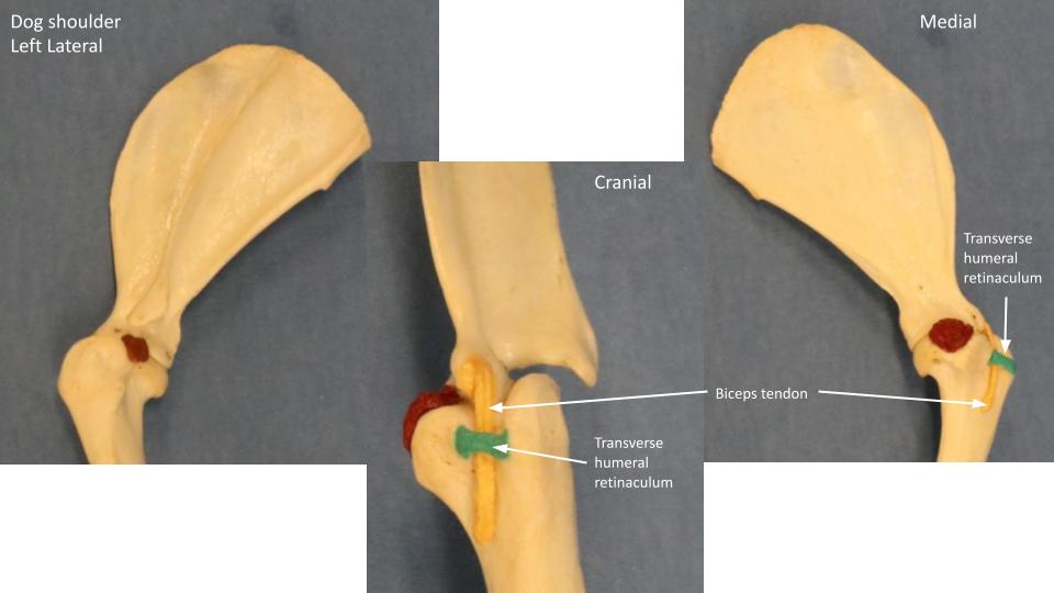
| Transverse humeral retinaculum | Carnivore |
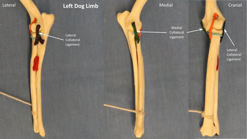
| Medial/lateral collateral ligaments of elbow | All |
| Medial/lateral collateral ligaments of carpus | All |
| Elastic dorsal ligaments – know concept | Cat |
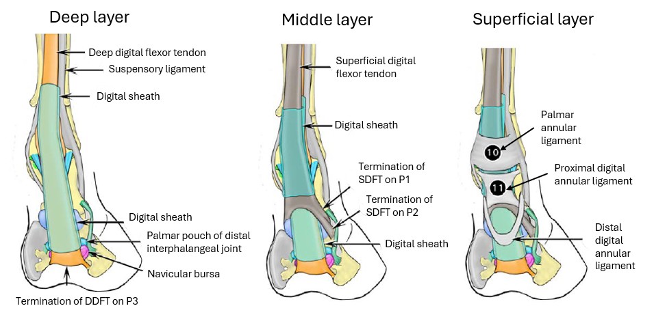
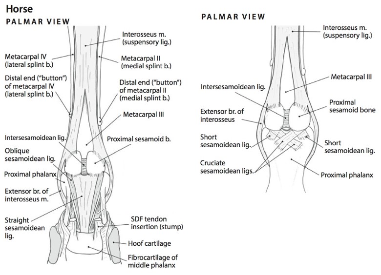
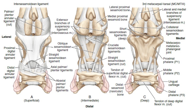
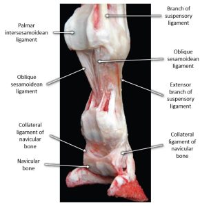
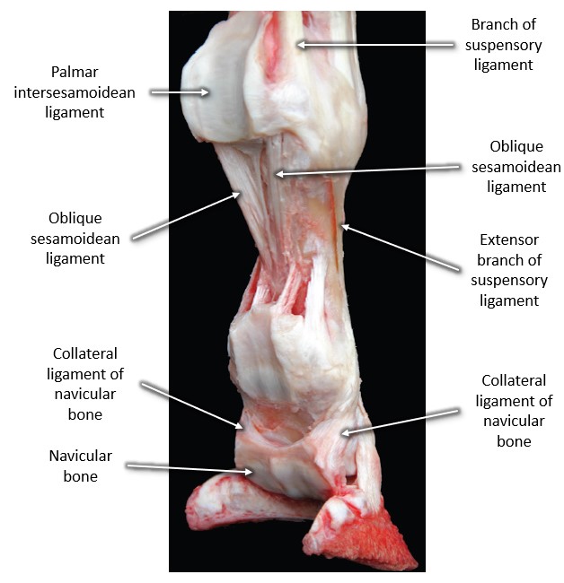
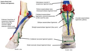
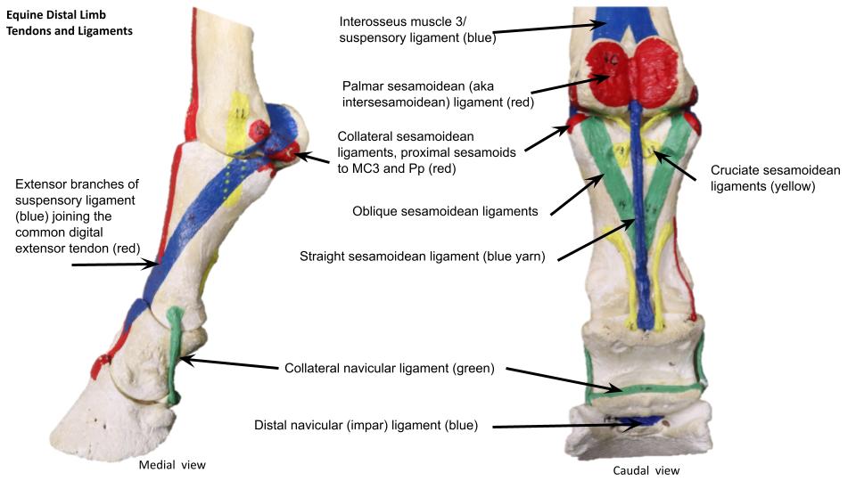
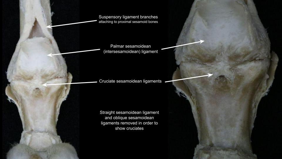
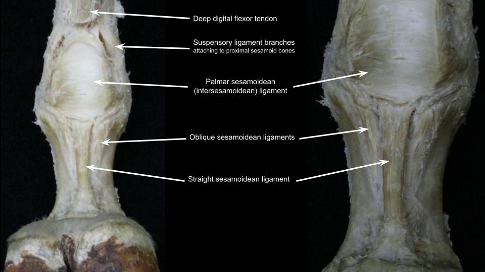
| Palmar (inter) sesamoidean ligament | Horse |
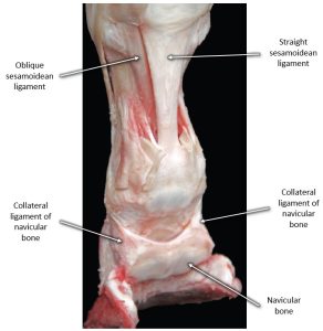
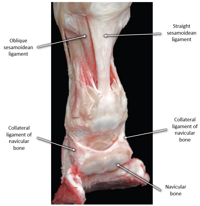
| Straight sesamoidean ligament | Horse – superficial, Y |
| Oblique sesamoidean ligament | Horse – middle, V |
| Cruciate sesamoidean ligament | Horse – deep, X |
| Collateral sesamoidean ligaments | Horse – proximal sesamoids to MC3 and Pp |
| Collateral (suspensory) ligg. of navicular bone | Horse |
| Distal navicular (impar) ligament | Horse |
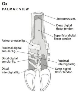
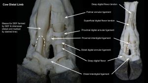
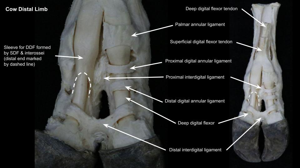
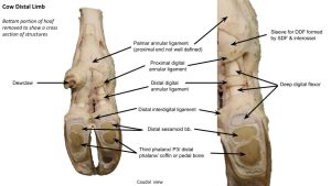
| Proximal and distal interdigital ligaments | Ruminant |
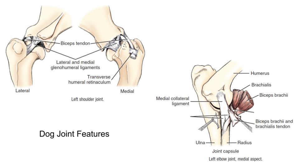
Carnivore
-

- Dog joint features 1
-
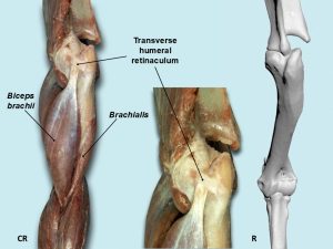
- Dog transverse humeral retinaculum. 40
-

- Dog shoulder
-
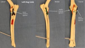
- Dog elbow ligaments
-
- Skeleton and joints of the left forepaw of the dog: (A) dorsal and (B) palmar aspect. 7
-
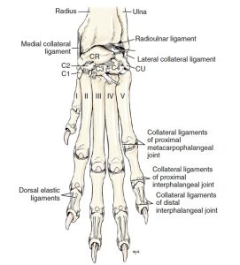
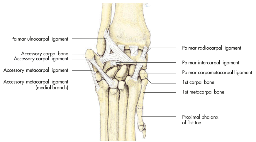
- Ligaments of left forepaw, dorsal aspect. 1
-

- Ligaments of the left carpus of the dog, lateral aspect. 7
-

- Ligaments of the left carpus of the dog, palmar aspect. 7
Ungulate
-
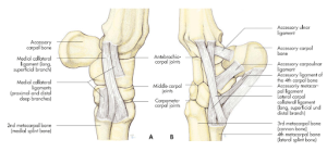
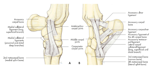
- Long collateral ligaments and ligaments of the accessory carpal bone of the left carpus of the horse: (A) medial and (B) lateral. 7
-
- Equine Suspensory Apparatus and Other Ligaments; Caudomedial View. 2

-
- Equine digital region, caudomedial view. 2
-

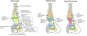
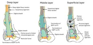
- Superficial and Deep Ligaments of the Left Digit of the Horse, Palmar View. 6
-
- Palmar plantar View of the Distal Sesamoidean Ligaments of the Equine Limb. A. Superficial. B. Intermediate. C. Deep. 9
-

- Palmar aspect of the right digital area of the horse after removal of the flexor tendons as well as straight and lateral oblique sesamoidean ligaments. 17

-
- Palmaromedial aspect of the right digital area of the horse after removal of the flexor tendons. 17
-
- Palmarolateral aspect of the right digital area of the horse after removal of the flexor tendons and straight sesamoidean ligament. 17
-
- Digits of the Ox, Palmar View. 6
-

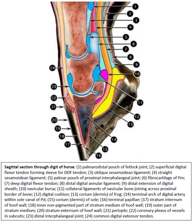
- Sagittal section through digit of horse. 2
-
- Horse tendons and ligaments
-
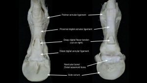
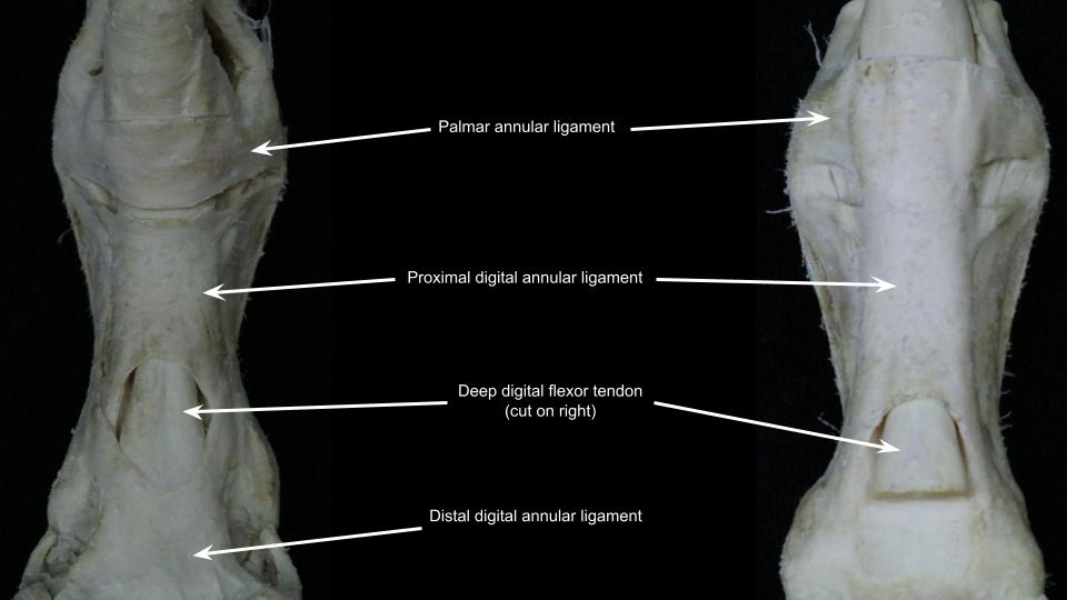
- Horse distal annular ligament
-
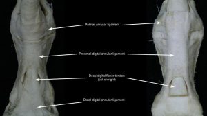
- Horse distal annular ligament
-

- Horse sesamoidean ligaments
-
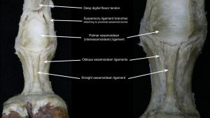
- Horse sesamoidean ligaments
-
- Ox distal limb ligaments
-
- Ox distal limb ligaments

Review Videos
Carnivore Thoracic Limb Regions and Bones – 6 minutes
Carnivore Thoracic Limb Osteology – 12 minutes
Horse Thoracic Limb Osteology – 22 minutes
Cow Thoracic Limb Osteology – 11 minutes
Horse and ruminant Thoracic Limb Ligaments – 17 minutes Watch after completing the Ungulate TL lab for the best understanding
Terms
| Regions of the thoracic limb (proximal to distal) | Bones of the region |
Common Name | Species |
| Withers | Highest point of spinous processes of thoracic vertebrae | Horse | |
| Shoulder | Scapula | All | |
| Brachium | Humerus | Arm | All |
| Antebrachium | Radius, ulna | Forearm | All |
| Manus | Bones of carpus, metacarpus, digits | All | |
| Carpus | Carpal bones | ‘Knee’ – particularly in horse | All |
| Metacarpus | Metacarpal bones | ‘Cannon’ – ungulate | All |
| Digit | Phalanges (proximal, middle, distal) | In ungulates, includes pastern and “foot” | All |
| Pastern | Proximal and middle phalanges | Ungulates | |
| Foot | Distal phalanx | Hoof | Ungulates |
| Joints of the thoracic limb (proximal to distal) | Common Name |
| Scapulohumeral or glenohumeral joint | Shoulder |
| Cubital joint | Elbow |
| Carpus | “Knee” in horse |
| antebrachiocarpal joint | |
| middle carpal joint | |
| carpometacarpal joint | |
| Metacarpophalangeal joint | Fetlock, Ankle (ungulates) |
| Proximal interphalangeal joint | Pastern joint – ungulates |
| Distal interphalangeal joint | Coffin joint – ungulates |
Osteology of Shoulder and Brachium |
||
| Osteology | Features | Species differences/comments |
| Scapula | Scapular spine | |
| Acromion | Two parts in cat; acromion not present in horse and pig | |
| Supraspinous fossa | ||
| Infraspinous fossa | ||
| Subscapular fossa | ||
| Supraglenoid tubercle | ||
| Serrated face | ||
| Glenoid cavity | ||
| Neck of scapula | ||
| Scapular cartilage (attached at dorsal margin of scapula) | Ungulate | |
| Clavicle | Near point of shoulder. | Dog – a fibrous clavicular intersection; Cat – has bony clavicle |
| Humerus | Head of humerus | |
| Greater tubercle | Horse – cranial and caudal parts | |
| Lesser tubercle | ||
| Intermediate tubercle | Horse | |
| Intertubercular (bicipital) groove | ||
| Neck of humerus | ||
| Shaft or body of humerus | ||
| Deltoid tuberosity | ||
| Teres major tuberosity | ||
| Teres minor tuberosity | ||
| Lateral supracondylar crest | ||
| Lateral/medial epicondyles | ||
| Humeral condyle | ||
| Supratrochlear foramen | Dog | |
| Supracondylar foramen | Cat | |
Osteology of Antebrachium |
||
| Osteology | Features | Species differences/comments |
| Radius | Head of radius | |
| Radial tuberosity | ||
| Shaft or body of radius | ||
| Medial styloid process | ||
| Ulna | Olecranon of ulna/tuber olecrani/olecranon tuberosity | |
| Trochlear notch | ||
| Anconeal process | ||
| Medial and lateral coronoid process | ||
| Ulnar tuberosity | ||
| Lateral styloid process | Fused to radius in horse | |
Osteology of Carpus |
|
| Osteology | Species differences/comments |
| Intermedioradial carpal bone | Carnivore only (= fused intermediate and radial carpal bones) |
| Radial carpal bone | Ungulate only |
| Intermediate carpal bone | Ungulate only |
| Ulnar carpal bone | |
| Accessory carpal bone | projects palmarly as very palpable prominence |
| Carpal bones I, II, III, IV | Carnivore – 1, 2, 3, 4
Pig – 1, 2, 3, 4 Ruminant – Fused 2/3, 4 Horse – [inconsistent 1], 2, 3, 4 |
| Sesamoid in abductor pollicis longus | Carnivore |
Osteology of Metacarpus and Digit |
||
| Osteology | Features | Species differences/comments |
| Metacarpal bones | Base (proximal end); Head (distal end) | |
| Metacarpal bone I | Carnivore only | |
| Metacarpal bone II | Horse – “button” of splint bone at distal end | Horse – MC2 is the medial splint bone |
| Metacarpal bone III
aka Cannon bone of ungulates |
Sagittal ridge of MC3 | Horse |
| Metacarpal tuberosity | Ungulate – Insertion of ECR m.
Ruminant – MC3+MC4 fused |
|
| Metacarpal bone IV | Horse – “button” of splint bone at distal end | Horse – MC4 is the lateral splint bone
Ruminant – MC3+MC4 fused |
| Metacarpal bone V | Carnivore present, and ruminant – vestigially present | |
| Proximal sesamoid bones | ‘Base’, ‘apex’, axial and abaxial surfaces | Ungulate only; at level of palmar side of metacarpophalangeal joint |
| Dorsal sesamoid bones | Carnivore only | |
| Distal sesamoid/Navicular bone | proximal border, distal border, flexor and articular surfaces, sagittal ridge of navicular bone | Ungulate at level of palmar distal interphalangeal joint. |
| Digits I-V – species variation | Consist of proximal, middle and distal phalanges; axial and abaxial side to each digit (or medial and lateral sides in horse since only one digit) | Carnivores – 5 digits (2-5 are weight bearing)
Ruminant and Pig – 4 digits (3 and 4 are weight bearing; 2 and 5 = dewclaws) Horse – 1 digit (3rd digit, bears all weight of limb!) |
| Proximal phalanx | Base (proximal end); Head (distal end) | Ungulate – Pp, P1, long pastern bone |
| Middle phalanx | Base (proximal end); Head (distal end) | Ungulate – Pm, P2, short pastern bone
Carnivore – Pm is absent in digit 1. |
| Distal phalanx | Ungulate – Pd, P3, coffin or pedal bone | |
| Extensor process | All (attachment of CDE) | |
| Flexor tubercle of Pd | Carnivore, Ruminant | |
| Solar border (margin) | Horse | |
| Crena marginis solearis | Horse | |
| Medial and lateral palmar processes (‘wings’) of Pd | Horse | |
| Solar surface | Horse (DDFT attaches to solar surface) | |
Ligaments related to bones and joints of the thoracic limb |
|
| Landmarks | Species differences/comments |
| Transverse humeral retinaculum | Carnivore |
| Medial/lateral collateral ligaments of elbow | |
| Medial/lateral collateral ligaments of carpus | |
| Elastic dorsal ligaments – know concept | Cat |
| Palmar (inter)sesamoidean ligament | Horse |
| Straight sesamoidean ligament | Horse – superficial, Y |
| Oblique sesamoidean ligament | Horse – middle, V |
| Cruciate sesamoidean ligament | Horse – deep, X |
| Collateral sesamoidean ligaments | Horse – proximal sesamoids to MC3 and Pp |
| Collateral (suspensory) ligg. of navicular bone | Horse |
| Distal navicular (impar) ligament | Horse |
| Proximal and distal interdigital ligaments | Ruminant |
Example Practical Exam Questions
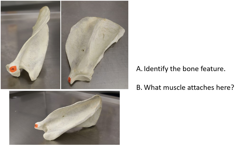
A. Identify the bone feature
B. What muscles attaches here? (Don’t expect to be able to answer this question until after lab 4)
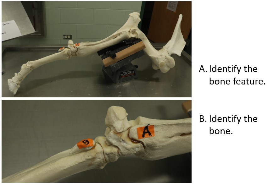
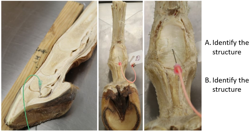
A. Identify the bone feature
B. Identify the bone
This question will be easiest to answer after lab 7.
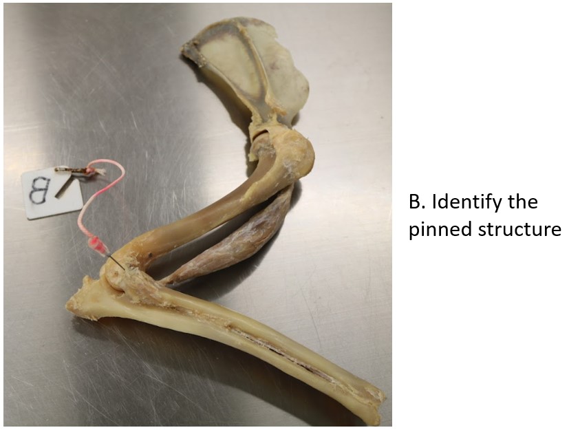
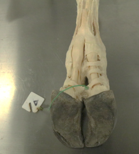
A. Identify the pinned structure – be specific.
B. True or False: The structure in A is transected during claw amputation.