
Clinical Application Answers
Lab 1 Answers
Clinical Application: Q0
Q0: You receive a phone call from a distraught owner who miniature horse “Viserion” has a laceration, as
described by the owner, between Viserion’s knees and ankles in her right front limb. What region is
injured?
A: Region injured = metacarpal region or cannon bone region.
Cartoons included so you don’t automatically see the next answer.

Clinical Application: Q1
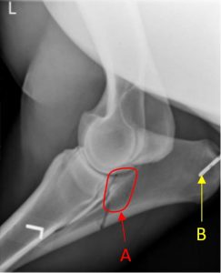 Q1. Review the equine radiograph and describe the fracture.
Your radiographic description should include the bone, the location of the fracture on the bone, the direction of the fracture line (e.g. transverse, oblique), is it comminuted or not (may be very hard to tell from one view), and does the fracture extend into a joint or not (articular or non-articular).
What else is present in the view?
A. This is an articular distal oblique fracture of the distal olecranon-proximal ulnar body.
An ‘island’ of bone (A – circled) associated with the mid aspect of the fracture line suggests a comminuted fracture and a second view would help confirm this.
Ulnar fractures are classified and this one is a Type 5 fracture.
A teat canula (B – arrow) is seen – it was inserted into the wound over the point of the elbow.

Clinical Application: Q2
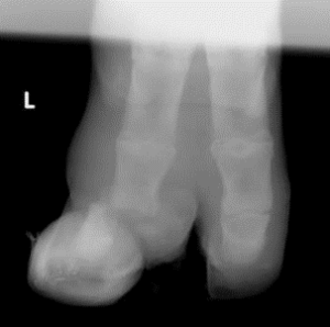 Q2. Review the radiograph. Perissodactyl or artiodactyl? What bone is fracture? (be specific). What joint is affected?
A. Artiodactyl (two digits noted).
Fracture of the distal phalanx of the left lateral (4th) digit involving the distal interphalangeal joint.
(Lateral because the L for left (or R if it was a right limb) by convention is placed on the lateral aspect of the limb).
Thoracic or pelvic limb is not discernable.
Clinical Application: Q3
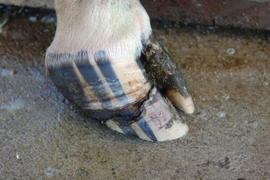
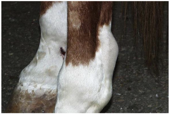
Q3: Diagnose the condition in the image. What soft tissue structure would a veterinarian considering
transecting first of all, to treat this condition?
A. Distal interphalangeal hyperflexion or ‘club foot’, left front.
Cut the distal check ligament first of all in a case of this severity.
Clinical Application: Q4
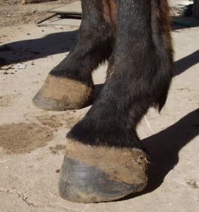
Q4: Review the image. Where is the swelling focused?
What is a common term for this swelling?
What structure is swollen? (Photo credit: Gary Baxter).
A. Swelling is palmar to the suspensory branch and just proximal to the fetlock, surrounding the path of the digital flexors. This is referred to as ‘windpuffs’ and is due to swelling of the digital flexor tendon sheath.

Clinical Application: Q5
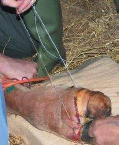
Q5: Review the image and ponder the following questions.
1) What common cutting device is being used to amputate the digit (claw)?
2) What ligament of the ruminant foot must be transected to allow removal of one digit, if amputation is through the mid shaft of the proximal phalanx?
3) Note the green butterfly catheter and orange torniquet – what vessel has likely been catheterized? What drug may have been injected?
A. 1) Gigli wire is the cutting device
2) the distal interdigital ligament
3) The dorsal common digital v. 3; local anesthetic (eg lidocaine or mepivicaine)
Lab 3 Answers
Thoracic Limb Amputation
It is at times necessary to perform a thoracic limb amputation. In doing so, all of the extrinsic muscles must be transected! Can you name all eight of them?
Extrinsic muscles of the thoracic limb are as follows:
- Superficial pectorals
- Deep pectoral
- Brachiocephalicus
- Omotransversarius
- Trapezius
- Rhomboideus
- Latissimus dorsi
- Serratus ventralis
- [Subclavius – well developed and only identified in the horse and pig]