MSK LAB 3A – Thoracic Limb Extrinsic muscles
Learning Objectives
- Identify extrinsic muscles of the thoracic limb.
- Name attachments and actions of extrinsic mm. of the thoracic limb.
- Define a “muscle action group,” and categorize individual mm. into these groups.
- Name the muscles which must be transected to perform a thoracic limb amputation in the carnivore.
Extrinsic muscles of the thoracic limb are those that have one attachment to the axial skeleton or “body” (e.g. the neck, head, or thorax) and the other attachment to a bone of the thoracic limb (i.e. a bone of the appendicular skeleton).
Lab instructions:
For this lab, teams will focus primarily on dissecting the muscles attaching the thoracic limb to the body, in canine and feline cadavers. This carnivore understanding can then be applied to the prosected large animal cadavers to identify the same muscles (and any extras) as directed.
Before we dissect:
What do we mean by muscle attachments?
All muscles have attachments. In most instances, the more proximal attachment, the part that moves the least, is considered the origin. The insertion is the more distal attachment, or the part that moves the most. The origin is usually a direct attachment of the muscle cells to the bone. For our purposes, you are not responsible for learning which attachments are the origin versus the insertion. At times, however, you may be asked for the distal or proximal attachment of a muscle or muscles. The insertion often is by a tendon or aponeurosis extending from the muscle cells to the bone. A tendon consists of dense, regularly arranged fibrous connective tissue organized into a small, well-defined bundle. An aponeurosis has the same consistency as a tendon, but the fibrous tissue is arranged as a thin sheet of tissue (imagine taking a tendon and flattening it out with a rolling pin, the end result is an aponeurosis). A ligament typically refers to dense fibrous connective tissue between bones, and the term is also used for a variety of thin fibrous connections between organs in the body or between an organ and the body wall.
MUSCLE CHARTS: You are responsible for knowing the attachments and action(s) of muscles as described in the muscle action charts – Muscle Chart.
Read before you continue dissection! In many instances, during the study of muscles, a description of a specific muscle will be given before the instructions for dissection. Follow instructions as carefully as possible. In each instance clean the exposed surface of the muscle being described, isolate its borders, and verify its attachments. If the muscle is to be “transected,” free it from underlying structures first. As each muscle is defined, try to visualize its attachments and position on the skeleton and understand its function.
Ready? Then lets go! All dissection commences on the left side of the body.
Cutaneus trunci m.
The trunk skin muscle (P.S. – not one of the extrinsic TL mm.)
The cutaneus trunci is a relatively thin sheet of muscle that covers most of the dorsal, lateral, and ventral walls of the thorax and abdomen. It has no direct bony attachments. It is more closely applied to the skin than to underlying structures and is often reflected with the skin before being observed. Like all cutaneous muscles, it is developed in the superficial fascia of the thorax and abdomen. Caudal to the shoulder the fibers sweep obliquely toward the axilla (armpit); farther caudally they are principally longitudinal and arise from the superficial fascia over the pelvic region.
The attachments of the cutaneus trunci are the superficial fascia of the trunk and the skin. The muscle sends a fasciculus to the medial side of the forelimb; caudal and ventral to this the fibers fray out over the deep pectoral muscle. The cutaneus trunci twitches the skin. It is innervated by the lateral thoracic nerve. In the male there is a distinct development of this muscle adjacent to the ventral midline, caudal to the xiphoid. This is the preputial muscle, which passes caudally and radiates into the prepuce, forming an arch with the muscle of the opposite side. It functions to support the cranial end of the prepuce during the non-erect state and to pull the prepuce back over the glans penis after erection and protrusion.
-

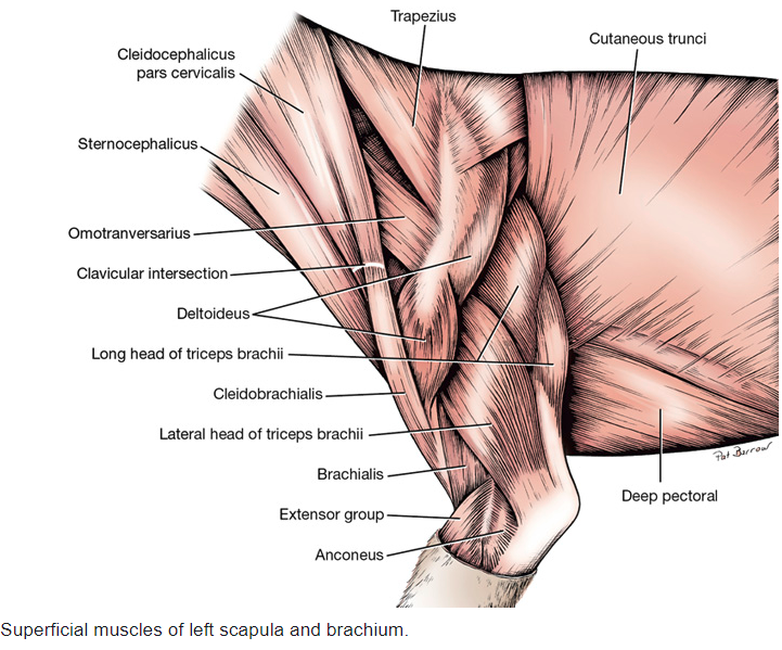
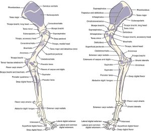
- Superficial muscles of the left scapula and brachium of the dog 1
-
- Dog cutaneous trunci
-

- Cat cutaneous trunci
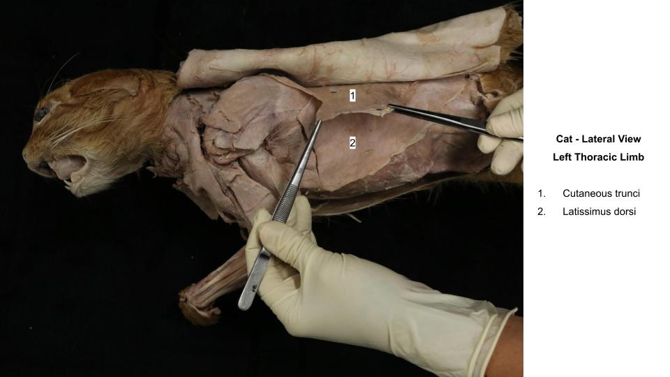
Dissect: Transect (cut across) the axillary and ventral attachments of the cutaneous trunci and reflect the muscle dorsally, taking care to separate it from the underlying latissimus dorsi m. Note, in your cadaver much of the cutaneous trunci m. may have been removed with the skin, making it more difficult to discern what is left behind to reflect…ask an instructor. And for the feline cadavers it is difficult to successfully isolate the CT m. – not to worry.
Now on to dissecting the actual extrinsic TL muscles!
Extrinsic muscles of the thoracic limb are as follows:
- Superficial pectorals
- Deep pectoral
- Brachiocephalicus
- Omotransversarius
- Trapezius
- Rhomboideus
- Latissimus dorsi
- Serratus ventralis
- [Subclavius – well developed and only identified in the horse and pig – see below].
Observe: The left limb will be dissected to visualize muscles. We are not specifically concerned with preserving vessels and nerves on this “muscle limb”.
In the ventral thoracic region are the superficial and deep pectoral muscles, which extend between the sternum and the humerus.
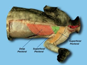
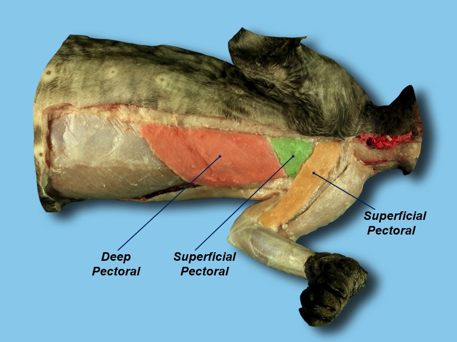
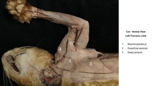
Superficial pectoral m.
The superficial pectoral lies between the cranial part of the sternum and the humerus. It has two parts, the transverse and descending pectoral muscles. While we will consider this muscle as a whole, it is helpful to differentiate between its two parts so as not to confuse them for different muscles. The smaller descending pectoral part is superficial to the transverse pectoral part, which obliquely crosses from its origin on the first sternebra to its insertion on the crest of the greater tubercle of the humerus. The transverse pectoral arises from the first two or three sternebrae and inserts over a longer distance on the crest of the greater tubercle of the humerus. In the cat, the superficial pectoral extends distally to an insertion on the ulna. The transverse part of the superficial pectoral muscle is related on its deep surface to the deep pectoral muscle (aka. ascending pectoral). The caudal border of the superficial pectoral muscle is thin; the cranial border is thick and rounded and forms the caudal border of a triangle at the base of the neck. At their humerus attachments these muscles lie between the brachiocephalicus m. in front and the biceps brachii m. and humerus behind.
AXIAL ATTACHMENT: Cranial sternum (1st-3rd sternebrae)
LIMB ATTACHMENT: Proximal cranial humerus (crest of greater tubercle)
-
-
- Cat difference: attachment extends to proximal ulna.
-
ACTION: Adduct limb
-
- Superficial muscles of neck and thorax of the dog, ventral aspect. 1
-
- Cat pectoral muscles. 40
-
- Cat superficial pectoral

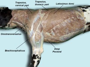
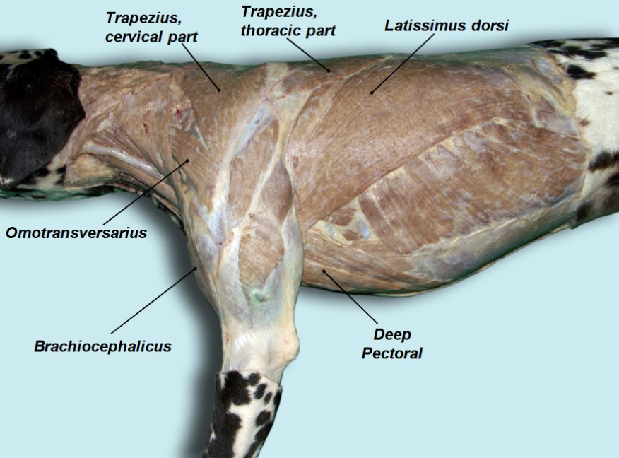
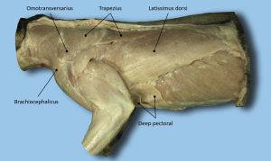
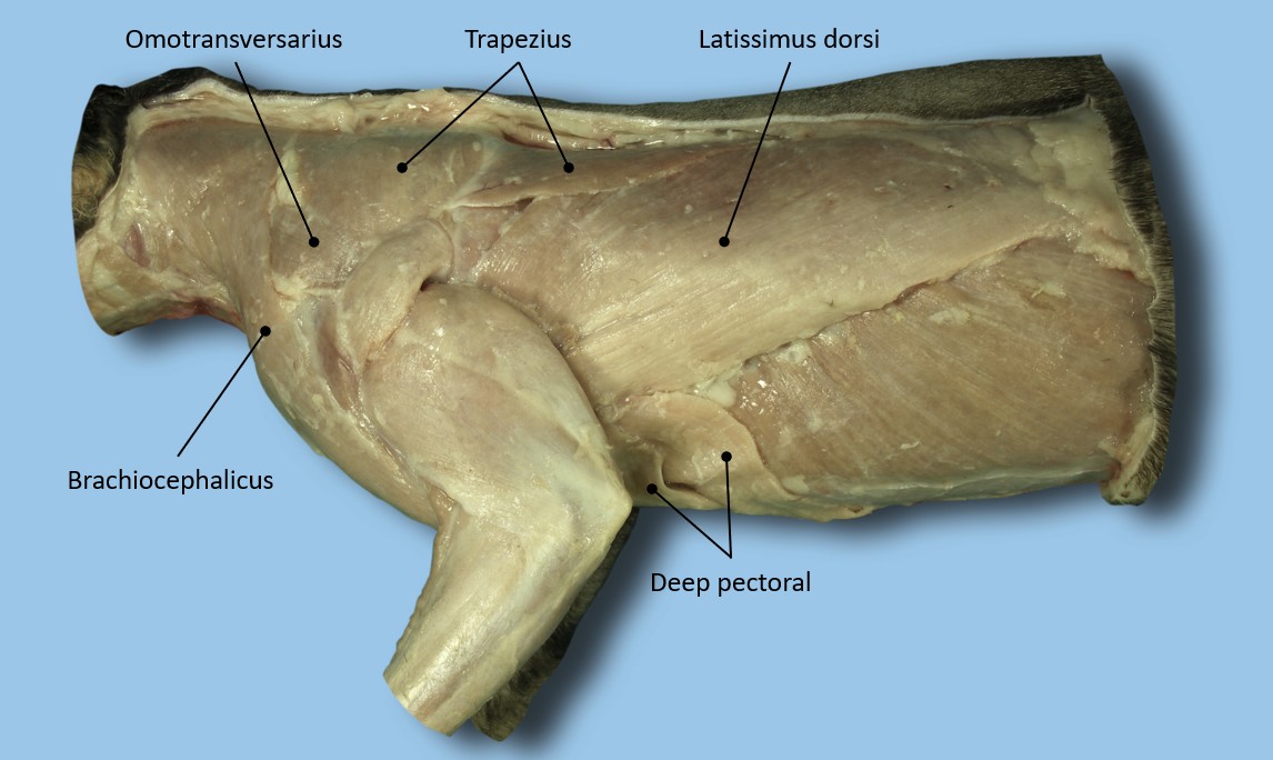
Deep pectoral m.
The deep pectoral extends from the sternum to the humerus and is larger and longer than the superficial pectoral m. It lies largely under the thoracic mammae, and the ventral portion of the cutaneous trunci. The papilla of the caudal thoracic mamma usually lies at the caudal border of the muscle. Only the cranial part is covered by the superficial pectoral muscles. An abdominal slip of this muscle is often present on the caudolateral border.
AXIAL ATTACHMENT: Ventral sternum
LIMB ATTACHMENT: Lesser and greater tubercles (of humerus)
ACTION: Adduct limb
-
- Superficial muscles of neck and thorax of the dog, ventral aspect. 1
-
- Superficial muscles of the left scapula and brachium of the dog 1
-
- Deeper muscles of the left scapula, brachium, and antebrachium, lateral view. 1

-
- Superficial musculature of the head, neck, and thoracic girdle, left lateral view. 4
-
- Dog cutaneous trunci
-
- Dog extrinsic muscles. 40
-
- Cat extrinsic muscles. 40
Dissect: Thoroughly clean both the superficial and deep pectoral muscles. Specific separation of the parts of the superficial pectoral m. is not required and is often difficult to do in the cat anyway. Fat tissue and mammary gland tissue may need to be removed to isolate the muscles. Palpate and visualize the muscle attachments. Visualize the muscle’s position and action on the skeleton and, if available, attempt to palpate it on a live dog or cat outside of lab.
Transect the superficial pectoral m. (transverse and descending parts together) ~1 cm from the sternum, and reflect it toward the humerus.
Transect the deep pectoral m. ~ 2 cm from, and parallel to, the sternum, and reflect it to as close to its insertion on the humerus as possible. There are a number of small nerves between the deep pectoral m. and body wall. These may be transected.
The superficial fascia of the neck is continued on the head as the superficial fascia of the various regions of the head. Caudally, it becomes continuous with the superficial brachial and pectoral fascia. Some of the fascia is also continued into the axillary space.
Notice that the external jugular vein is completely wrapped by superficial fascia. Note this vein for future orientation. Reflecting the superficial pectoral m. should have uncovered where the external jugular vein enters the thoracic inlet.
The cutaneous muscles of the neck are completely enveloped by superficial fascia. The platysma is the best developed of the cutaneous muscles of the neck and head. Its fibers sweep cranioventrally over the dorsal part of the neck and over the lateral surface of the face. The muscle may have been reflected with the skin. Its insertion will be seen when the head is dissected.
Continuing with extrinsic muscles of the thoracic limb:
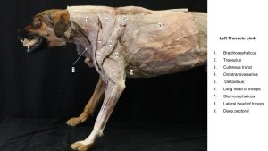
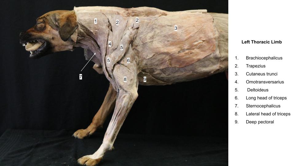
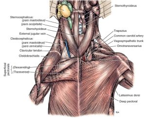
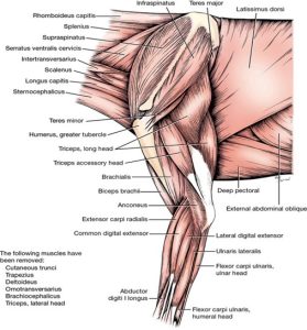
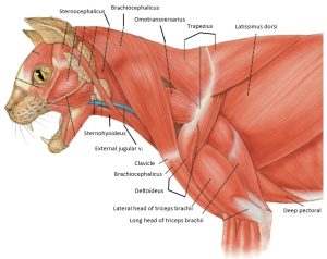
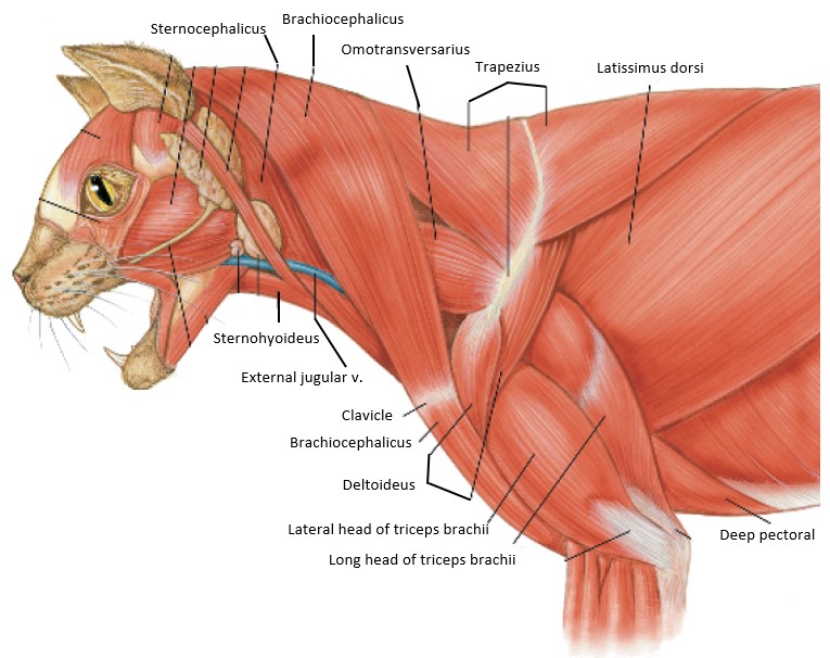
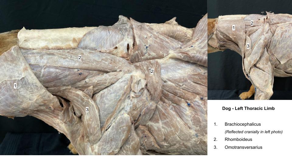
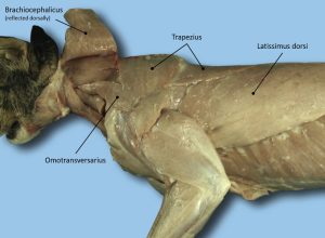
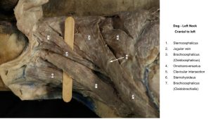
Brachiocephalicus m.
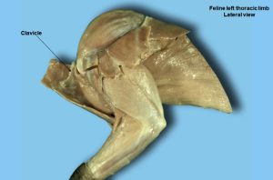
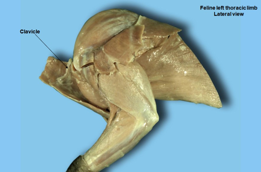
The brachiocephalicus is a compound muscle developmentally, although it appears as one muscle that extends from the arm to the head and neck. One end attaches on the distal third of the humerus, where it lies between the biceps brachii medially and the brachialis laterally. In the cat, this attachment is further distal, on the ulna. Proximally on the humerus it partly covers the pectoral muscles at their insertions and lies craniomedial to the deltoid muscle. It crosses the cranial surface of the shoulder, divides into two parts, and obliquely traverses the neck. At the shoulder a faint line crosses the muscle. This is the edge of a fibrous plate, the clavicular intersection, on the deep surface of which the clavicle (collarbone) is connected. In the cat, the clavicle is a bony remnant, palpable, and seen on radiographs. In the dog, the clavicle is vestigial. A band of connective tissue can be felt extending from this vestigial clavicle to the manubrium of the sternum and to the scapula. Although the clavicle has lost its functional significance in the dog and cat, it is still considered the origin of the components of the brachiocephalicus muscle.
Dissect: Clean the brachiocephalicus m.
Identify the clavicular intersection, about halfway between the proximal and distal attachments of the brachiocephalicus muscle. Palpate the clavicle in the cat on the deep side of the brachiocephalicus m. about halfway between its proximal and distal attachments.
The muscle distal to the clavicular intersection that attaches to the humerus in the dog, and ulna in the cat, is the cleidobrachialis. The muscle that extends from the clavicular intersection to the neck and head is the cleidocephalicus. The cervical part of the cleidocephalicus is bounded caudally by the trapezius and cranially by the occipital part of the sternocephalicus.
Note: The cleidocephalicus itself also has two parts. You are not responsible for knowing the subparts of the cleidocephalicus, but it can be useful to understand them when learning attachments. These parts are the thin pars cervicalis, which attaches to the dorsal midline of the neck; and a thicker pars mastoidea, which attaches to the mastoid process of the temporal bone of the skull (same location as the pars mastoidea of the sternocephalicus m.).
AXIAL ATTACHMENT: Cranial midline neck and temporal bone (mastoid process)
Clavicular intersection – represents approximate midpoint of brachiocephalicus m.
LIMB ATTACHMENT: Cranial, distal humerus
Cat difference: distal attachment on proximal ulna
ACTION: Extend shoulder joint; draw head and neck to the side.
-
- Superficial muscles of the left scapula and brachium of the dog 1
-
- Superficial muscles of neck and thorax of the dog, ventral aspect. 1
-
- Superficial musculature of the head, neck, and thoracic girdle, left lateral view. 4
-
- Superficial and deep musculature of the head and neck of the cat, ventral view. 4

-
- Dog cutaneous trunci
-
- Cat extrinsic muscles. 40
-
- Cat clavicle. 40
Omotransversarius m.
The omotransversarius is in a deeper plane than the cleidocephalicus (cranial part of the brachiocephalicus). It is straplike and extends from the distal end of the spine of the scapula to the atlas. It is related to the deep cervical fascia medially. Its caudal part is subcutaneous, but cranially it is covered by the cervical part of the cleidocephalicus.
AXIAL ATTACHMENT: Wing of atlas (C1 vertebra)
LIMB ATTACHMENT: Acromion of scapula and scapular spine
ACTION: Advance the limb; flex neck laterally
Dissect: Clean the omotransversarius m.
-
- Superficial muscles of the left scapula and brachium of the dog 1
-
- Superficial musculature of the head, neck, and thoracic girdle, left lateral view. 4
-

- Dog shoulder
-
- Cat omotransversarius. 40

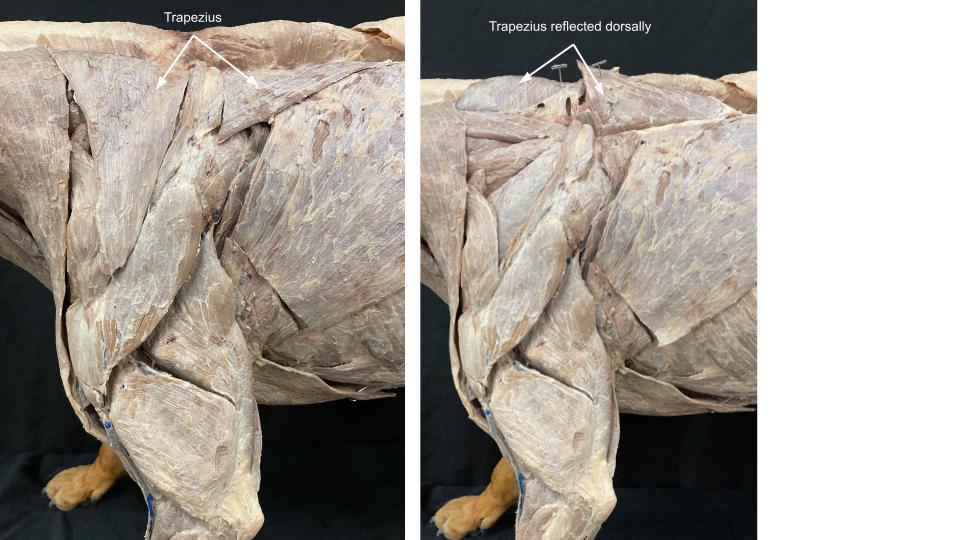
Trapezius m.
The trapezius is thin and triangular. It is divided into cervical and thoracic parts, separated by an aponeurosis. There is no need to separate these parts. The muscle as a whole extends from the median raphe of the neck and the supraspinous ligament to the spine of the scapula. The cervical part is overlapped by the cervical part of the cleidocephalicus whereas the thoracic part overlaps the latissimus dorsi.
AXIAL ATTACHMENT: Dorsal midline – neck to cranial thorax
LIMB ATTACHMENT: Scapular spine
ACTION: Draw scapula dorsally (i.e. elevate scapula/limb like ‘shrugging shoulders’)
Dissect: Clean the cranial and caudal borders of the trapezius m. Then incise the trapezius parallel to and along both sides of its attachment to the spine of the scapula. Reflect the muscle dorsally.
-
- Superficial muscles of the left scapula and brachium of the dog 1
-
- Superficial musculature of the head, neck, and thoracic girdle, left lateral view. 4
-

- Dog brachium
-
- Cat extrinsic muscles. 40
Rhomboideus m.
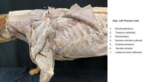
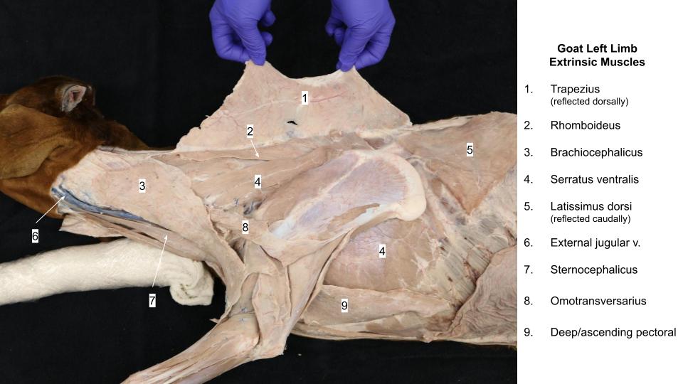
The rhomboideus lies deep to the trapezius and holds the dorsal border of the scapula close to the body. It has capital, cervical, and thoracic parts in carnivores (you will not be responsible for these individually). The narrow rhomboideus capitis attaches the cranial dorsal border of the scapula to the nuchal crest of the occipital bone. The rhomboideus cervicis runs from the dorsal border of the scapula to the median raphe of the neck. The rhomboideus thoracis is short and thick, connecting the dorsal border of the scapula to the spines of the first seven thoracic vertebrae. Its caudal border is deep to the latissimus dorsi. The cervical and thoracic parts of the rhomboideus are contiguous on the dorsal border of the scapula.
AXIAL ATTACHMENT: Skull, and dorsal midline – neck to cranial thorax
LIMB ATTACHMENT: Dorsal border of scapula
ACTION: Draw scapula dorsally (elevate scapula/limb)
Dissect: Clean the rhomboideus m.
-
- Deeper muscles of the left scapula, brachium, and antebrachium, lateral view. 1
-
- Dog shoulder
-
- Dog rhomboideus. 40

Latissimus dorsi m.
The latissimus dorsi is large and roughly triangular. It lies caudal to the scapula and covers much of the dorsal and dorsolateral thoracic wall.
AXIAL ATTACHMENT: Thoracolumbar fascia (mostly)
LIMB ATTACHMENT: Teres major tuberosity (of humerus)
ACTION: Flex shoulder joint; draw free limb caudally as in digging.
Dissect: Clean the latissimus dorsi m., and then 2-3 cm caudal to the forelimb, where the muscle converges towards the axilla, transect it at a right angle to its fibers.
The thoracolumbar fascia is the deep fascia of the trunk. It arises from the supraspinous ligament and spines of the thoracic and lumbar vertebrae; covers the muscles of the vertebrae, ribs, and abdomen; and fuses with the opposite fascia on the ventral midline median fibrous raphe, i.e. the linea alba (considered in detail later). The thoracolumbar fascia serves as an attachment for numerous muscles. It will be further considered with back and abdominal muscles.
-
- Deeper muscles of the left scapula, brachium, and antebrachium, lateral view. 1
-
- Superficial musculature of the head, neck, and thoracic girdle, left lateral view. 4
-
- Cat cutaneous trunci
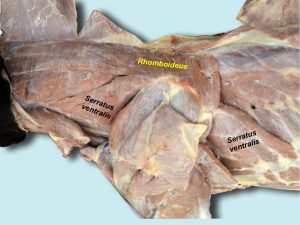
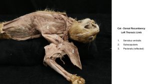
Serratus ventralis m.
The serratus ventralis cervicis and serratus ventralis thoracis form a continuous large fan-shaped muscle that passes from the cervical vertebrae and ribs to the dorsomedial aspect of the scapula and acts as a sling to support the body between the limbs. The attachment of the slips of muscle to the cervical vertebrae and ribs have the appearance akin to the cutting edge of a serrated knife, hence the muscle’s name.
AXIAL ATTACHMENT: Cervical vertebrae and ribs.
LIMB ATTACHMENT: Serrated face of scapula
ACTION: Support the trunk
Dissect: Reflect the latissimus dorsi m. as needed to view the thoracic part of the serratus ventralis. Clean the borders of the thoracic serratus ventralis caudal to the forelimb. Clean the cranial border of the cervical serratus ventralis where accessible between the more superficial extrinsic TL muscles.
Observe: Visualize the attachments of the serratus ventralis in a standing animal to appreciate how the trunk is suspended between the limbs.
-
- Deeper muscles of the left scapula, brachium, and antebrachium, lateral view. 1
-
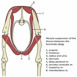
- Muscles suspending the thorax. University of Minnesota
-
- Dog serratus ventralis
-
- Dog rhomboideus. 40
-
- Cat serratus ventralis
Clinical Application:
It is at times necessary to perform a thoracic limb amputation. In doing so, all of the extrinsic muscles must be transected! Can you name all eight of them?
Ventral neck muscles
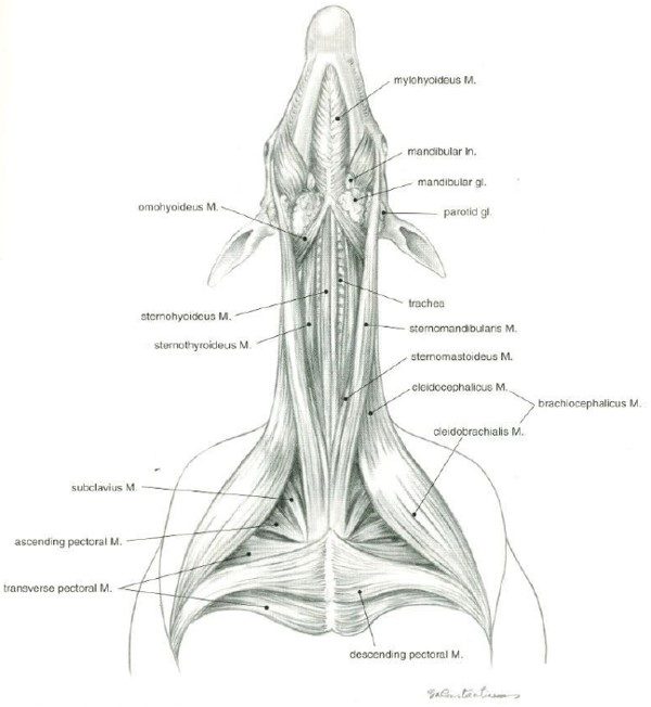
The following three muscles are not extrinsic mm. of the thoracic limb. However, they are conveniently identified at this time due to their proximity and relationship to muscles already studied. ‘Strap’ is a reference, typically to the sternohyoid and sternothyroid muscles, because of being long, relatively thin bands of muscle running the length of the neck.
Note: During embalming (on whatever side), the incision and dissection made to access the blood vessels frequently disrupts the strap muscles. This can make finding midline challenging and portions of the strap muscles may be disrupted.
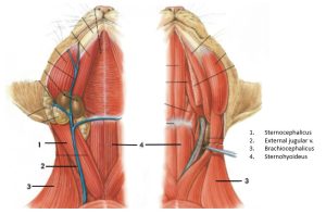
Sternocephalicus m.
The sternocephalicus arises on the sternum and inserts on the head. At the cranial end of the sternum the muscle is thick, rounded, and closely united with its fellow on the opposite side. Even after the main parts of the paired muscle diverge, there may be considerable crossing of fibers between the two on the ventral surface of the neck. Note: The cranial part of the muscle divides into two parts, in the carnivore, ie. the occipital and mastoid parts. You are not responsible for naming the parts, but don’t confuse them for another muscle. The dorsal border of the sternocephalicus is adjacent to the ventral border of the cleidocephalicus (part of the brachiocephalicus). The external jugular vein crosses its lateral surface obliquely.
CAUDAL ATTACHMENT: 1st sternebrae (aka manubrium)
CRANIAL ATTACHMENT: Occipital bone and mastoid process of temporal bone
ACTION: To draw the head and neck to the side
-
- Superficial muscles of neck and thorax of the dog, ventral aspect. 1
-
- Superficial and deep musculature of the head and neck of the cat, ventral view. 4
-
- Dog brachiocephalicus
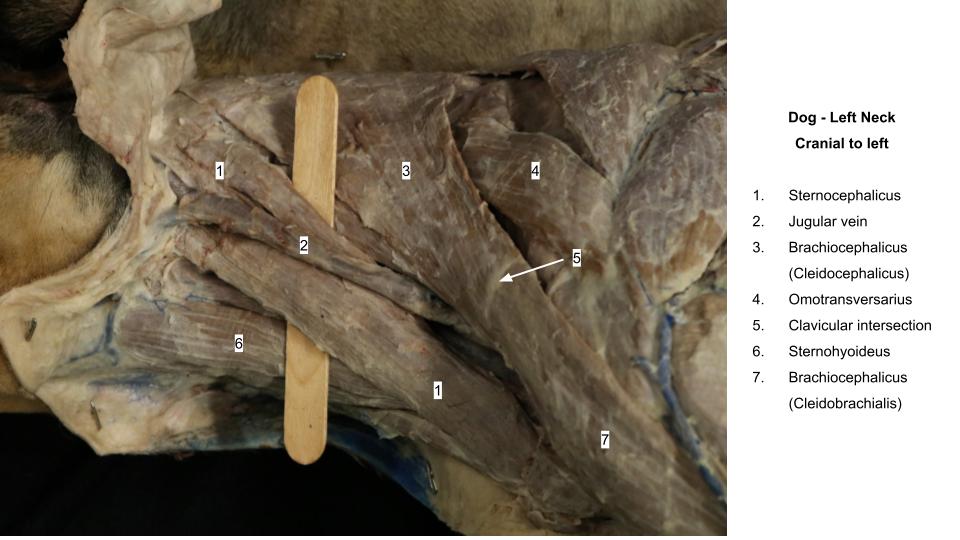
Observe: The sternohyoideus and sternothyroideus muscles are both covered by the deep fascia of the neck and lie dorsal (deep) to the sternocephalicus at their origin. Sternohyoideus pairs run in a straight cranial/caudal direction, while sternothyroideus pairs form more of a “V” shape (when viewed ventrally) because of their lateral attachment to the thyroid cartilage of the larynx.
Sternohyoideus m.
The sternohyoideus lies on the trachea, covered by the sternocephalicus caudally. A midventral groove indicates the separation of right and left sternohyoideus muscles.
CAUDAL ATTACHMENT: 1st sternebrae & 1st costal cartilage
CRANIAL ATTACHMENT: Hyoid apparatus
ACTION: Pull tongue & larynx caudally
-
- Superficial muscles of neck and thorax of the dog, ventral aspect. 1
-
- Superficial and deep musculature of the head and neck of the cat, ventral view. 4
-

- Dog sternohyoideus.40
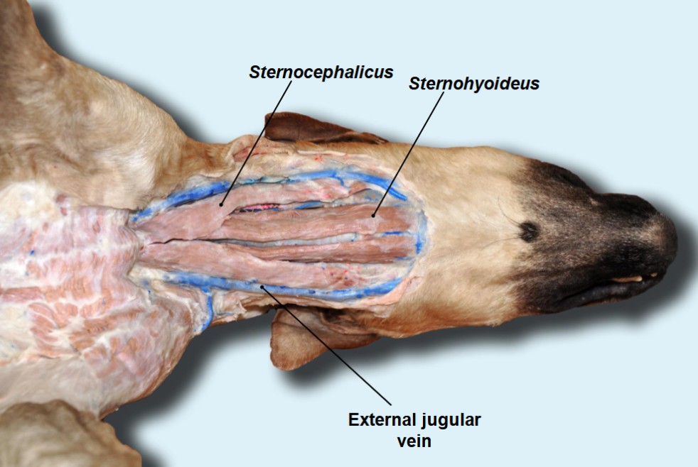
Sternothyroideus m.
The sternothyroideus is covered at its origin by the sternohyoideus. The sternothyroideus inserts on the lateral surface of the thyroid cartilage. The left muscle is bounded dorsally by the esophagus and medially by the trachea.
CAUDAL ATTACHMENT: 1st costal cartilage
CRANIAL ATTACHMENT: Thyroid cartilage of larynx.
ACTION: Pull tongue & larynx caudally
-
- Superficial muscles of neck and thorax of the dog, ventral aspect. 1
-
- Superficial and deep musculature of the head and neck of the cat, ventral view. 4
-

- Dog sternothyroideous. 40
Ungulate Comparative Anatomy
Cutaneous trunci m. and extrinsic thoracic limb mm.
Observe: When you have completed the dissection on a carnivore – as time and space around the large animal cadavers permits, examine these prosected animals and identify the same muscles. Note the specific comparative features described below, per the large animal you are examining.
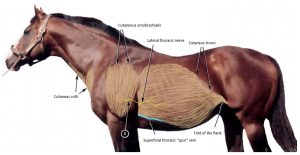
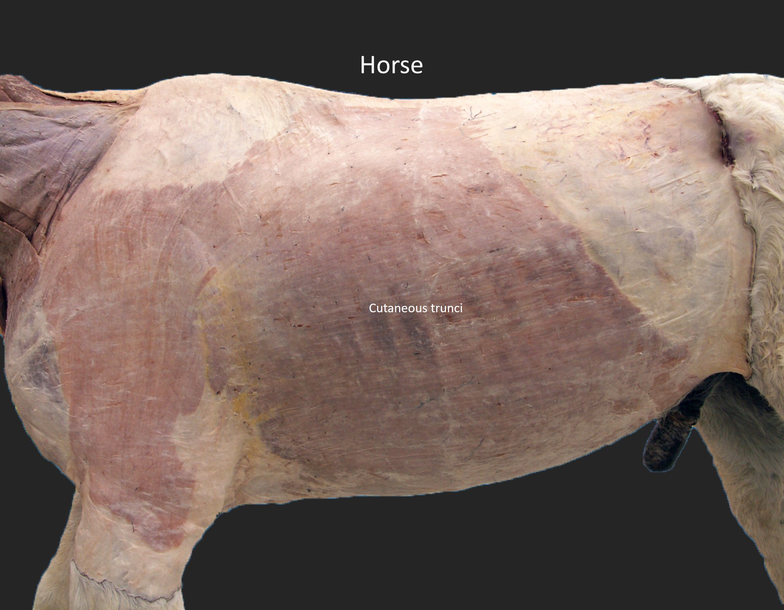
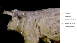
Cutaneous trunci m. – horse and ruminant
During skin reflection, the large cutaneous trunci m. may be partially removed with the skin. Its thick caudoventral border forms the fold of the flank cranial to the thigh/stifle region.
Clinical relevance:
Thick cutaneous muscle, well developed in horse and ox, fibers converge caudally into fold of flank, provides for vigorous skin twitching.
-
- Cutaneous muscles of the neck, shoulder and trunk of the horse. 2

-

- Horse cutaneous trunci 40
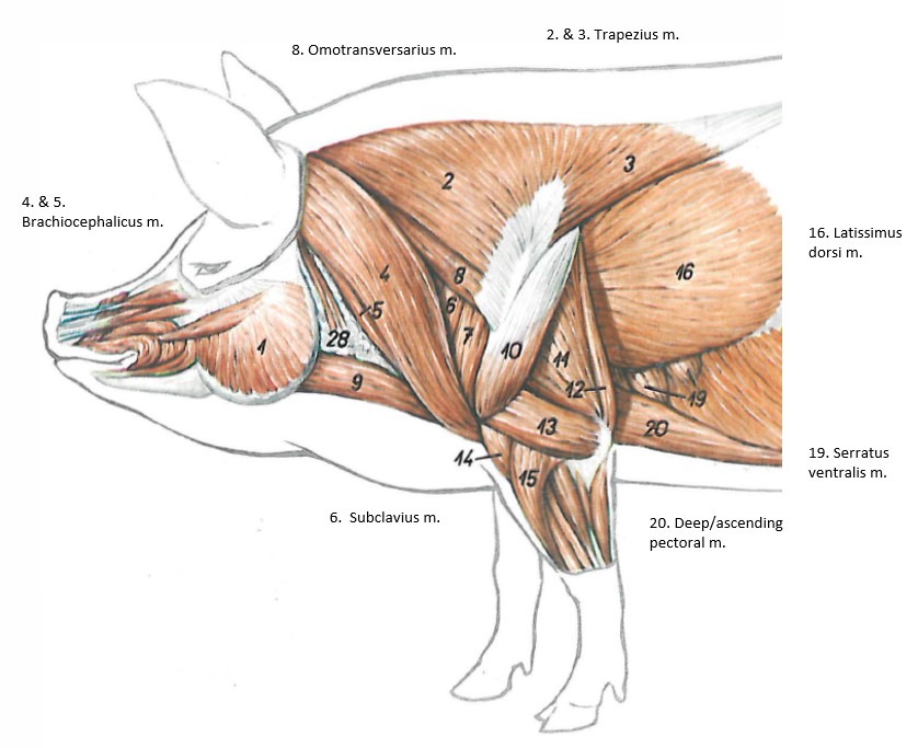
Extrinsic Muscles to Observe in Ungulates:
Click the link to jump back to the carnivore muscle description
- Cutaneous trunci (not a true extrinsic)
- Superficial pectoral
- Deep pectoral
- Brachiocephalicus
- Omotransversarius
- Trapezius
- Rhomboideus
- Latissimus dorsi
- Serratus ventralis
- Subclavius
Horse anatomy
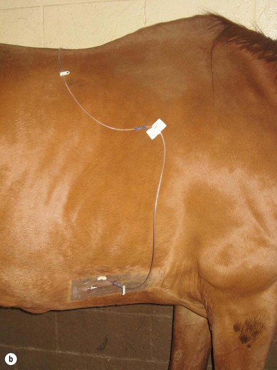
In the horse, at the level of the elbow, a large vein courses horizontally between the margins of the cutaneous trunci and deep (ascending) pectoral mm. This is the superficial thoracic vein, which because of its position is sometimes called the “spur vein.”
Observe: Observe the superficial thoracic v. It may have been reflected with the skin i.e. look for it on the reflected skin, as available.
Clinical relevance:
The superficial thoracic v. is a site for IV catheterization in the horse.
-

- Lateral thoracic v. catheter. veteriankey.com
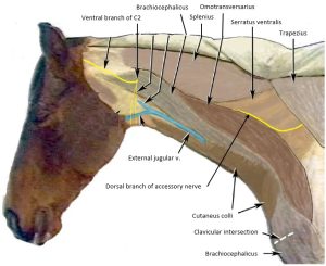
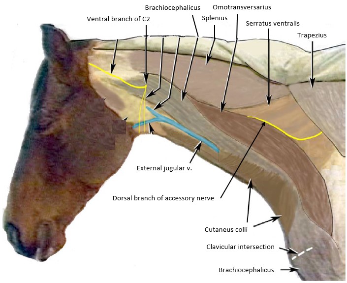
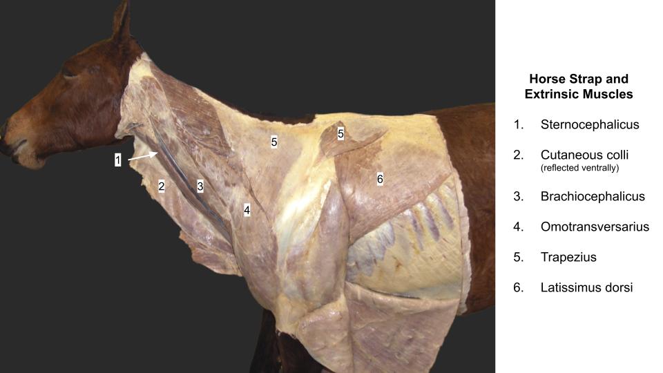
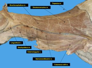
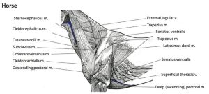
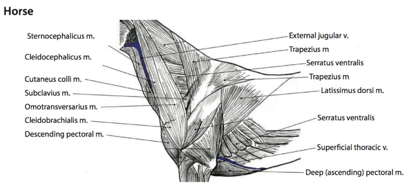
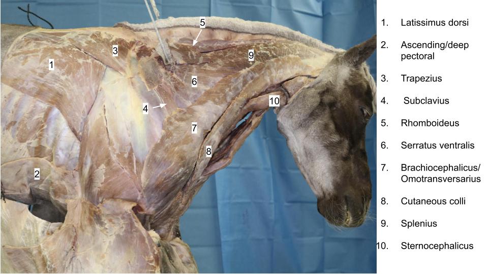
The ventral border of the omotransversarius m. is fused to the brachiocephalicus m. in the horse. We don’t separate them during dissection. The dorsal ‘half’ is omotransversarius and the ventral ‘half’ (forming the border of the jugular groove) is the brachiocephalicus m.
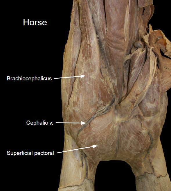
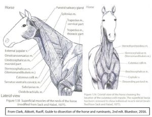
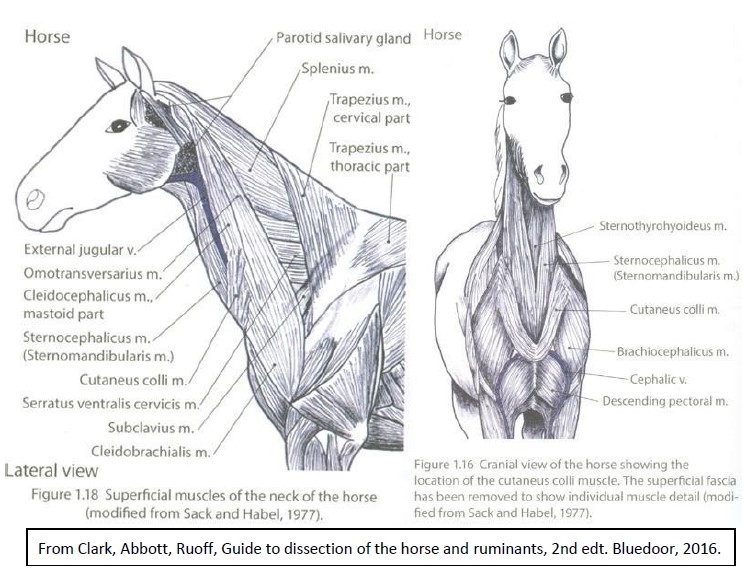
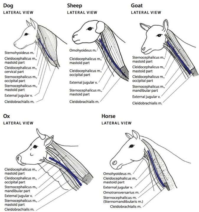
The brachiocephalicus m. subparts include the cleidobrachialis and the cleidocephalicus (mastoid part). The sternocephalicus m. is one muscle in the horse (often called the sternomandibularis) (FYI – see Appendix Table A1-1 and Fig A1-1 for a comparative understanding of the muscle subparts). The superficial pectoral m. has descending and transverse parts. The descending part (‘chest muscle’) forms a thick muscle belly in the horse for IM injections.
-
- Cutaneous muscles of the neck, shoulder and trunk of the horse. 2
-
- Superficial structures of horse neck. 2
-
- Deeper structures of horse neck. 2
-

- Horse strap and extrinsic mm. 40
-

- Horse superficial pectoral 40
Clinical relevance:
The brachiocephalicus and sternocephalicus mm. form the dorsal and ventral boundaries of the jugular groove respectively. This groove accommodates the external jugular vein which is commonly used for venipuncture in the horse and ruminants. The superficial pectoral m. is an intramuscular injection site in the horse.
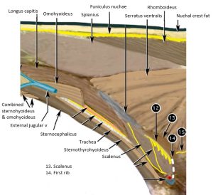
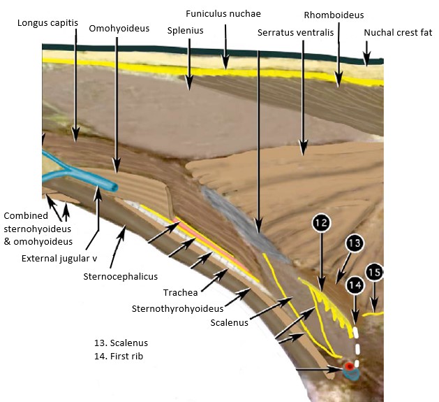
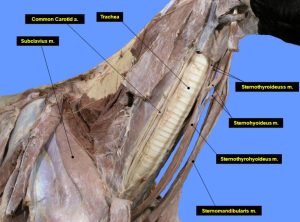
There is also a medial boundary (‘backstop’) of the jugular groove in the cranial half of the neck in the horse and this muscle is the omohyoideus m. (not present in carnivores). The omohyoideus. m. is deceptively long, extending from deep fascia near the shoulder and passing cranially to insert on the hyoid apparatus with its opposite number (and the sternohyoideus mm.)
Observe: Retract the external jugular v. and other muscles as needed in the cranial half of the neck of the horse to observe the omohyoideus m.
-
- Horse neck muscles. 40

-
- Horse strap muscles
Clinical relevance:
Observe: Identify and note the relationship of the cutaneus colli m. to the external jugular vein. Observe that the muscle covers the caudal (distal) part of the external jugular vein and becomes progressively thicker as the vein approaches the thoracic inlet.
The relationship of the cutaneus colli m. to the external jugular vein is another reason why the preferred site for venipuncture in the horse is in the cranial part of the neck.
-
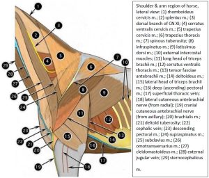
- Shoulder & arm region of horse, lateral view. 2

-
- Superficial muscles of the neck of the horse 6
-
- Extrinsic muscles of the thoracic limb of the horse. 6
-

- Horse shoulder
-
- Horse neck muscles. 40
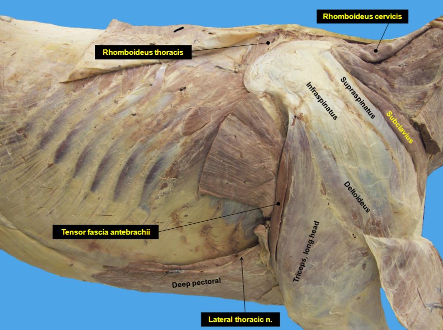
Well developed in the horse (and pig), the subclavius m. is like an extra pectoral muscle and can be considered an extrinsic muscle of the thoracic limb. The subclavius m. is vestigial in the ox and sheep (where it consists of a narrow band of muscle extending from the first costal cartilage to blend with the deep face of the brachiocephalicus muscle) and it is a little more developed in the goat. The subclavius m. of the horse can be palpated in the live animal.
The rhomboideus m. in the horse and ruminants differs from carnivores in not having a capitis (head) belly. The cervicis (neck) and thoracis (thorax) bellies are present.
-
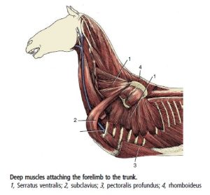
- Deep muscles attaching the forelimb to the trunk of the horse. 8

-
- Horse subclavius. 40

-

- Horse subclavius 40
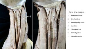
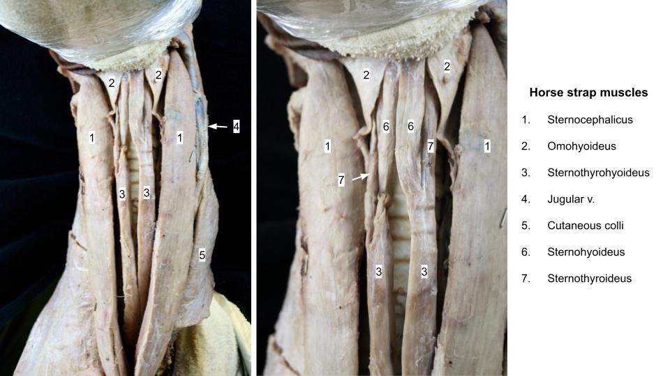
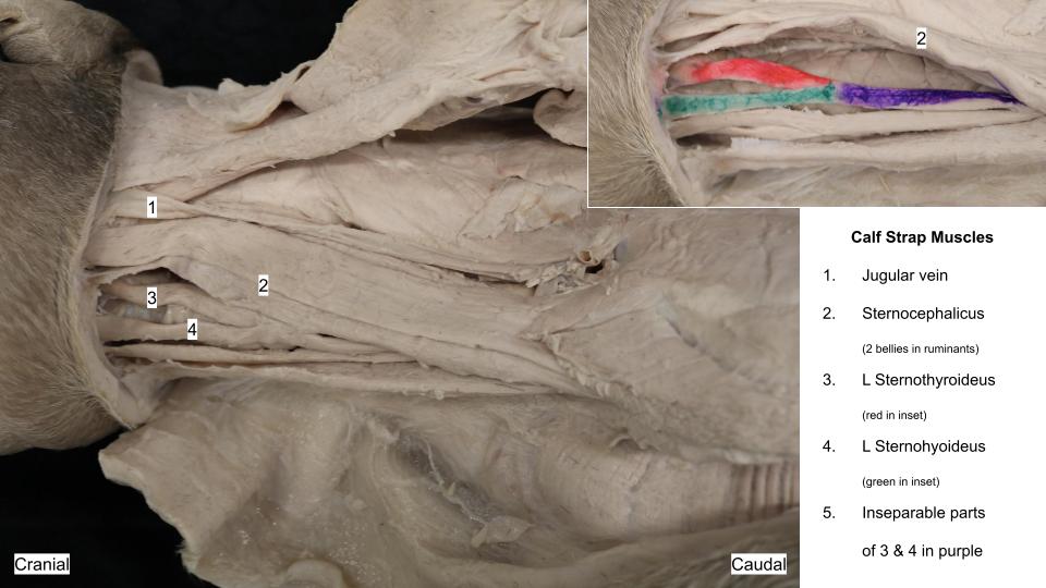
The ‘strap muscles’
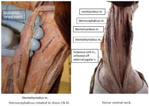
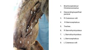
Identify the ventral (‘strap’) muscles of the neck lying on the surface of the trachea. As in the carnivore, there is a left and right sternothyroideus m. and a left and right sternohyoideus m. i.e. 4 muscles. Caudally, the left set fuse with each other, and the right set do the same, and these fused muscles are referred to as the sternothyrohyoideus mm. Left and right sternothyrohyoideus mm. are also very closely joined on midline, caudally. In the horse, on the left side, the ventral branch of the Accessory n. (CN XI) entering the belly of the sternocephalicus m. at its cranial myotendinous junction may be noted, by rotating the muscle to look at its medial (inner) surface. We will revisit this nerve in the nervous system.
-
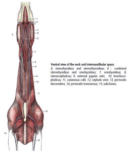
- Ventral view of the neck and intermandibular space of the horse. 8

-
- Horse strap muscles 2

-
- Horse strap muscles
-
- Horse strap muscles
Clinical relevance:
Hypertrophy of the ventral neck muscles occurs in horses that crib (crib-biting or “wind-sucking”). The Modified Forssell’s procedure involves excising a segment of the “strap muscles” (combined name is the sternothyrohyoideus mm. at their caudal fused portion) and omohyoideus m., and transecting the innervation of the sternocephalicus m. Partial myectomy of the strap muscles or a sternothyroideus tenectomy may also be performed to help manage dorsal displacement of the soft palate (DDSP) in horses. The sternohyoideus mm. are separated on midline during a tracheotomy.
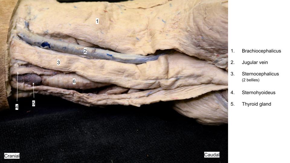
Ruminant anatomy
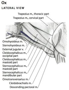
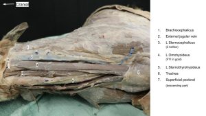
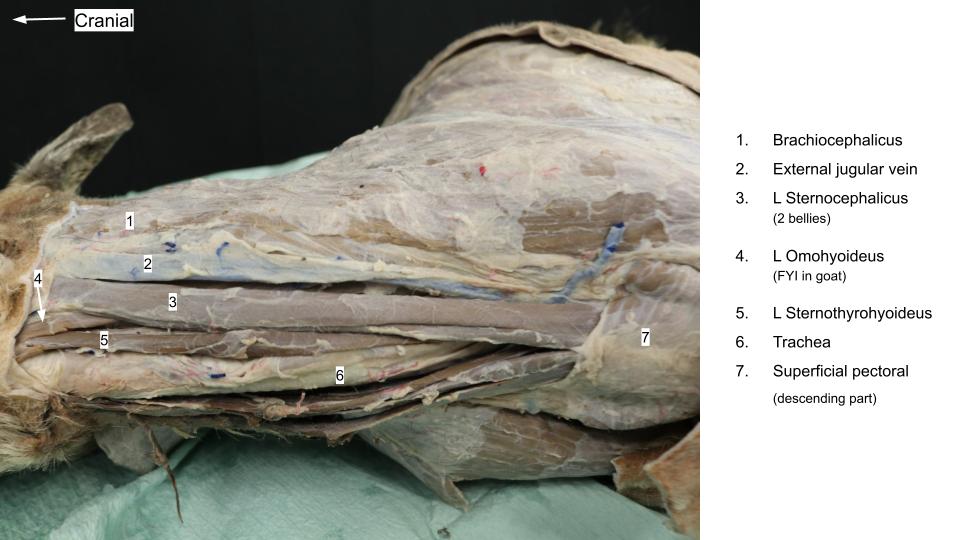
The brachiocephalicus m. subparts include the cleidobrachialis and the cleidocephalicus (mastoid part). Ruminants also have an occipital part to their cleidocephalicus m.. The sternocephalicus m. has two parts in the ox and goat (sternomandibularis and sternomastoideus). In the goat ‘sternozygomaticus’ is often used in place of sternomandibularis because the muscle inserts on the zygomatic arch in this species. FYI – In sheep, there is only one part to the sternocephalicus m., the mastoid part. Without a mandibular part of the sternocephalicus the ventral border of the jugular groove in sheep is less well defined and represented only by the mastoid part, which also forms the medial border cranially.
As for the horse and carnivore, it is not necessary to memorize the names of the subparts of the brachiocephalicus and sternocephalicus mm., unless you wish to. (FYI – see Appendix Table A1-1 and Fig A1-1 for a comparative understanding of the muscle subparts).
-
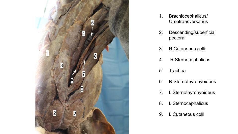
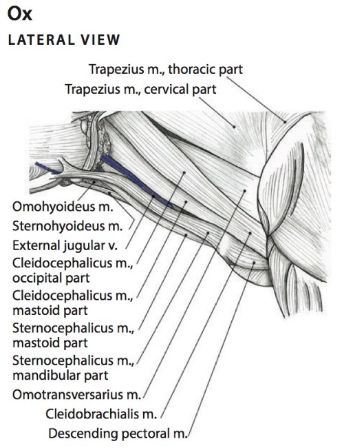
- Extrinsic Muscles of the Thoracic Limb of the Ox. 6
-
-
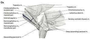
Superficial muscles of the neck of the ox.
5
-
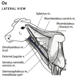
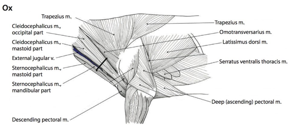
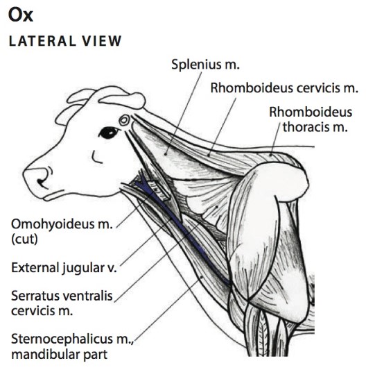
- Deep muscles of the neck of the ox, lateral view. The occipital part of cleidocephalicus and trapezius muscles have been removed. 5
-
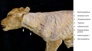
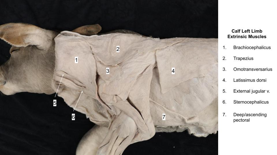
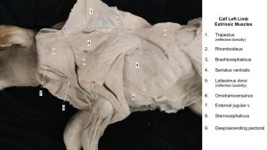
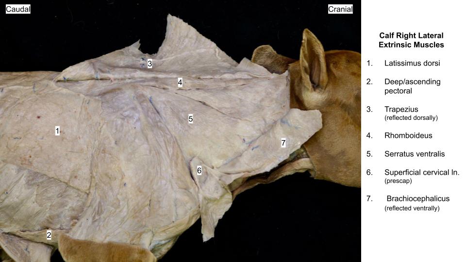
- Calf shoulder

-
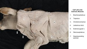
- Calf extrinsic limb mm.
-
- Calf extrinsic limb mm.

-

- Calf shoulder
-
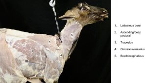
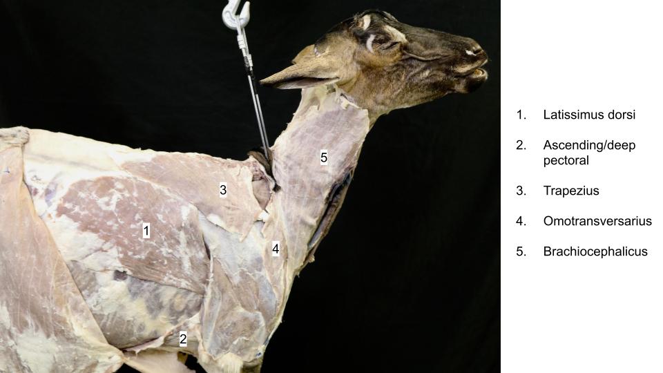
- Goat shoulder
-
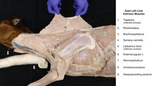
- Goat extrinsic mm.
-
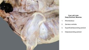
- Cow deep extrinsic muscles. 40
Clinical relevance:
The brachiocephalicus and sternocephalicus mm. form the dorsal and ventral boundaries of the jugular groove respectively. This groove accommodates the external jugular vein which is commonly used for venipuncture in the horse and ruminants.
There is also a medial boundary (‘backstop’) of the jugular groove in the cranial half of the neck, and in the ruminant this a subpart of the sternocephalicus m. (i.e. the sternomastoideus m.).
Observe: To view this “backstop” to venipuncture in the ruminant, retract the cranial third of the external jugular vein from its groove. This should expose a subpart of the sternocephalicus m. i.e. the sternomastoideus m. FYI – the omohyoideus m. is also readily seen in the goat and is identifiable in the ox.
Clinical relevance:
The sternocephalicus (sternomastoideus part – ruminant) offers some protection to the structures of the carotid sheath lying more deeply when venipuncture is performed in the cranial half of the neck (how might you determine if you have performed an intracarotid stick i.e. before injecting anything!?).
The rhomboideus m. in the horse and ruminants differs from carnivores in not having a capitis (head) belly. The cervicis (neck) and thoracis (thorax) bellies are present. The rhomboideus m. contributes to the large hump of Brahman cattle.
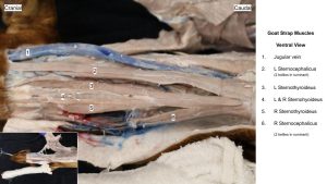
Identify the sternohyoideus and sternothyroideus mm. lying on the surface of the trachea. These thin straps of muscle are fused caudally.
-
- Goat ventral neck 22
-
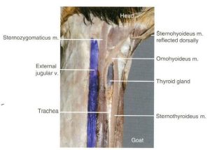
- Goat strap muscles 12

-
- Goat strap muscles

-
- Goat strap muscles
-

- Calf strap muscles
-

- Calf strap muscles
-
- Calf shoulder
-
- Calf extrinsic limb mm.
-
- Goat extrinsic mm.
Pig anatomy
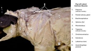
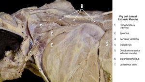
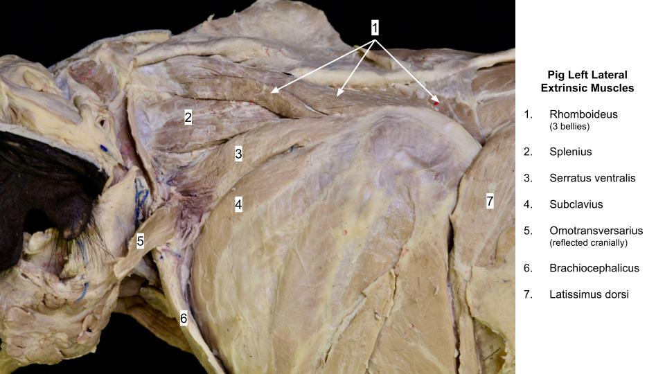
Well developed in the pig (and horse), the subclavius m. is like an extra pectoral muscle and can be considered an extrinsic muscle of the thoracic limb.
Observe: Identify the subclavius m. and the remaining extrinsic muscles of the thoracic limb in the prosected pig specimens.
-

- Extrinsic muscles of the pig. 11
-
- Pig shoulder

-
- Pig shoulder

-
- Pig shoulder
Appendices
Table A1-1: Comparison of brachiocephalicus and sternocephalicus muscles. Modified from deLahunta, A., and Habel, R. E.: Applied Veterinary Anatomy. W. B. Saunders, Philadelphia, 1986, p. 67.
| Brachiocephalicus Muscle | Sternocephalicus Muscle |
| cleidobrachialis – all species | sterno-occipitalis – dog, cat |
| cleidomastoideus – all species | sternomandibularis – horse, ox, goat |
| cleido-occipitalis – pig, ruminants | sternomastoideus – ruminants, pig, dog, cat |
| cleidocervicalis – dog, cat |
Review Videos
Muscle Chart/Terms List
|
Muscle/structure |
Muscle attachments/comments |
Muscle actions/comments |
| CUTANEUS TRUNCI M. |
|
Twitches skin |
| SUPERFICIAL PECTORAL M. |
|
Adduct limb |
| DEEP PECTORAL M. |
|
Adduct limb |
| BRACHIOCEPHALICUS M. |
|
Extend shoulder joint; draw head and neck to the side |
| Clavicular intersection/clavicle in cat | The ‘center’ of the brachiocephalicus m. from which parts are named. | Note in carnivores only. |
| OMOTRANSVERSARIUS M. |
|
Advance the limb; flex neck laterally |
| TRAPEZIUS M. |
|
Draw scapula dorsally (elevate scapula/limb) |
| RHOMBOIDEUS M. |
|
Draw scapula dorsally
(elevate scapula/limb) |
| LATISSIMUS DORSI M. |
|
Flex shoulder joint; draw free limb caudally, as in digging action. |
| SERRATUS VENTRALIS M. |
|
Support the trunk |
| STERNOCEPHALICUS M. |
|
To draw the head and neck to the side |
| STERNOHYOIDEUS M. |
|
Pull tongue & larynx caudally |
| STERNOTHYROIDEUS M. |
|
Pull tongue & larynx caudally |
| OMOHYOIDEUS M. |
Note in horse only; no need to know attachments |
‘Backstop muscle’ of jugular groove in cranial neck. |
| CUTANEUS COLLI M. |
Note in horse only; no need to know attachments |
Covers external jugular v. in caudal neck. |
| SUBCLAVIUS M. |
Horse, pig – no need to know attachments. |
Extra pectoral muscle. |
| Superficial thoracic v. |
Horse only feature. |
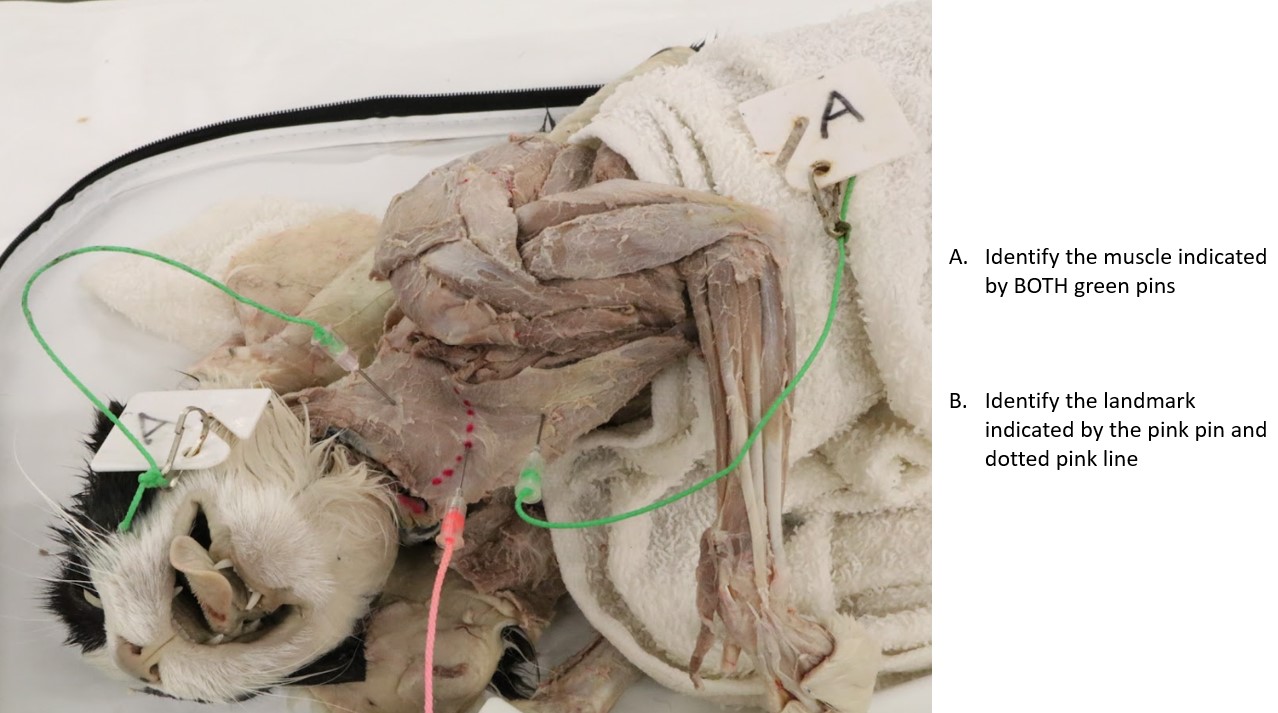
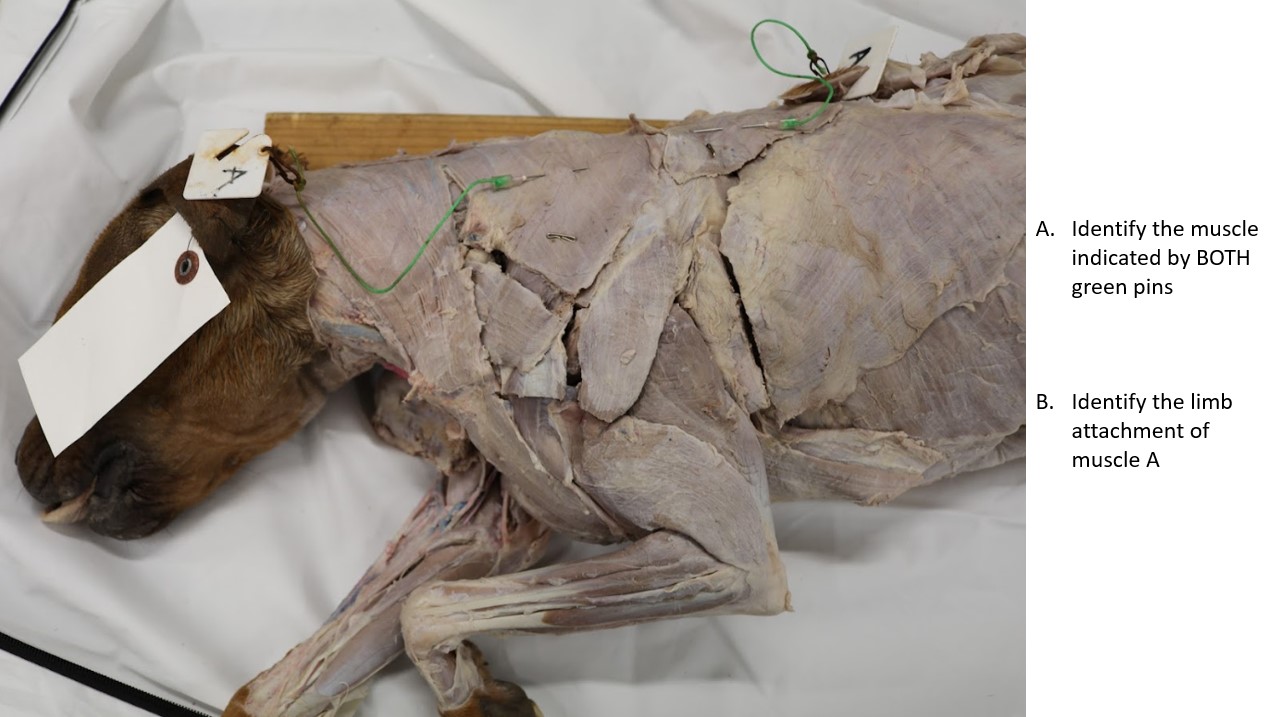
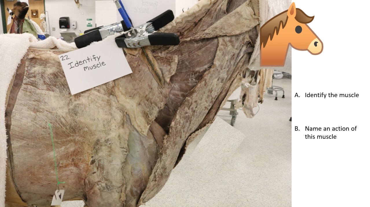
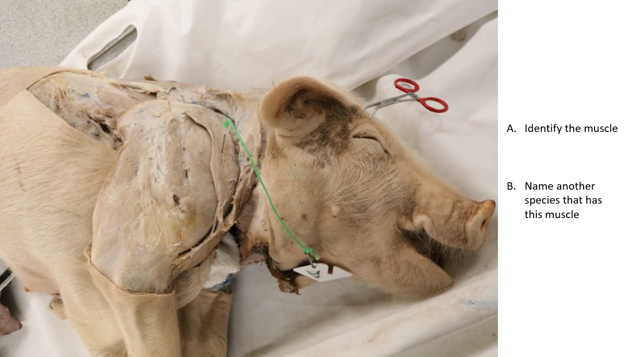
Example Practical Exam Questions