MSK LAB 4A – Thoracic Limb Shoulder/Brachium muscles
Learning Objectives
- Identify intrinsic mm. of the shoulder and brachium and associated bursae
- Name attachments of the shoulder and brachium mm.
- Name the actions of the shoulder and brachium mm.
- Categorize shoulder and brachium mm. into “muscle action groups”
- Explain the anatomy of “sweeny”, the anatomical causes of a ‘dropped elbow’ appearance, the key relationship between the radial n., the ulnar, and the triceps brachii m.
We are studying intrinsic muscles of the thoracic limb in this lab. ‘Intrinsic’, a general term, in this instance refers to muscles that attach only between bones of the thoracic limb. Put another way, intrinsic muscles of the thoracic limb are solely associated with the thoracic limb structure. Recall that extrinsic muscles attach between the thoracic limb and somewhere else on the body, i.e. these muscles are not solely associated with the thoracic limb, they also affect other parts of the body.
Lab instructions:
For this lab, teams will focus on dissecting the intrinsic shoulder and brachial muscles, in canine and feline cadavers. This carnivore understanding can then be applied to the prosected large animal cadavers to identify the same muscles (and any extras) as directed. A link to the muscle/structure terms chart for this lab is provided below.
Shoulder and Brachium Muscle Chart
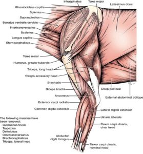
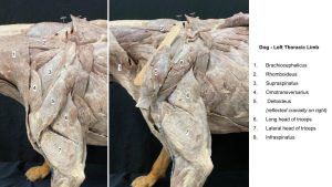
The dissection begins on the lateral shoulder and scapula (left limb) to identify the following muscles:
- Deltoideus
- Infraspinatus
- Teres minor
- Supraspinatus
Observe: Read the muscle description and then clean and identify each muscle.
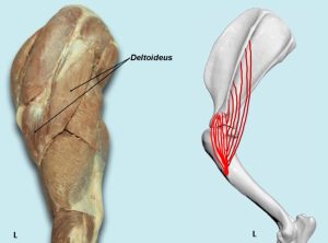
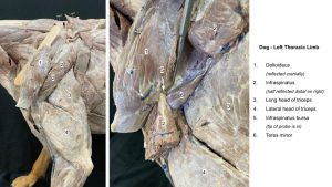
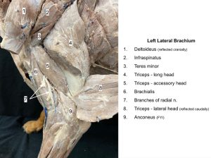
Deltoideus m.
The deltoideus is composed of two portions that fuse and act in common across the shoulder. The scapular part arises as a wide aponeurosis from the length of the scapular spine and covers the infraspinatus m. The latter muscle can be seen through this aponeurosis and should not be confused with the deltoideus. The acromial part of the deltoideus arises from the acromion and has a fusiform (elongated and tapering) shape. Both portions of the muscle fuse before they insert on the deltoid tuberosity of the humerus.
PROXIMAL ATTACHMENT: Acromion of scapula and scapular spine
DISTAL ATTACHMENT: Deltoid tuberosity (of humerus)
ACTION: Flex shoulder joint
Dissect: Transect the combined muscle ~1-2 cm distal to the acromion and reflect the stumps. Free the scapular part from the infraspinatus and work under the aponeurosis of origin to verify its attachment to the spine of the scapula.
-
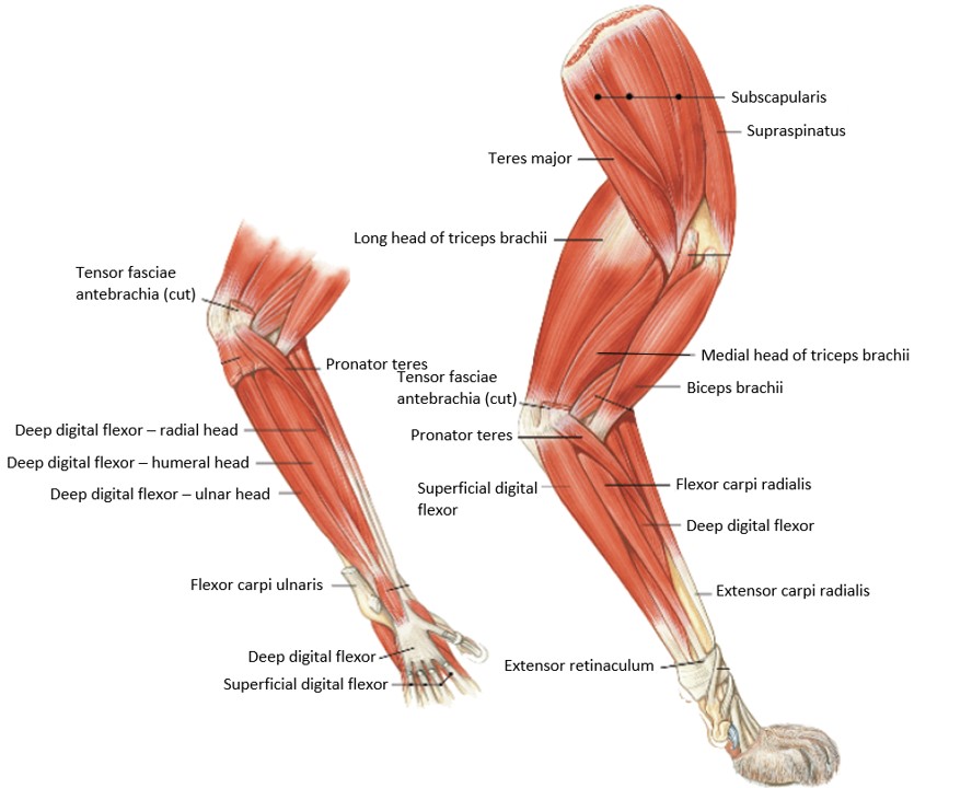
- Deeper muscles of the left scapula, brachium, and antebrachium, lateral view. 1

-

- Intrinsic muscles of the cat thoracic limb. 4
-
- Dog deltoideus. 40

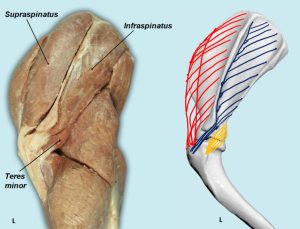
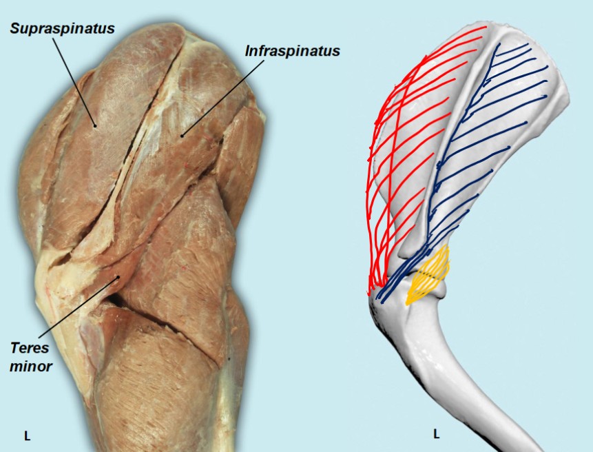
Infraspinatus m.
The infraspinatus is fusiform and lies principally in the infraspinous fossa, deep to deltoideus.
PROXIMAL ATTACHMENT: Infraspinous fossa
DISTAL ATTACHMENT: Greater tubercle (of humerus)
ACTION: Extend shoulder joint/shoulder stabilization
Dissect: Completely transect the infraspinatus across its distal third. Undermine and free the distal part from the scapula by elevating the fibers away from the spine and fossa with the handle of the scalpel. Reflect this part to its insertion on the side of the greater tubercle. This will expose the (subtendinous synovial) infraspinatus bursa between the tendon of insertion and the greater tubercle of the humerus.
A bursa is a closed connective tissue sac containing a fluid (usually synovial fluid) – imagine a pillow case closed at both ends and containing a small amount of fluid. This cushion like structure is often located between a tendon or ligament and its pathway across a bone. The bursa reduces friction as the tendon or ligament passes/glides over the bone, protecting the soft tissue structure. Bursa are constant in nature (normal anatomic structures) or acquired (develop in response to increased wear and tear over a region e.g. the point of the elbow can acquire a subcutaneous bursa). Appendix on synovial structures.
-
- Deeper muscles of the left scapula, brachium, and antebrachium, lateral view. 1
-
- Intrinsic muscles of the cat thoracic limb. 4
-
- Dog lateral shoulder. 40
-
- Dog shoulder
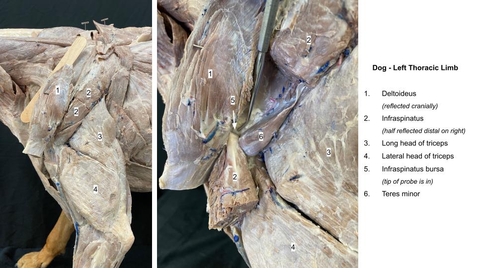
Teres Minor m.
The teres minor is a small, wedge-shaped muscle, just caudal to the shoulder joint. It is covered superficially by the deltoideus and is abutted craniodorsally by (and runs roughly parallel to) the distal extent of the infraspinatus.
PROXIMAL ATTACHMENT: Caudal border of scapula
DISTAL ATTACHMENT: Teres minor tuberosity of humerus
ACTION: Flex shoulder, rotate shoulder laterally
Dissect: Identify and clean the margins of the teres minor, tucked tightly against the distal caudal border of the infraspinatus m.
-
- Deeper muscles of the left scapula, brachium, and antebrachium, lateral view. 1
-
- Dog lateral shoulder. 40
-
- Dog shoulder
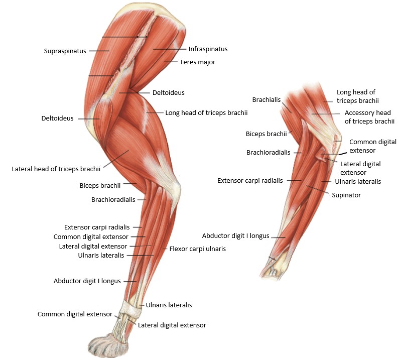
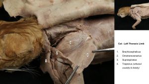
Supraspinatus m.
The supraspinatus which is wider and larger than the infraspinatus is largely covered by the cervical part of the trapezius and the omotransversarius. It lies in the supraspinous fossa and extends over the cranial border of the scapula so that a part of the muscle is closely united with the subscapularis medially.
PROXIMAL ATTACHMENT: Supraspinous fossa
DISTAL ATTACHMENT: Greater tubercle (of humerus)
ACTION: Extend shoulder joint/shoulder stabilization
Dissect: Identify and observe the insertion of this muscle on the greater tubercle of the humerus
-
- Deeper muscles of the left scapula, brachium, and antebrachium, lateral view. 1
-
- Intrinsic muscles of the cat thoracic limb. 4
-
- Dog brachium

-
- Cat shoulder

Continue to the dissection on the medial shoulder and scapular to identify the following muscles:
- Subscapularis
- Teres major
- Coracobrachialis (FYI m., not dissected)
Dissect: To view the medial muscles of the shoulder and scapula, firstly, further reflect the pectoral muscles, then transect (sever) the nerves of the brachial plexus and related vessels (thick bundle of nerves and vessels passing between limb and body). Take care not to tear the serratus ventralis completely from its attachment to the dorsal medial scapula (i.e. the serrated face), as you abduct the limb.
Observe: Read the muscle description and then clean and identify each muscle.
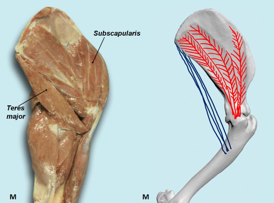
Subscapularis m.
The subscapularis occupies the entire subscapular fossa, the boundaries of which it overlaps slightly. The supraspinatus is closely associated with it cranially, whereas the teres major has a similar relation caudally. Considerable support is provided to the shoulder joint by the subscapularis muscle medially, the teres minor and infraspinatus muscles laterally, and the supraspinatus cranially. Luxation (joint dislocation) will not occur without injury to the joint capsule and its glenohumeral ligaments.
PROXIMAL ATTACHMENT: Subscapular fossa
DISTAL ATTACHMENT: Lesser tubercle (of humerus)
ACTION: Extend shoulder joint/shoulder stabilization
Dissect: Identify the muscle and clean/observe its attachment to the humerus.
-
- Muscles of left thoracic limb, medial view, canine.1

-

- Intrinsic muscles of the cat thoracic limb, medial view. 4
-

- Dog subscapularis. 40
-
- Cat brachium
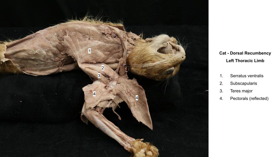
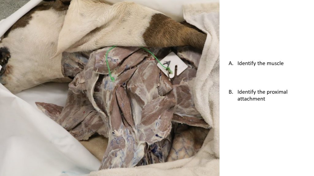
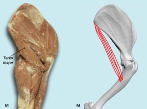
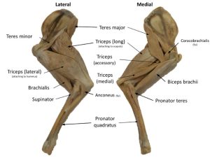
Teres Major m.
The teres major, directly caudal to the subscapularis, belies its descriptive name (teres [L] = round) because it is not round, rather, it has three surfaces. Its proximal end arises from the subscapularis and the proximal caudal border of the scapula. Fibers extend distally to join the tendon of insertion of the latissimus dorsi on to the teres major tuberosity.
PROXIMAL ATTACHMENT: Caudal border of scapula
DISTAL ATTACHMENT: Teres major tuberosity (of humerus)
ACTION: Flex shoulder joint
Dissect: Work between the distal half of the muscle and the subscapularis. Observe the close relationship between the teres major and the stump of latissimus dorsi as they approach their insertion on the proximal medial surface of the body of the humerus at the teres major tuberosity.
-
- Muscles of left thoracic limb, medial view, canine.1
-
- Intrinsic muscles of the cat thoracic limb, medial view. 4
-
- Dog teres major.40

-
- Cat brachium
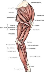
The coracobrachialis (FYI) crosses the medial surface of the shoulder obliquely. It is a small spindle-shaped muscle that arises from the coracoid process of the scapula by a relatively long tendon that courses caudodistally across the lesser tubercle. There it crosses the tendon of insertion of the subscapularis. The tendon of the coracobrachialis is provided with a synovial sheath as it crosses the lesser tubercle of the humerus. The muscle belly is distal to the lesser tubercle and medial to the origin of the medial head of the triceps. The conjoined tendon of the teres major and latissimus dorsi crosses the insertion of the coracobrachialis. The coracobrachialis inserts distally on the crest of the lesser tubercle of the humerus proximal to the teres major tuberosity. The muscle acts to adduct, extend, and stabilize the shoulder joint.
Now move to the caudal aspect of the brachium to identify:
- Tensor fasciae antebrachii
- Triceps brachii (long, lateral, accessory and medial heads)
- Anconeus (FYI m., not dissected)
This group is a large, muscular mass that almost completely fills the space between the caudal border of the scapula and the olecranon. It consists of three muscles: the triceps brachii, the tensor fasciae antebrachii, and the anconeus. By far the largest of these muscles is the triceps brachii. All of the caudal muscles of the arm are extensors of the elbow joint.
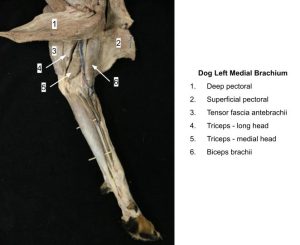
Tensor fasciae antebrachii m.
The tensor fasciae antebrachii is a broad, flat strap that extends from fascia covering the latissimus dorsi to the medial fascia of the forearm and the olecranon. It almost completely covers the medial aspect of the long head of the triceps brachii. In the cat, the tensor fasciae antebrachii m. is relatively wider and covers more of the medial surface of the triceps brachii m. than in the dog.
PROXIMAL ATTACHMENT: Fascia covering lateral side of latissimus dorsi
DISTAL ATTACHMENT: Olecranon of ulna
ACTION: Extend elbow joint (weak)
-
- Muscles of left thoracic limb, medial view, canine.1
-
- Intrinsic muscles of the cat thoracic limb, medial view. 4
-
- Dog brachium

-
- Cat brachium
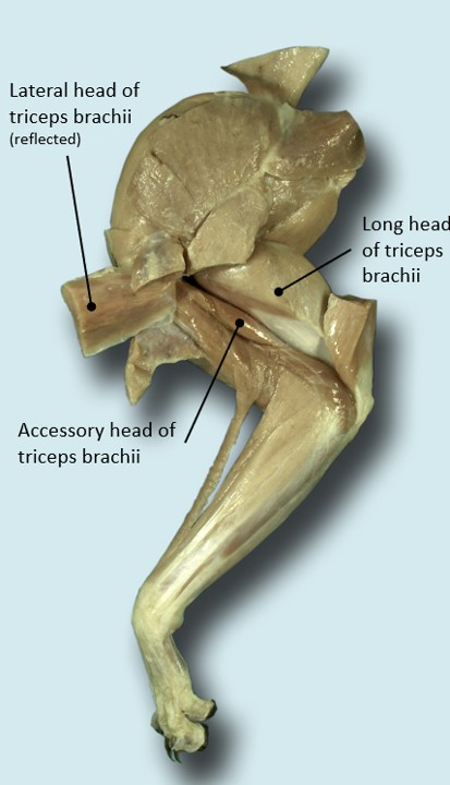
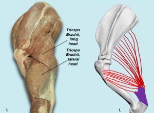
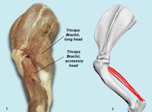
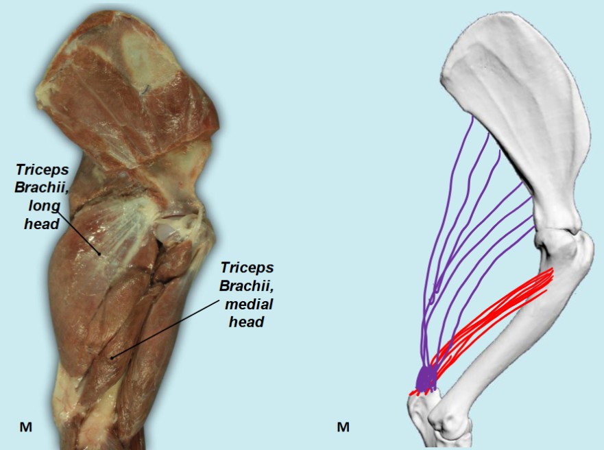
Triceps Brachii m.
The triceps brachii (named for being a 3-headed muscle in humans) consists of four heads in the dog and cat, with a common tendon to the olecranon tuber (point of the elbow). Only the long head arises from the scapula. The other three arise from the proximal end of the humerus.
The long head completely bridges the humerus. It arises from the caudal border of the scapula and inserts on the olecranon tuber. It appears to have two bellies.
Long head of the triceps brachii
PROXIMAL ATTACHMENT: Caudal border of scapula
DISTAL ATTACHMENT: Olecranon of ulna
ACTION: Extend elbow joint (secondary: flex shoulder joint)
Dissect: Caudal to the shoulder, on the lateral side, palpate a groove between the long and lateral heads of the triceps. The long head can appear to have a false separation more proximally. The groove between the long and lateral heads is closer to the humerus; separate these two heads along this groove. Expose the tendon of the long head. A subtendinous bursa exists between the tendon of the long head of the triceps brachii and the groove of the olecranon (see bursa description under infraspinatus m.). You need not visualize the triceps subtendinous bursa, but understand that it is present here and what function it has. If needed to fully visualize the triceps brachii muscle bodies, transect the tensor fascia antebrachii m. ~1 cm from its distal attachment.
Notice how the tendons of the other heads blend with that of the long head.
The lateral head of the triceps brachii lies distal to the long head, caudal to the acromial part of the deltoideus and lateral to the accessory head which it covers.
PROXIMAL ATTACHMENT: Proximal lateral humerus
DISTAL ATTACHMENT: Olecranon of ulna
ACTION: Extend elbow joint
Dissect: Transect the lateral head at its attachment to the humerus and reflect it to expose the underlying accessory and medial heads. This also exposes the brachialis muscle which will be revisited shortly.
The accessory head is small and lies between the lateral and medial heads.
PROXIMAL ATTACHMENT: Neck of the humerus
DISTAL ATTACHMENT: Olecranon of ulna
ACTION: Extend elbow joint
The medial head lies caudally on the humerus lateral and caudal to the biceps brachii. In the cat, the medial head of the triceps brachii m. has an additional short part on the distal medial brachium (i.e. it has two sub bellies, and no need to separate).
PROXIMAL ATTACHMENT: Proximal medial humerus (crest of lesser tubercle)
DISTAL ATTACHMENT: Olecranon of ulna
ACTION: Extend elbow joint
Dissect: From the medial side of the limb, separate the medial head from the long head caudally and from the accessory head laterally. The long tendon of the accessory head is closely bound to its lateral surface.
-
- Muscles of left thoracic limb, medial view, canine.1
-
- Intrinsic muscles of the cat thoracic limb, medial view. 4
-
- Dog brachium

-
- Dog brachium
-
- Dog brachium

-

- Cat triceps brachii 40
-
- Dog triceps brachii. 40

-
- Dog triceps brachii. 40

-

- Dog triceps brachii. 40
The anconeus m. (FYI) is a small muscle located almost completely in the olecranon fossa. It lies deep to the distal portion of the lateral head of the triceps brachii m. and will not be dissected. It attaches to the lateral supracondylar crest and the lateral and medial epicondyles of the humerus and spans the space to insert on the lateral surface of the olecranon of the ulna. Its action is to extend the elbow joint.
Conclude with the cranial muscles of the brachium:
- Biceps brachii
- Brachialis
Dissect: Clean and identify the following two muscles. Detailed dissection of their distal attachments is not necessary.
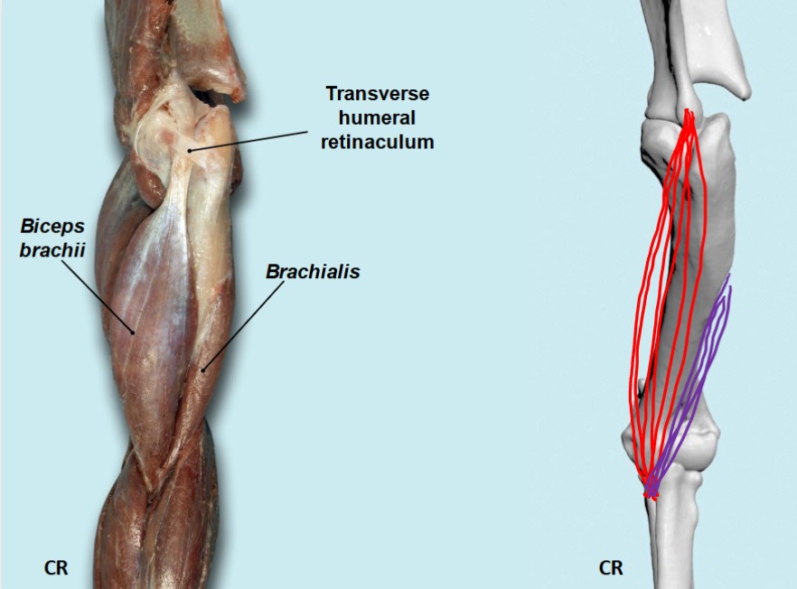
Biceps Brachii m.
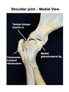
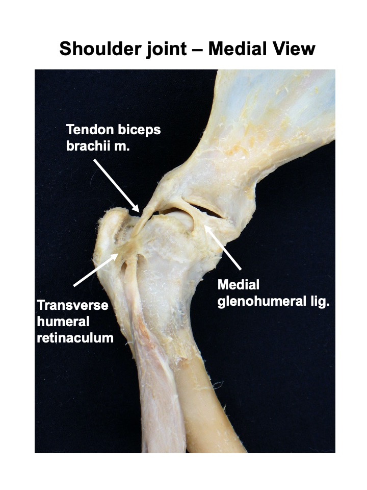
The biceps brachii has only one head in the dog and cat. It is a long, fusiform muscle that lies on the medial and cranial surfaces of the humerus. It completely bridges this bone. The muscle arises on the supraglenoid tuberosity of the scapula via a long tendon that courses distal through the intertubercular groove of the humerus. Distally, in the dog, the muscle inserts via a tendon that splits into two parts to attach to the proximal ends of the radius and ulna. The larger of the two parts inserts on the ulnar tuberosity and the smaller inserts on the radial tuberosity. In the cat, the biceps brachii terminates on the radius only. It is covered superficially by the pectoral muscles. The tendons of insertion lie on the elbow joint capsule, covering its medial collateral ligament.
PROXIMAL ATTACHMENT: Supraglenoid tubercle
DISTAL ATTACHMENT: Ulnar and radial tuberosities
Cat difference: distal attachment on radial tuberosity only
ACTION: Flex elbow joint (secondary: extend shoulder joint)
-
- Muscles of left thoracic limb, medial view, canine.1
-
- Intrinsic muscles of the cat thoracic limb, medial view. 4
-
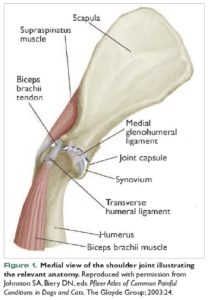
- Medial view of the canine shoulder joint illustrating the relevant anatomy.

-
- Dog brachium
-
- Dog brachium
-
- Dog shoulder
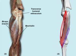
Brachialis m.
The brachialis is best viewed from the lateral side. It is a long, thin muscle that lies in the brachialis groove of the humerus. From the proximal third of this groove, the brachialis curves laterally and cranially as it courses distally, crosses the elbow, and, in the dog, attaches via a tendon to the proximal ulna and radius. This tendon inserts between the two tendons of the biceps brachii. In the cat, the brachialis terminates on the ulna only. A large part of its lateral surface is covered by the lateral head of the triceps. Distally, it runs medial to the origin of the extensor carpi radialis.
PROXIMAL ATTACHMENT: Proximal third of lateral surface of humerus
DISTAL ATTACHMENT: Ulnar and radial tuberosities
Cat difference: distal attachment on ulnar tuberosity only
ACTION: Flex elbow joint
-
- Deeper muscles of the left scapula, brachium, and antebrachium, lateral view. 1
-
- Intrinsic muscles of the cat thoracic limb. 4
-
- Dog brachium
-
- Dog brachium
-
- Dog biceps and brachialis 40
Ungulate Comparative Anatomy
Intrinsic muscles of the shoulder and brachium to identify on ungulates: |
|
|
Lateral muscles of the shoulder and scapula |
|
|
Medial muscles of the shoulder and scapula |
|
|
Caudal muscles of the brachium (arm) |
|
| Cranial muscles of the brachium |
|
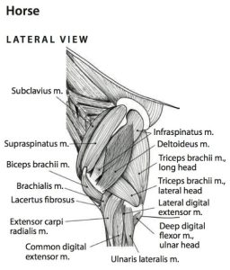
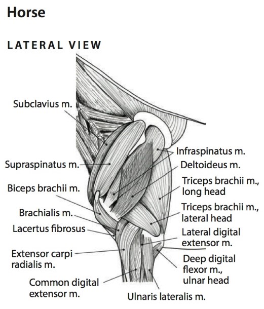
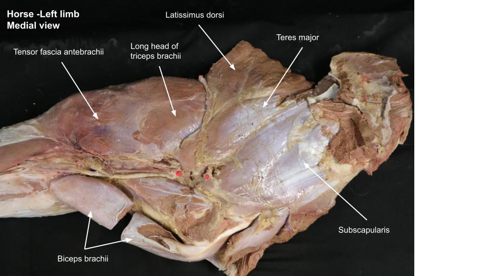
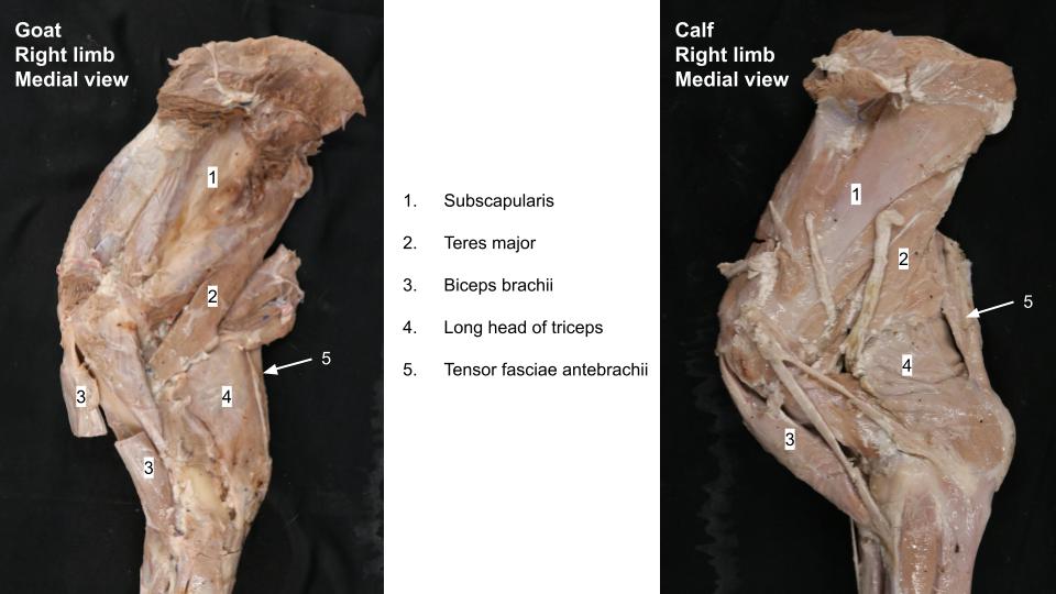
Horse and Ruminant
Observe: Referring to the provided table of muscles, the text below, and the figures, identify the intrinsic muscles of the shoulder and brachium in the horse and ruminant prosected models. Medial muscles will be best identified on detached thoracic limbs. Note the clinical relevance of structures as highlighted in this guide.
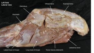
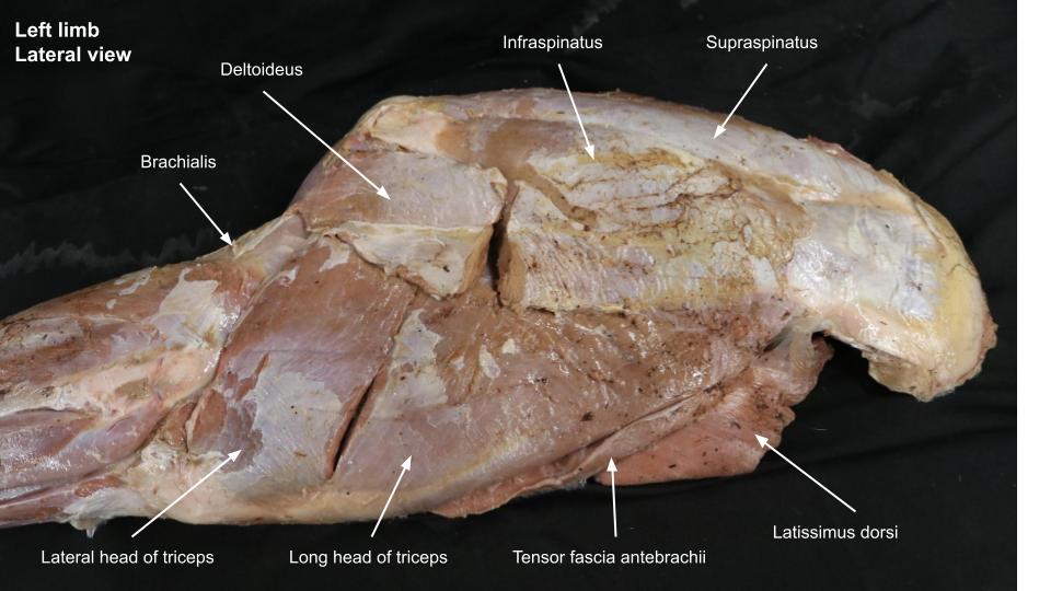
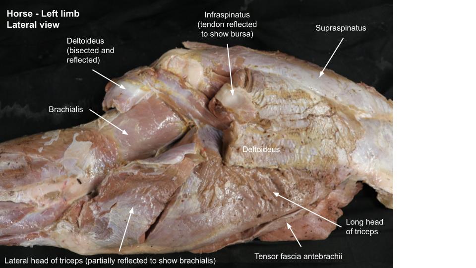
Identify the transected deltoideus m. (the two parts are more obvious in ruminants because an acromion is present). The supraspinatus and infraspinatus muscles are supplied by the same nerve and if this nerve is injured, neurogenic atrophy of these muscles leads to characteristic clinical signs of a condition called Sween(e)y (see clinical application box). The infraspinatus m. has superficial and deep tendons of insertion. The infraspinatus (subtendinous) bursa lies deep to the superficial tendon as it passes over the lateral aspect of the greater tubercle of the humerus. Reflect the distal part of the transected superficial tendon of the infraspinatus m. to view its bursa. This bursa may be normally absent in the goat/sheep. Lying caudally, tucked against the distal part of the infraspinatus muscle, identify the teres minor m.
Clinical relevance:
-
- Superficial intrinsic muscles of the proximal thoracic limb of the horse, lateral view. The following muscles have been removed, trapezius, latissimus dorsi, omotransversarius, brachiocephalicus, and serratus ventralis. 6
-
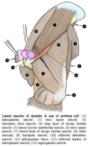
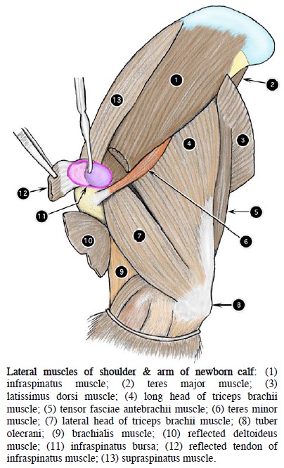
- Lateral muscles of shoulder and arm of newborn calf. 2
-
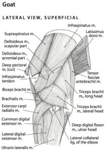
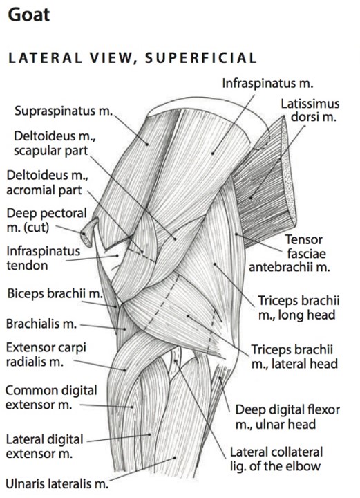
- Superficial intrinsic muscles of the proximal thoracic limb of the goat, lateral view. 6
-
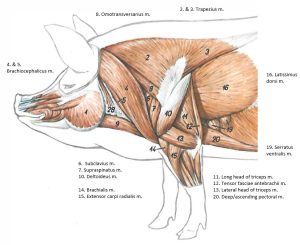
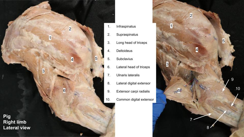
- Intrinsic muscles of the pig thoracic limb. 11

-
- Horse shoulder
-

- Horse shoulder
-

- Goat shoulder
-

- Pig shoulder
Clinical Application
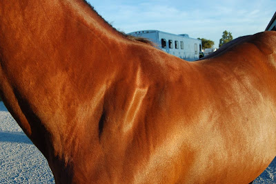 |
This horse has “sween(e)y”.
Atrophy of scapular muscles makes the scapular spine prominent and visible. Name the muscles that are atrophied. What nerve innervates the lateral scapular muscles? (ponder for now and/or look it up…nerves will be studied in the Nervous System unit)
|
| Image credit: http://arthurveterinaryclinic.blogspot.com/2009/10/sweeney-in-horses.html
|
|
Observe: On the medial side of a plastinated limb identify the subscapularis m. and teres major m.
The tensor fasciae antebrachii m. is well developed in the horse and pig with wide cranial and narrow caudal parts, and is present only as a small narrow caudal part in ruminants.
-
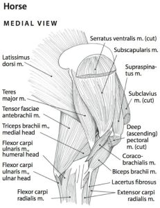
- Superficial intrinsic muscles of the proximal thoracic limb of the horse, medial view. 6
-
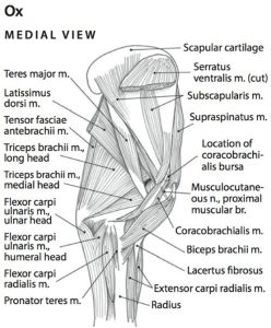
- Superficial intrinsic muscles of the proximal thoracic limb of the ox, medial view. 6
-

- Horse shoulder
-

- Goat and calf shoulder
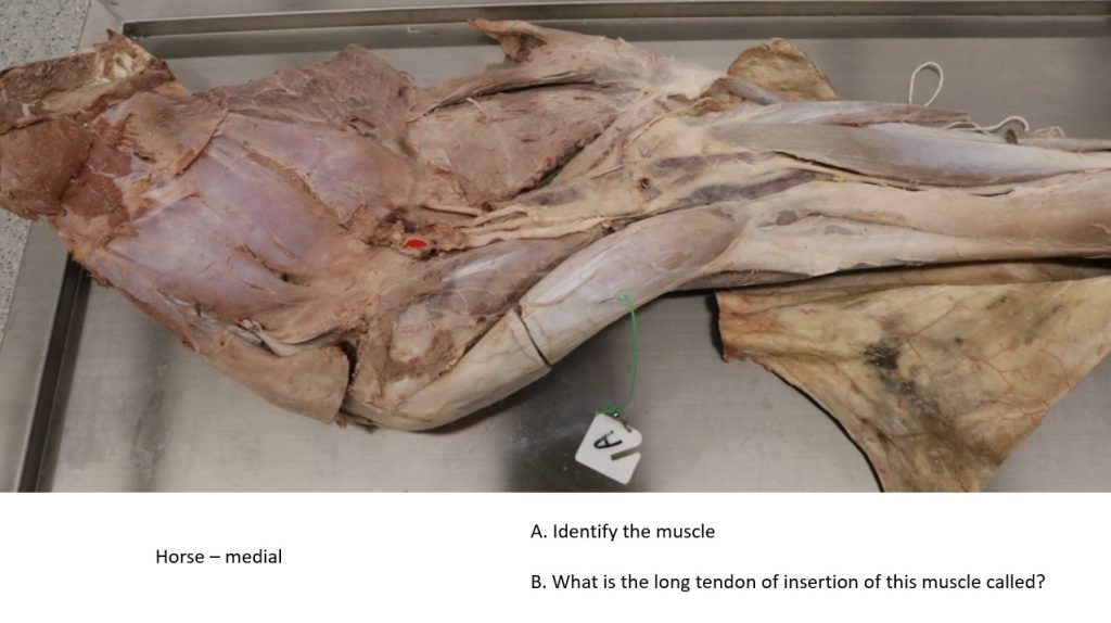
Observe: Identify the biceps brachii m. (and for FYI only, the coracobrachialis m.). Observe the tendon of origin of the biceps.
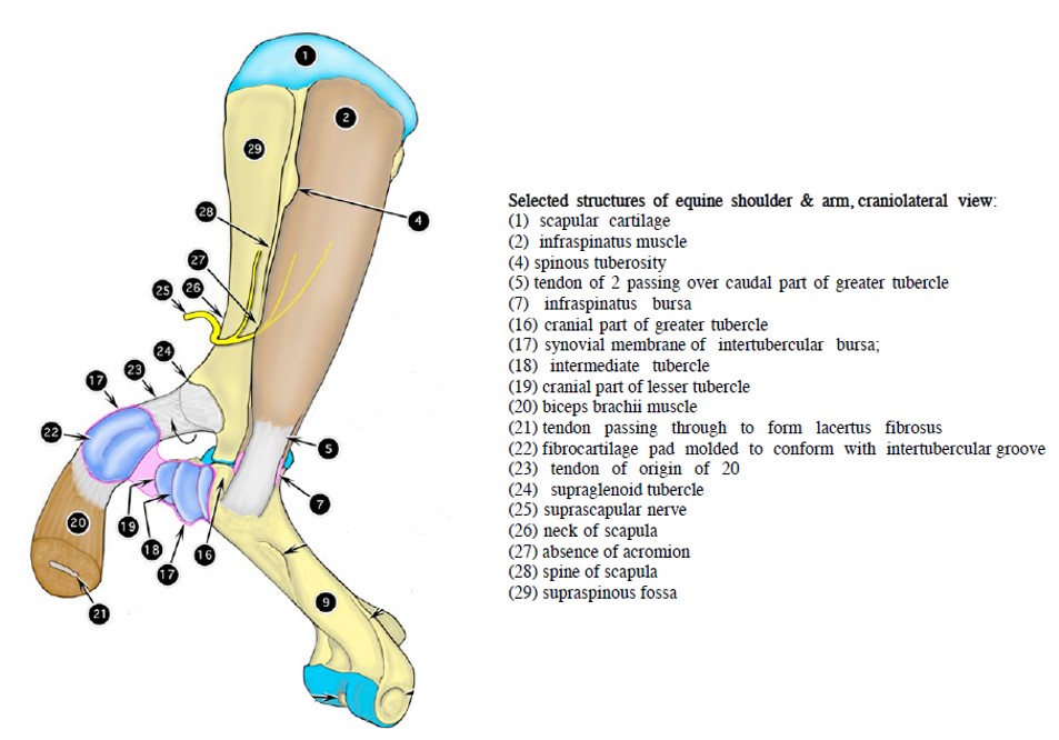
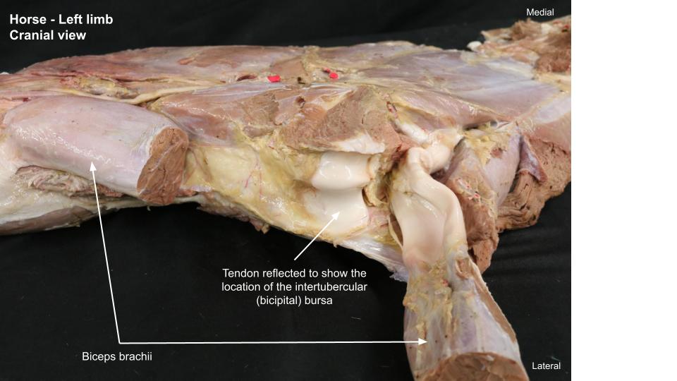
The tendon passes through the intertubercular (bicipital) groove of the humerus. Here it is lubricated by a large bursa, the intertubercular (bicipital) bursa (ruminants and horse, not present in pigs). Unlike the situation in the dog and pig, where the shoulder joint capsule extends distally to lubricate the biceps tendon, the intertubercular (bicipital) bursa of the horse and ruminants does not typically communicate with the shoulder joint capsule. This is an important difference from a clinical perspective.
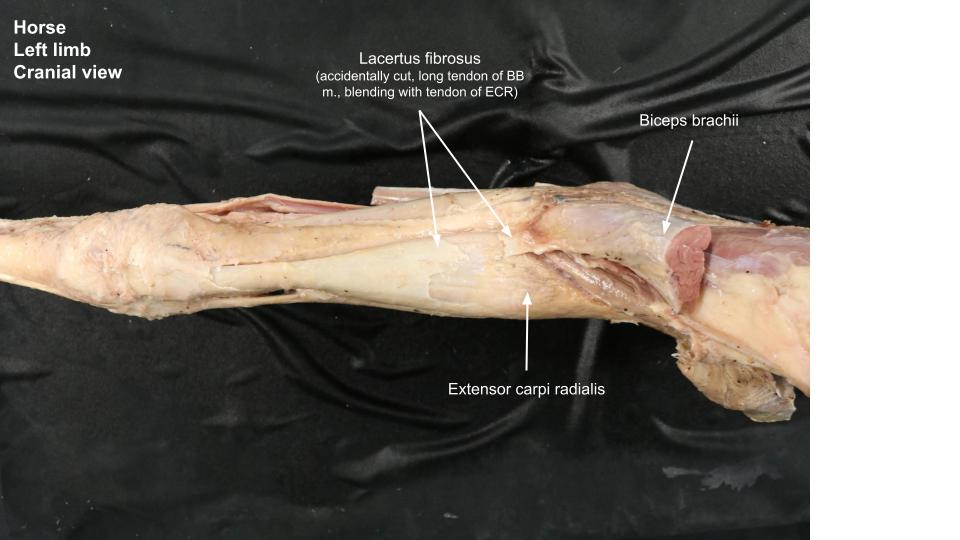
Observe: Reflect the proximal stump of the transected biceps brachii m. to open the large intertubercular (bicipital) bursa in the horse and ruminant. In the horse note the fibrocartilaginous mold of the biceps brachii tendon origin and how it fits over the intermediate tubercle.
This strong pad in the tendon replaces the need for a sesamoid bone over this area of significant pressure. Also note the tendinous band in the cut surface of the biceps brachii muscle. This band splits distally into the long tendon (i.e. lacertus fibrosus) and short tendon of the biceps brachii.
In the horse, focus on the distal extent of the biceps brachii m. and identify its long tendon (lacertus fibrosus) leaving the biceps brachii and joining the extensor carpi radialis. The lacertus fibrosus is present in ruminants but is not as well developed. The short tendon of the biceps inserts on the radial tuberosity.
-
-
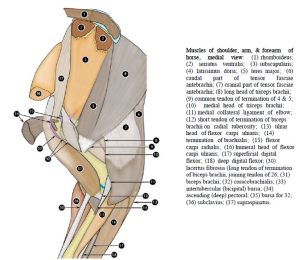
Muscles of shoulder, arm, & forearm of
horse, medial view. 2

-
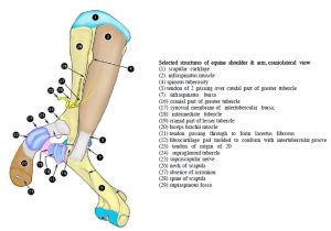
- Selected structures of equine shoulder & arm, craniolateral view. 2
-

- Horse brachium
-

- Horse antebrachium
Clinical relevance:
Observe: Identify the triceps brachii muscle and its individual heads, which vary according to the species (recall, long, lateral, medial and accessory in carnivores; no accessory head in the horse and pig; accessory head difficult to demonstrate in ox, and a little less so in goat, if present! Accessory head may be absent in goat, so don’t worry about it at all in the ruminant cadavers).
On the lateral side, elevate the transected lateral head of the triceps brachii and reflect it to the olecranon tuberosity. This should expose the brachialis m. and radial n.. Also from the lateral perspective portions of the medial head may be viewed, crossed over by the radial n., with the lateral head reflected.
-
- Superficial intrinsic muscles of the proximal thoracic limb of the horse, lateral view. The following muscles have been removed, trapezius, latissimus dorsi, omotransversarius, brachiocephalicus, and serratus ventralis. 6
-
- Superficial intrinsic muscles of the proximal thoracic limb of the goat, lateral view. 6
-
- Superficial intrinsic muscles of the proximal thoracic limb of the horse, medial view. 6
-
- Superficial intrinsic muscles of the proximal thoracic limb of the ox, medial view. 6
-
-
Muscles of shoulder, arm, & forearm of
horse, medial view. 2
-
- Lateral muscles of shoulder and arm of newborn calf. 2
-
- Horse shoulder
-
- Horse shoulder
-
- Goat shoulder
-
- Pig shoulder
-
- Horse shoulder
-
- Goat and calf shoulder
Clinical relevance:
Pig cubital joint
Observe: Take a look at the opened porcine elbow from the lateral side. Examine the anconeal process of the ulna and the humeral condyle. Note the presence of the normal synovial fossae, which should not be confused with pathologic erosions.
Clinical relevance:
| Humeral condyle is a common site of OCD in pigs | 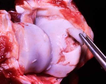
Pig humeral condyle OCD. Image from UGA Vet Path. |
Review Videos
Muscle Chart/Terms List – Intrinsic muscles of shoulder and brachium
| Muscle Action Groups | |
| Extensors of the shoulder |
|
| Flexors of the shoulder |
|
| Extensors of the elbow |
|
| Flexors of the elbow |
|
|
Muscle/structure
|
Attachments/comments
|
Action/comments
|
| SUPRASPINATUS M. |
|
Extend shoulder joint/shoulder stabilization |
| INFRASPINATUS M. |
|
Extend shoulder joint/shoulder stabilization |
| Infraspinatus m. bursa |
Deep to superficial tendon of m. ie it is a subtendinous bursa |
Protect tendon as passing over bone |
| SUBSCAPULARIS M. |
|
Extend shoulder joint/shoulder stabilization |
| DELTOIDEUS M. |
|
Flex shoulder joint |
| TERES MINOR M. |
|
Flex shoulder joint; rotate shoulder laterally |
| TERES MAJOR M. |
|
Flex shoulder joint |
| TENSOR FASCIAE ANTEBRACHII M. |
|
Extend elbow joint (weak) |
| TRICEPS BRACHII M. (LONG HEAD) |
|
Extend elbow joint (secondary: flex shoulder joint) |
| TRICEPS BRACHII M. (LATERAL HEAD) |
|
Extend elbow joint |
| TRICEPS BRACHII M. (ACCESSORY HEAD) |
|
Extend elbow joint |
| TRICEPS BRACHII M. (MEDIAL HEAD) |
|
Extend elbow joint |
| BICEPS BRACHII M. |
|
Flex elbow joint (secondary: extend shoulder joint) |
| Intertubercular (bicipital) bursa |
Horse and ruminant structure |
Protects tendon passing over point of shoulder |
| Lacertus fibrosus |
Horse; long tendon of insertion of biceps into Extensor Carpi Radialis m. |
|
| BRACHIALIS M. |
|
Flex elbow joint |