MSK LAB 16A – Pelvic limb osteology and arthrology
Learning Objectives
- Identify the regions of the pelvic limb, the bones and key bone features in each region, the joints and joint structures.
- Start to recognize and state sites of attachments for muscles, ligaments and tendons.
- Identify flexor and extensor sides of joints.
Lab instructions:
Part A: An instructor will provide an overview of this lab and highlight a few examples of comparative bone features across the species being studied.
Part B: Use ‘bone boxes’ (containing carnivore skeletons), bone models, articulated skeletons and images, to study the regions, bones and joints of the pelvic limb. Refer to the terms tables for focused studying. Be able to identify pelvic limb regions, the bones and their features, and joints and structures related to joints, in any species (i.e. dog, cat, horse, ruminant and pig), as applicable to that species. It is understood that not all material will be covered in lab and it is anticipated that continued learning and review of this content will continue as self-study effort.
Pelvic Limb Regions
-
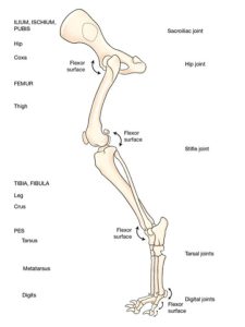
- Dog PL regions. 1
-
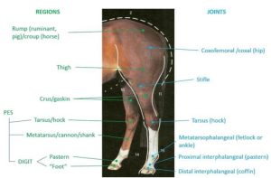
- Regions and joints (anatomical and lay terms) of the ungulate pelvic limb – based on the horse. 10

-
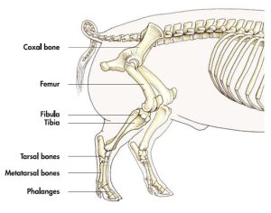
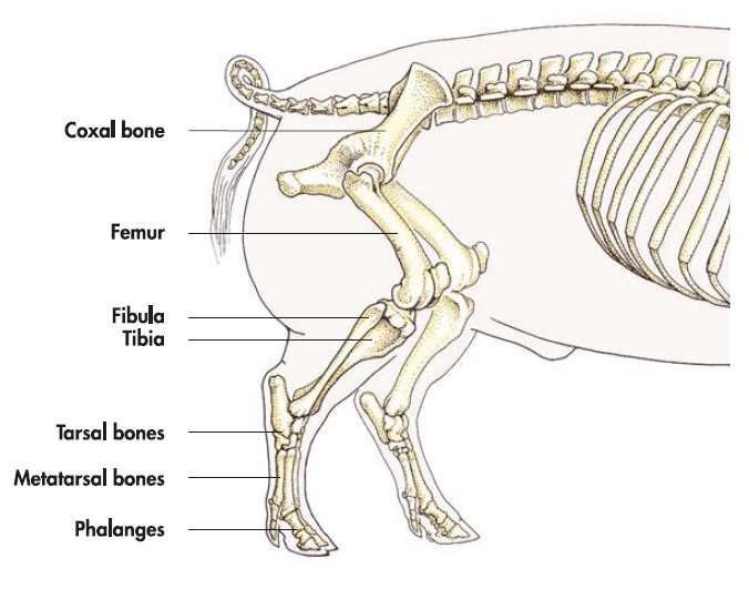
- Pig Pelvic Limb Bones. 7
Clinical Application: Q1
Q1: A horse has a deep laceration across its gaskin. Between what two joints is the gaskin?
A. coxofemoral and stifle
B. stifle and hock
C. tarsus and fetlock
D. pastern and coffin
Interactive practice question – Horse joints/regions
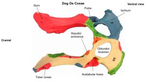
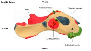
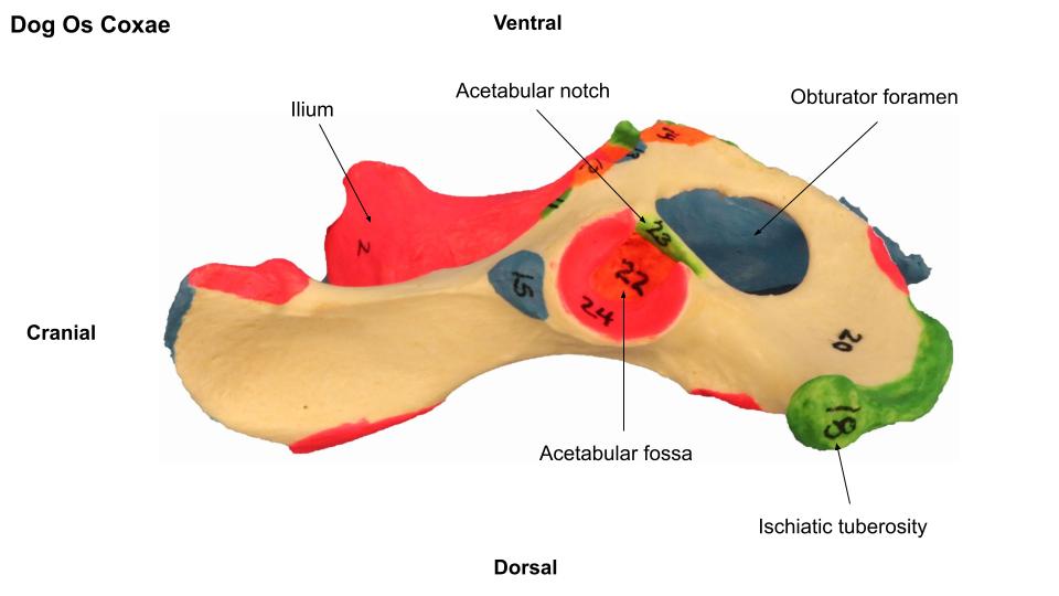
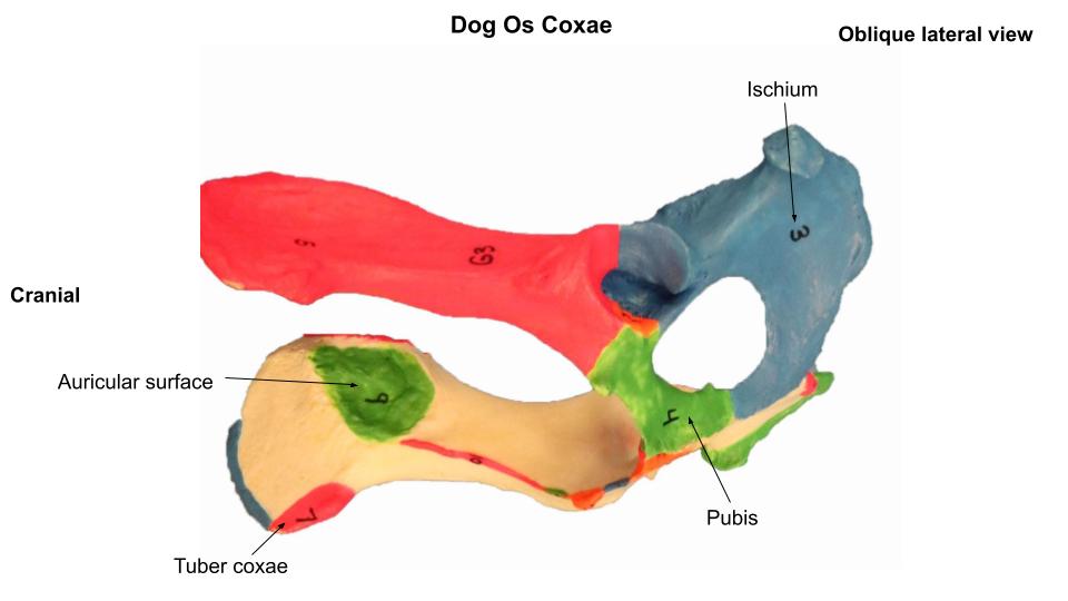
Os Coxae (pl: ossa coxae)
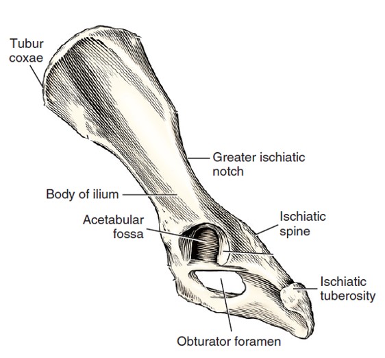
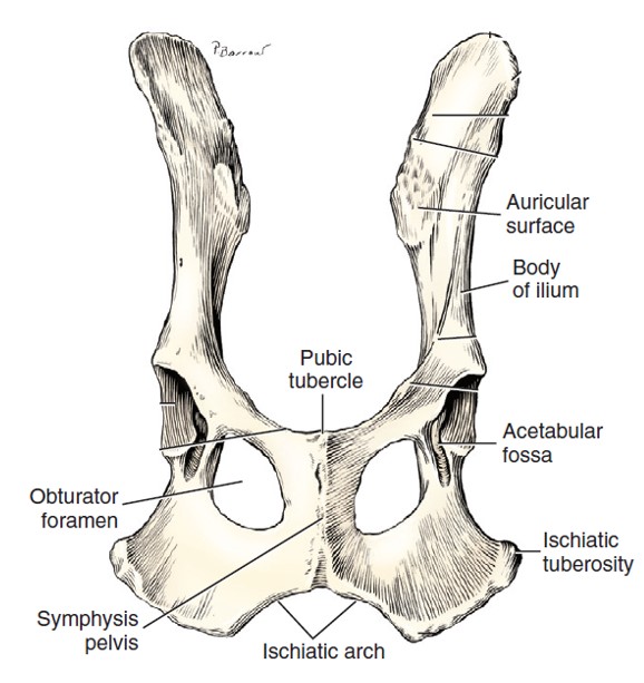
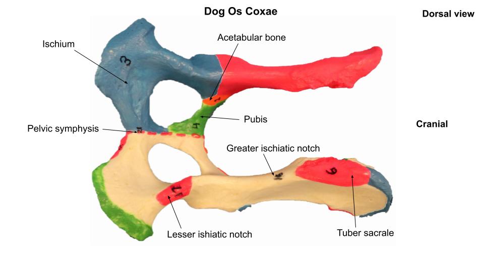
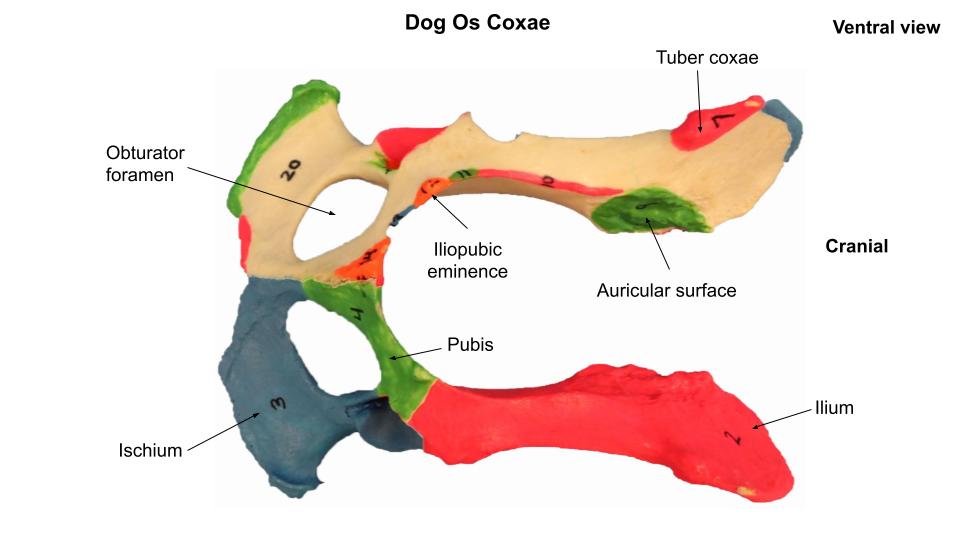
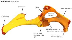
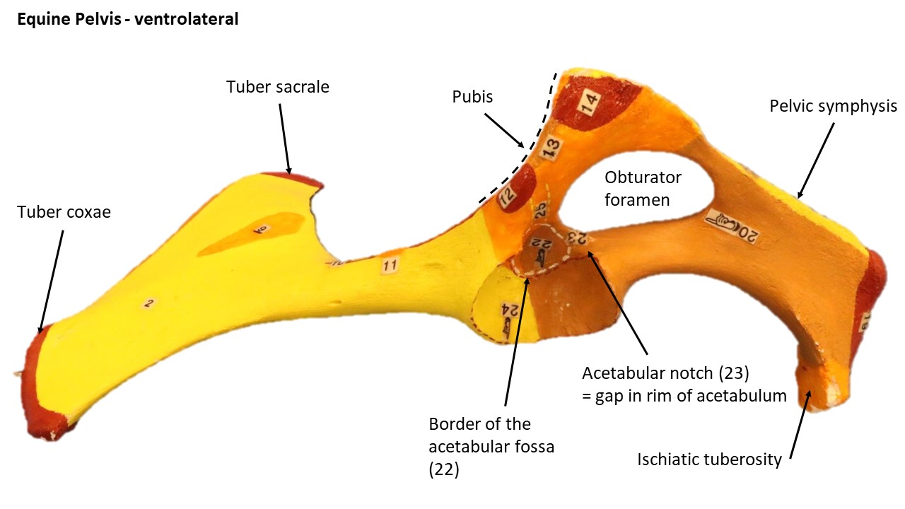
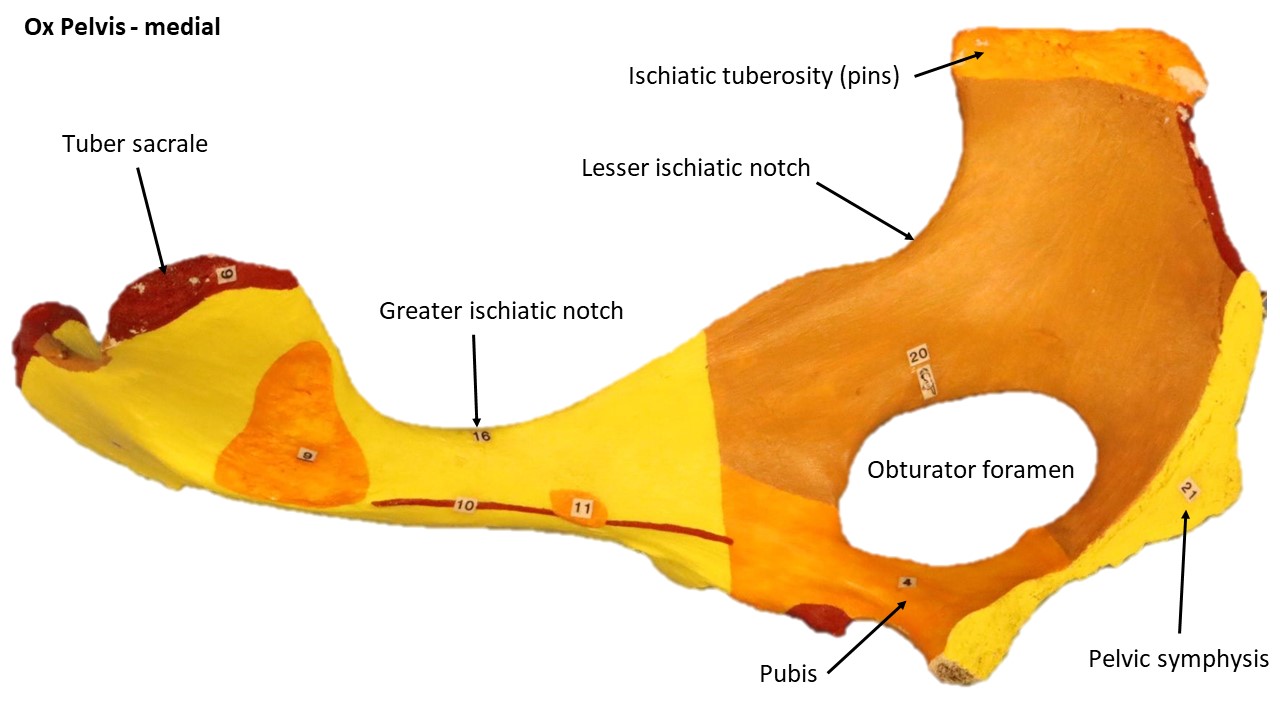
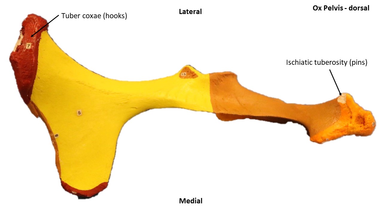
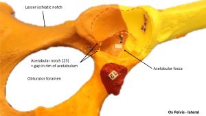
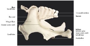
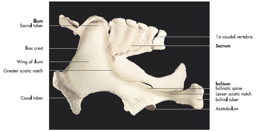
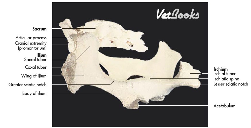
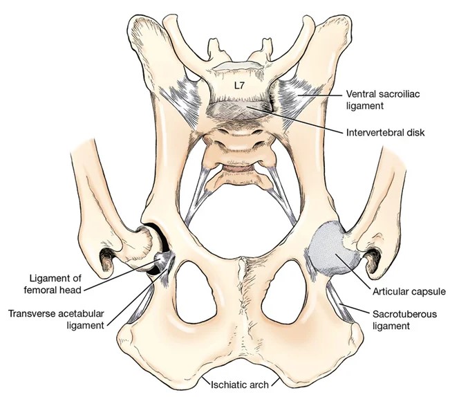
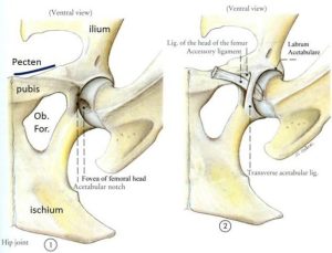
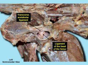
The pelvic girdle, or pelvis, consists of two hip bones (ossa coxae), which are united at the symphysis pelvis midventrally and join the sacrum dorsally. The pelvis shape and angles vary between species. Each hip bone, or os coxae, is composed of four distinct bones developmentally. These are the ilium, ischium, pubis, and acetabular bone. They fuse during the twelfth postnatal week, and form the socket, i.e. the acetabulum (from [L] = vinegar cup), that receives the head of the femur in creation of the ball and socket hip joint. The small acetabular bone, which helps form the acetabulum, is incorporated with the ilium, ischium, and pubis when they fuse so it is not identified in the mature animal. The articular surface of the acetabulum is indented by the acetabular fossa and the ligament of the head of the femur attaches to this fossa. The circumference of the articular surface is disrupted at the caudomedial part by the acetabular notch. The two sides of the notch are connected by the transverse acetabular ligament. The largest and most cranial of the os coxae bones is the ilium, which articulates with the sacrum (forming the sacroiliac joint). The ischium is the most caudal, whereas the pubis is located ventromedial to the ilium and cranial to the large obturator foramen. The obturator foramen is closed in life by the obturator membrane and the external and internal obturator muscles that the membrane separates. Vessels and nerves pass through the foramen.
-
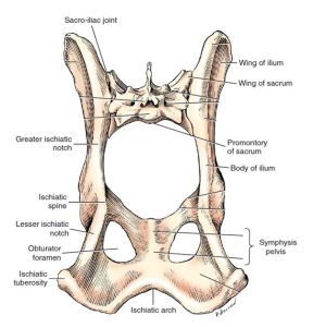
- Pelvis and sacrum, caudodorsal view. 1

-
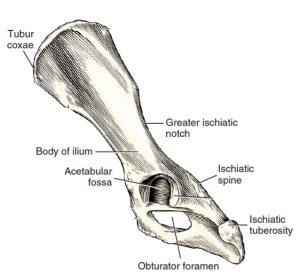
- Left os coxae, lateral aspect. 1
-
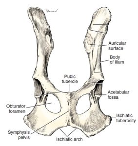
- Ventral aspect of ossa coxae. 1
-

- Cat Os Coxae. 5

-
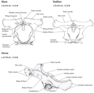
- Horse pelvis. 6
-
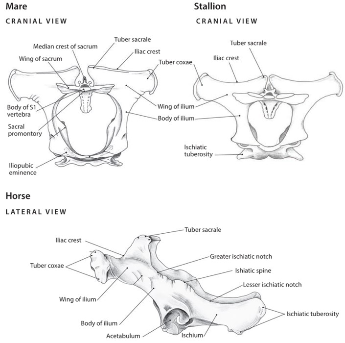
- Cow pelvis. 6
-
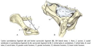
- Canine sacrotuberous ligament and bovine sacrosciatic ligament. 8
-

- Dog pelvis
-

- Dog pelvis
-
- Dog pelvis

-
- Dog pelvis
-

- Dog pelvis
-
- Horse pelvis
-
- Horse pelvis

-

- Ox pelvis
-

- Ox pelvis
-
- Ox pelvis
-
- Hip bones and sacrum of a horse (oblique left lateral aspect). 7
-

- Hip bones and sacrum of an ox (oblique left lateral aspect). 7
Interactive Review Content:
Femur and Patella
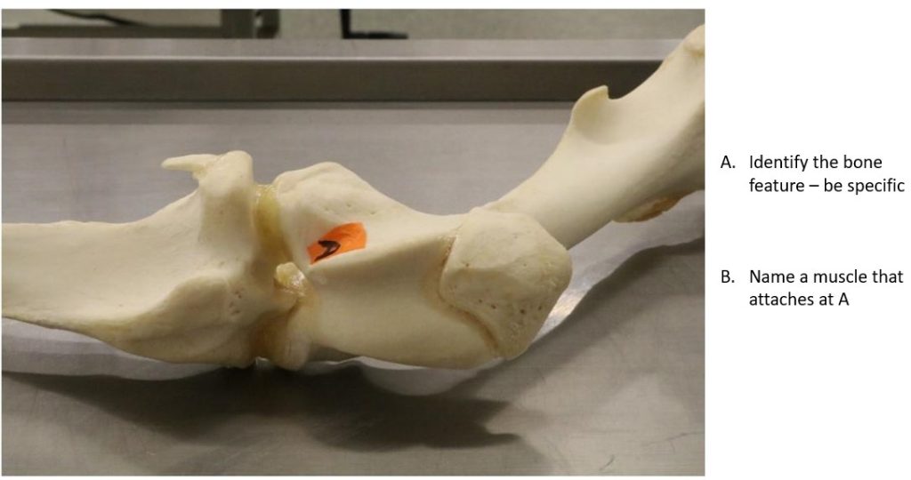
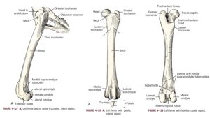
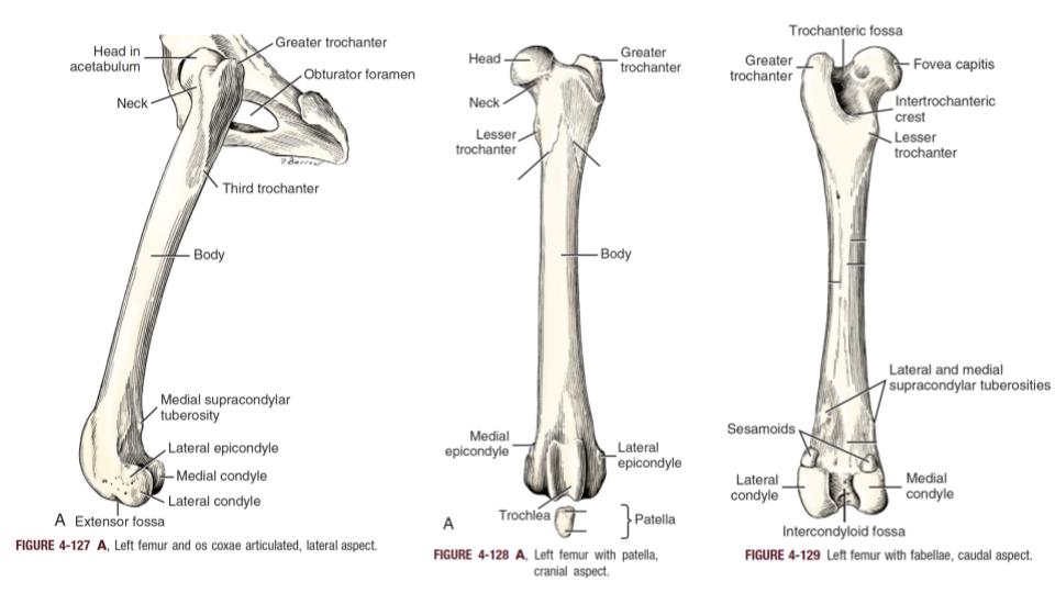
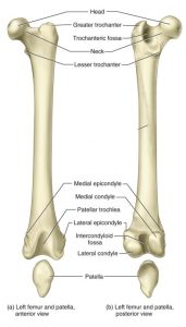
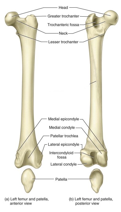
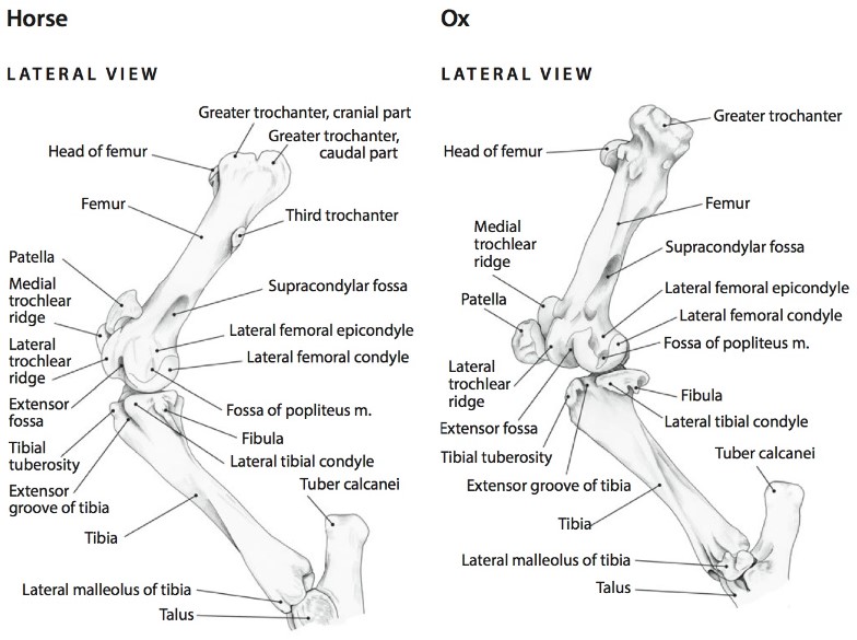
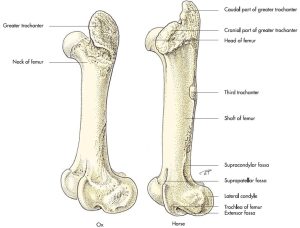
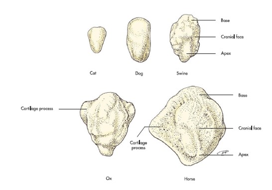
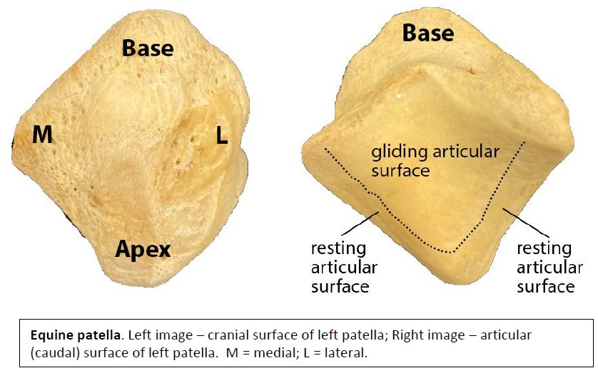
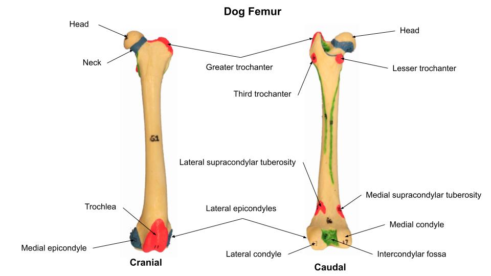
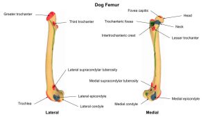
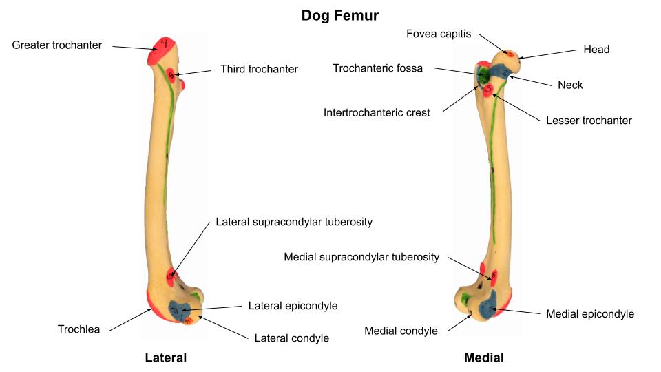
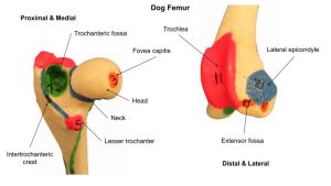
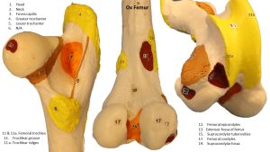
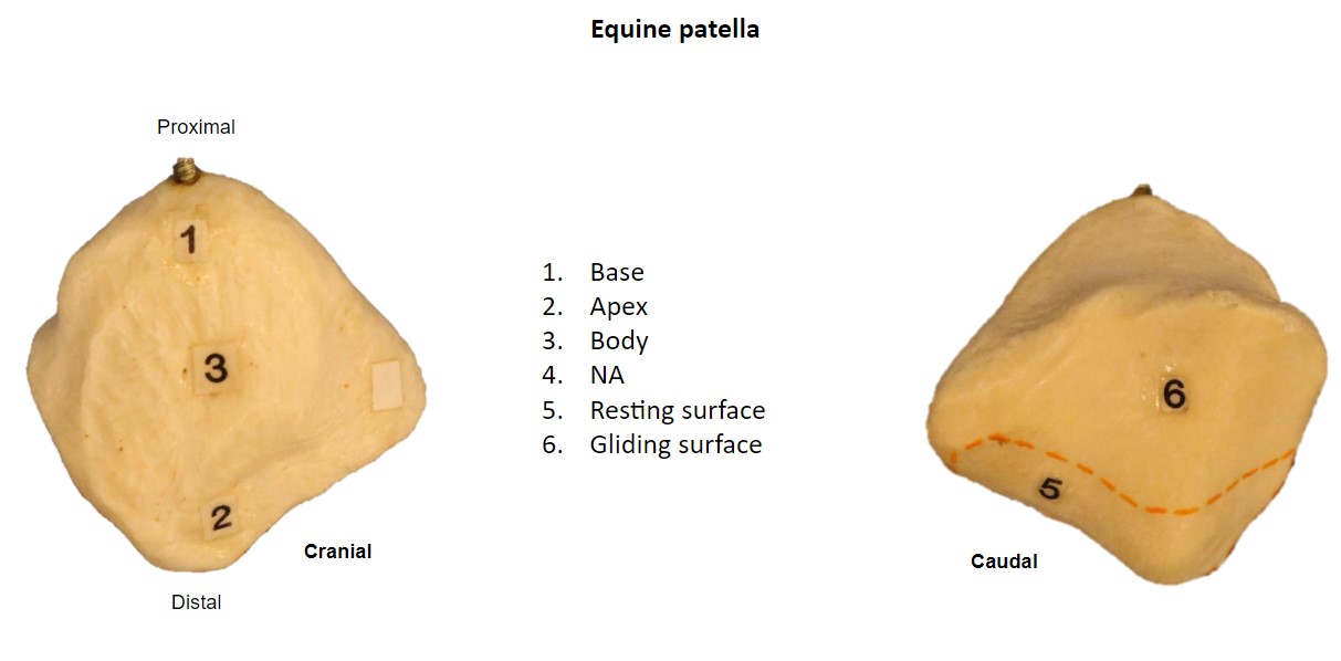
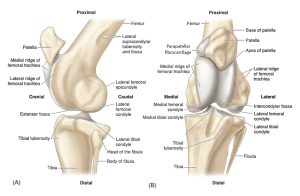
The proximal end of the femur consists of a head, neck and two processes, the greater trochanter and lesser trochanter. The horse greater trochanter is subdivided into cranial and caudal parts. A very prominent third trochanter is present in the horse. The fovea capitis is a circular shallow pit on the medial part of the femoral head. The fovea capitis serves for the attachment of the ligament of the head of the femur (ligamentum capitis ossis femoris) (and the accessory ligament in the horse, see arthrology section). Distally the femur consists of a trochlea with medial and lateral ridges and a trochlear groove, and medial and lateral condyles and epicondyles. The extensor fossa of the femur is an important muscle attachment site for cranial crus muscles. The patella articulates with the articular surface of the trochlea of the femur. This is the sesamoid bone associated with the tendon of the m. quadriceps femoris. Additional features for the femur and patella are noted in the terms list.
-
- Dog femur. 1
-
- Car femur. 5
-
- Horse & Cow thigh, lateral view. 6
-
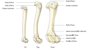
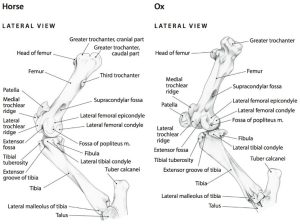
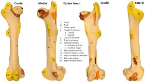
- Comparative femurs. 7
-
- Comparative femurs. 7
-
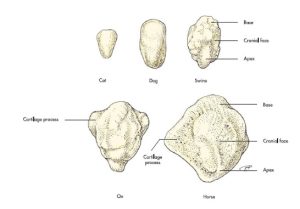
- Comparative patellae. 6
-
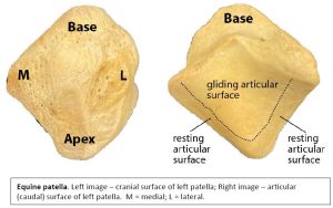
- Horse patella, resting and gliding surfaces. Gerard, M., 2020
-

- Dog femur
-
- Dog femur
-
- Dog femur

-
- Horse femur

-
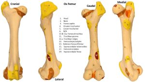
- Ox femur
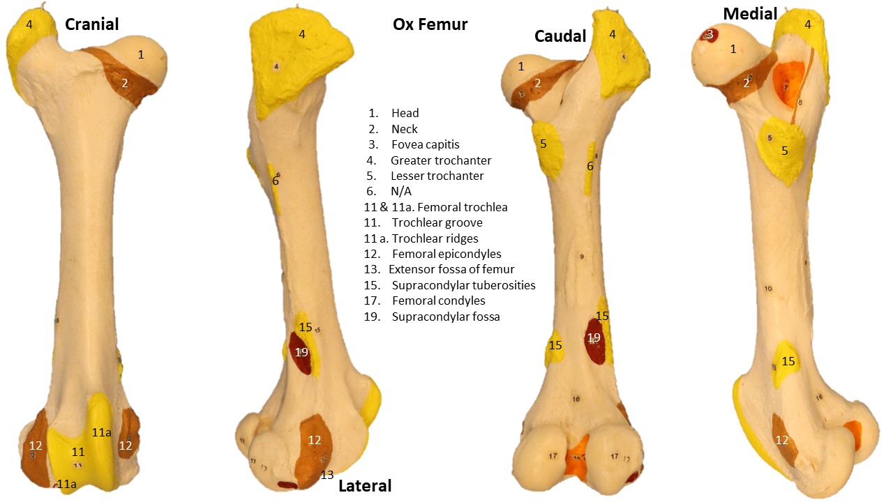
-
- Ox femur

-

- Horse patella
Interactive review content:
Clinical Relevance: Patellar Luxations
Patellar luxation (dislocation) is a condition where the patella rides outside the femoral groove when the knee is flexed. It can be further characterized as medial or lateral, the former being much more common. Patellar luxation also may be graded based on the severity of the condition. It is one of the most common orthopedic conditions in dogs, and small breeds (Boston and Yorkshire terriers, Chihuahuas, Pomeranians, and miniature poodles) are predisposed. Most dogs affected by this disease will suddenly carry the limb up for a few steps (“skip”), and may be seen shaking or extending the leg prior to regaining its full use. However, symptoms and consistency of clinical signs will vary based on the severity (grade) and duration of the condition. Left untreated, the condition may worsen and eventually cause painful osteoarthritic changes in some dogs, but some patients will fare well with conservative medical management.
LEARN MORE: Patellar Luxations
WATCH (pay special attention to included videos of dogs with this condition): Medial Patellar Luxation
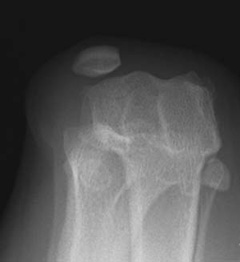
Pre-operative X-ray (skyline) view of the knee: the femoral groove is shallow with a knee cap riding outside of its groove.
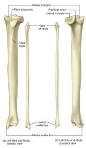
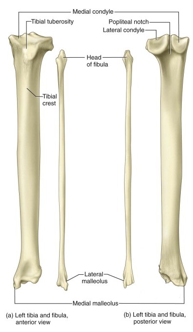
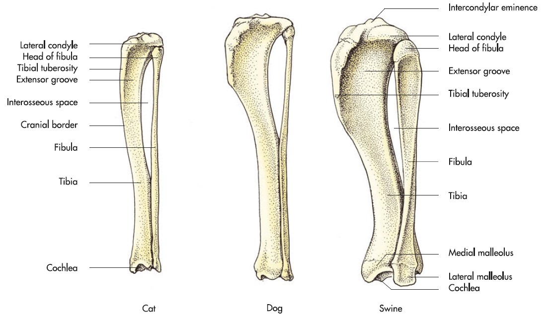
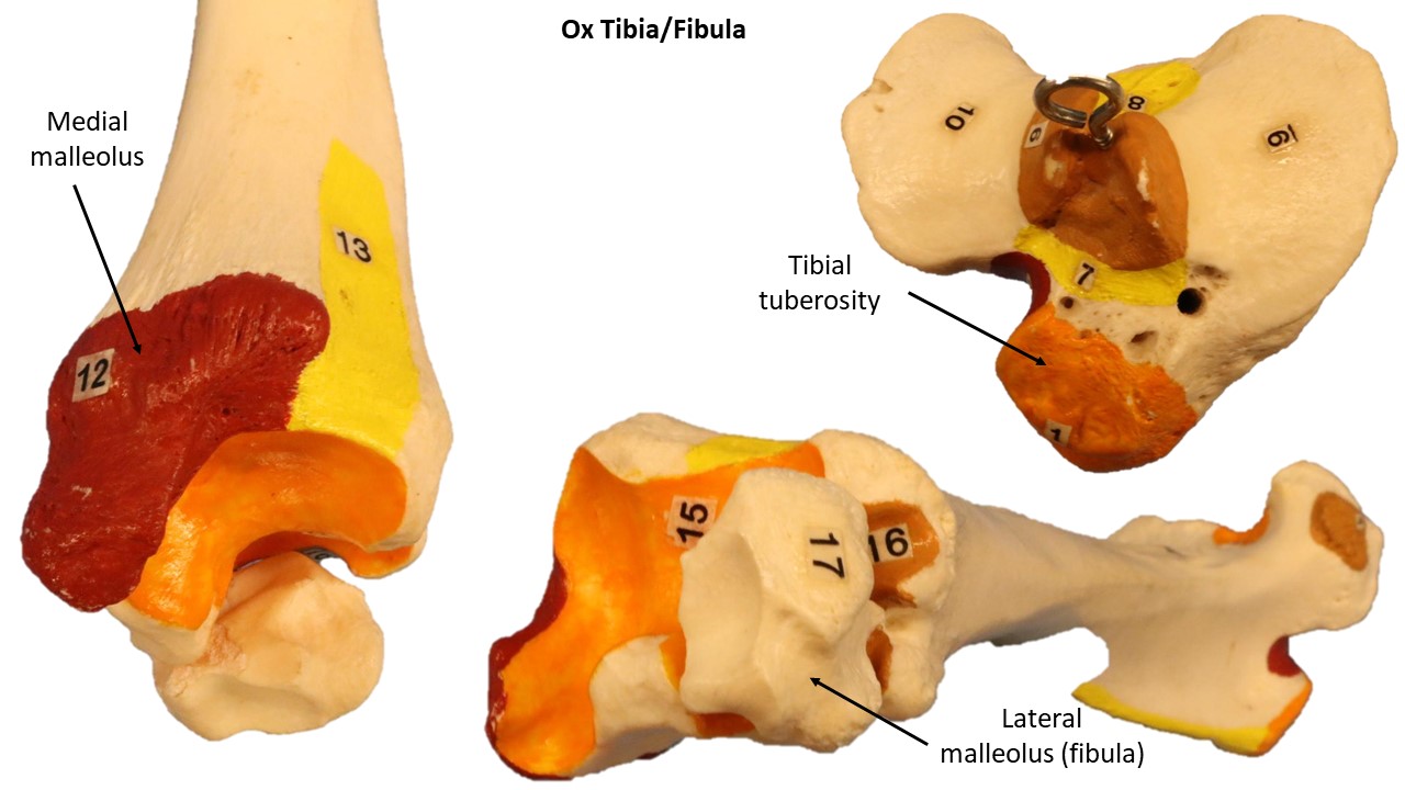
Tibia & Fibula
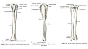
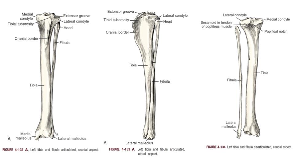
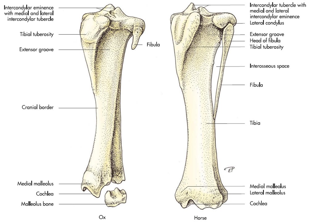
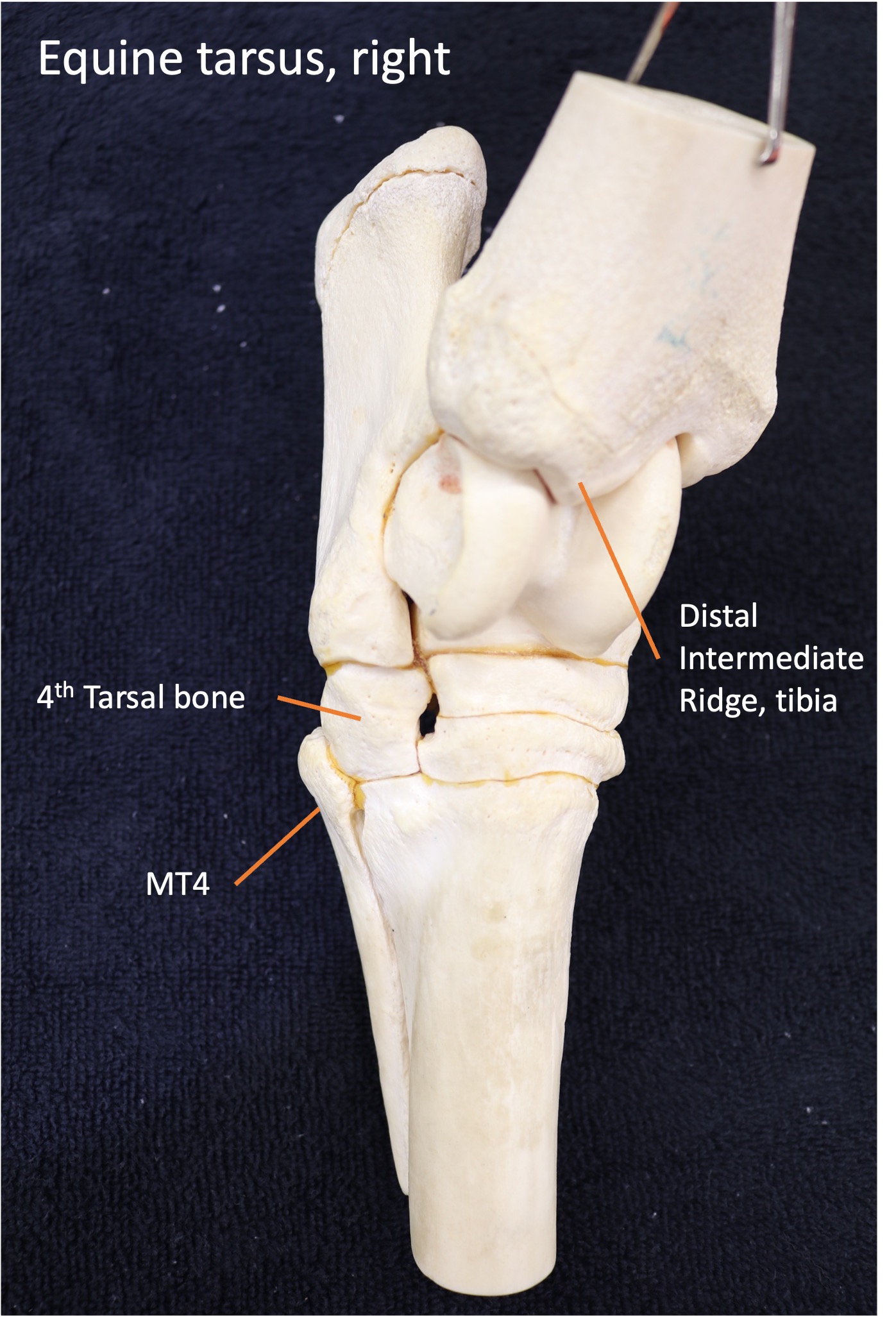
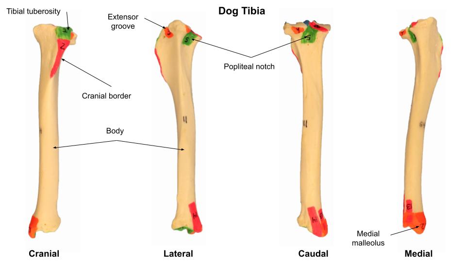
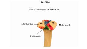
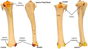
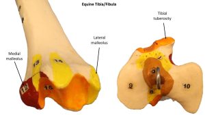
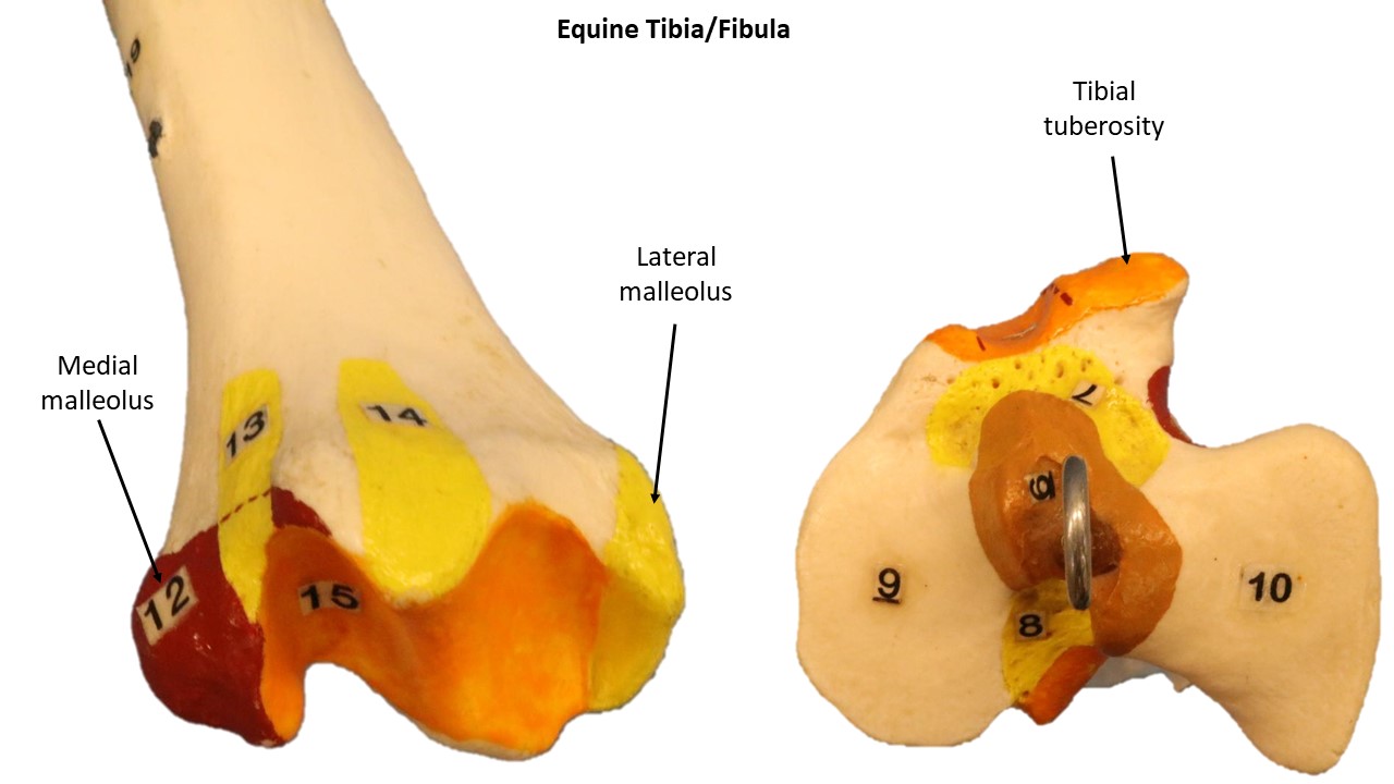
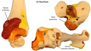
The tibia, the shin or leg bone, has a proximal surface that articulates with the femur and that is formed largely by two relatively flat condyles (medial and lateral). An intercondylar eminence separates the two condyles. The fibula articulates with the lateral condyle, and cranial tibial and fibularis longus mm. arise from here. A sesamoid bone in the tendon of origin of the popliteus (seen in radiographs) articulates with the caudolateral condyle of the tibia. Two biconcave fibrocartilages, the menisci, fill part of the space between the apposed condyles of the femur and tibia, making the joint congruent (see stifle joint below). The cruciate ligaments of the stifle are named according to their cranial or caudal attachment to the tibial intercondylar areas (see stifle joint below). The tibial tuberosity is the large quadrangular process on the proximal cranial surface of the tibia. The quadriceps femoris, the biceps femoris, and the sartorius mm. attach to this tuberosity by means of the patella and patellar ligament. Distally, the tibia articulates with bones of the tarsus. The prominent medial projection of the tibia is the medial malleolus. Centrally, the distal tibial articular surface (the cochlea) has an intermediate ridge that locks into the trochlear groove of the talus. In equine clinical jargon, this ridge is referred to as the distal intermediate ridge of the tibia (DIRT). It is an important bone feature because it is a very common location for osteochondritis dessicans (OCD, a developmental bone disease) to develop, and DIRT lesions are a thing :).
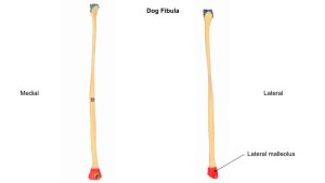
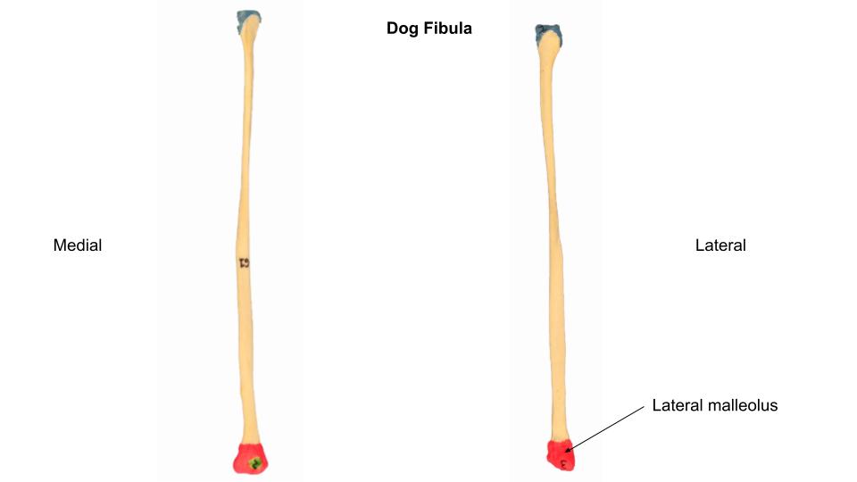
The fibula construct varies considerably between species, as shown in the images. It is a full length bone in the carnivore and pig and is missing much of its body in the ox and horse. A proximal extremity (head of fibula) is consistent and articulates with the lateral condyle of the tibia. The distal extremity, the lateral malleolus, articulates with the lateral distal tibia in the carnivore, swine, and ruminant. In the ruminant the lateral malleolus of the fibula is a solitary articulating bone, with no body attached to it (this is one key feature to keep in mind when looking at radiographs of the ruminant tarsus). The horse has a vestigial fibular body of varying length and distally the lateral malleolus of the fibula has fused completely to become a part of the tibia (recall this is a similar set up to the horse ulna and radius story, where the lateral styloid process of the ulna has fused to the radius).
-
- Dog tibia. 1
-
- Cat tibia. 5
-

- Comparative tibia & fibula, Left craniolateral aspect. 7
-
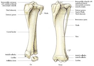
- Comparative tibia & fibula, Left craniolateral aspect. 7
-

- Equine DIRT – dorsolateral view
-
- Radiographic DIRT anatomy. (VRU 2020;61:497-506)

-

- Dog tibia
-
- Dog tibia

-
- Dog fibula
-
- Horse tibia

-
- Horse tibia
-

- Ox tibia
-
- Ox tibia
Interactive review content:
Tarsus
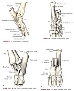
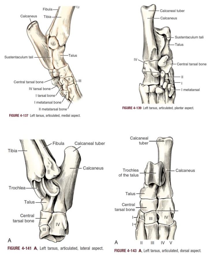
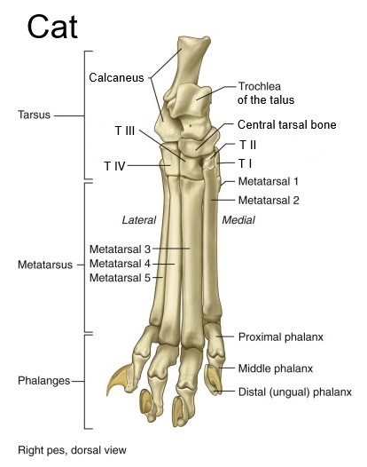
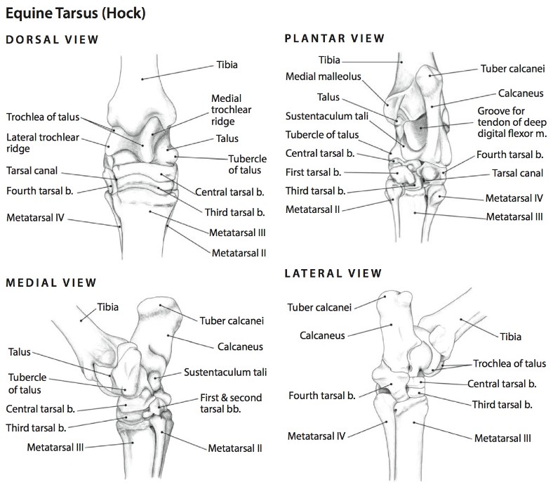
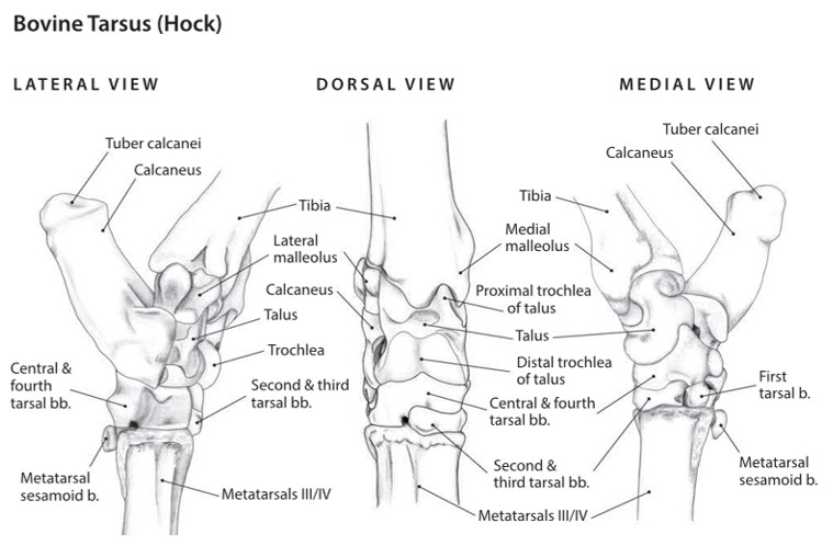
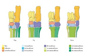
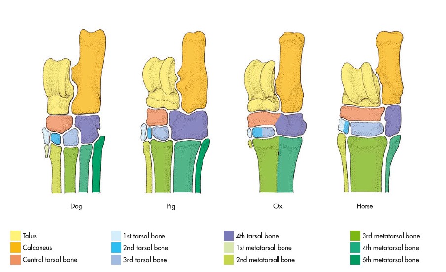
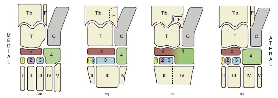
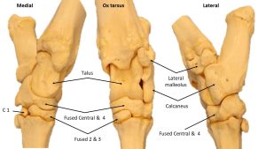
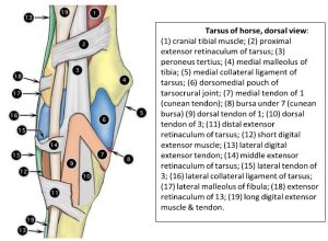
The tarsus, situated between the metatarsals and the crus (leg), is composed of six to seven tarsal bones and the related soft tissues. It is also called the hock. The bones are arranged in three irregular rows with variation between species in fusion of the bones with each other. The proximal row is composed of a long, laterally located calcaneus and a shorter, medially located talus. The talus has a trochlea on its proximal end in the carnivore and horse. In the artiodactyla there is a proximal and distal trochlea, a defining feature of animals in this order. The trochlea has medial and lateral trochlear ridges separated by a groove for articulation with the tibial cochlea. The tuber calcanei (aka point of the hock) is a traction process of the calcaneus that projects proximally and caudally. The extensor muscles of the tarsus insert on this process via the common calcanean tendon. On the medial side of the calcaneus is a bony process, the sustentaculum tali. A tendon of the deep digital flexor glides over the plantar surface of this process.
There is a central tarsal bone occupying the ‘middle row’. The distal tarsal row consists of up to four separate bones or a combination of these bones fused to each other. Species variation is notable in this part of the tarsus as the images show. The large fourth tarsal bone, which completes the distal row laterally, articulates with the calcaneus proximally. The fourth tarsal is as long as the combined lengths of the third and central tarsal bones against which it lies.
Dr. Spradley has a helpful memory tool for ungulate tarsal bones: 23 cows are playing with C4 (explosives). 12 horses run away. In the cow, tarsal bones 2 and 3 are fused, along with the central and 4th. In the horse, tarsal bones 1 and 2 are combined.
-
- Dog tarsus. 1
-

- Cat tarsus. 5
-
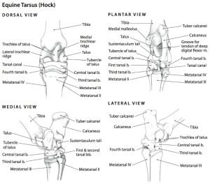
- Right tarsus of the horse; dorsal, plantar, medial, and lateral views. 6
-

- Cow tarsus. 6
-
- Skeleton of the tarsus in the domestic mammals. 7
-
-
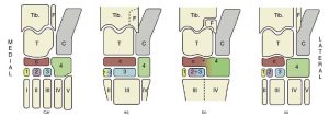
The bones of the tarsal skeleton in the carnivores (Car), horse (eq), cattle (bo), and pig (su), schematic. Roman numerals identify the metatarsal bones, Arabic numerals the distal tarsal bones. Tib., Tibia; F, fibula; T, talus; C, calcaneus;
c, central tarsal bone. 8
-
- Ox tarsus

Interactive review content:
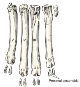
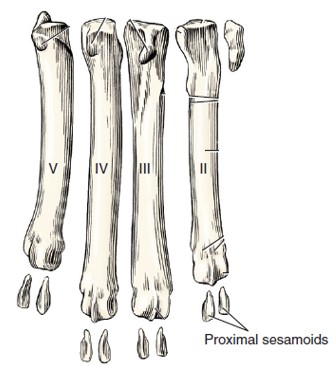
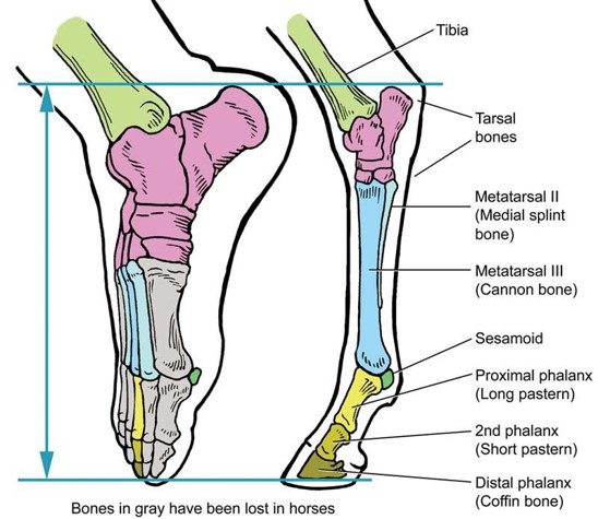
Metatarsals & Phalanges
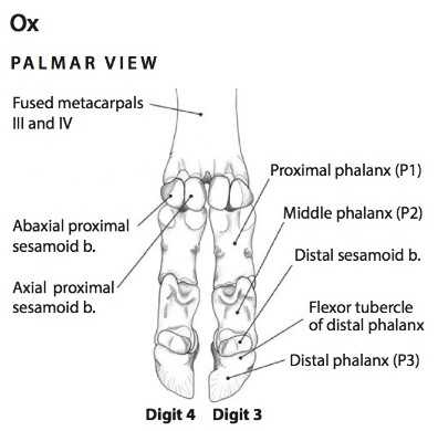
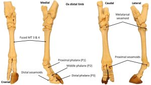
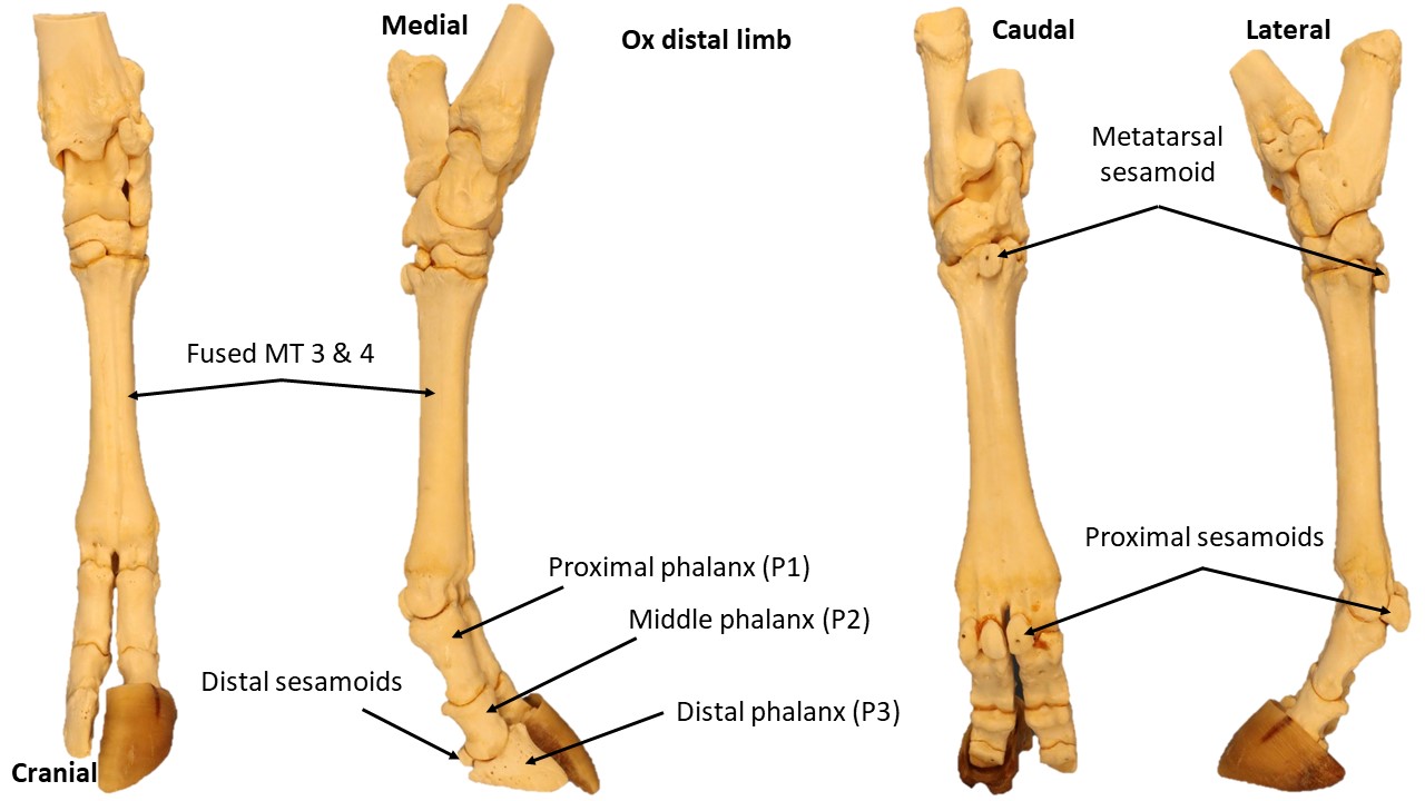
The metatarsal bones of the carnivore resemble the thoracic limb metacarpal bones except for the first metatarsal, which may be divided, rudimentary, or absent. Similarly, the metatarsal bones of the ungulates resemble their metacarpal counterparts. However in the ruminant, there is a metatarsal sesamoid bone in place of a 5th metatarsal bone. The phalanges and sesamoids form the skeleton of the digit. Those of the pes are similar to those of the manus.
-
- Dog Left metatarsal and sesamoid bones, disarticulated, plantar aspect. 3
-
- Cat tarsus. 5
-
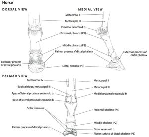
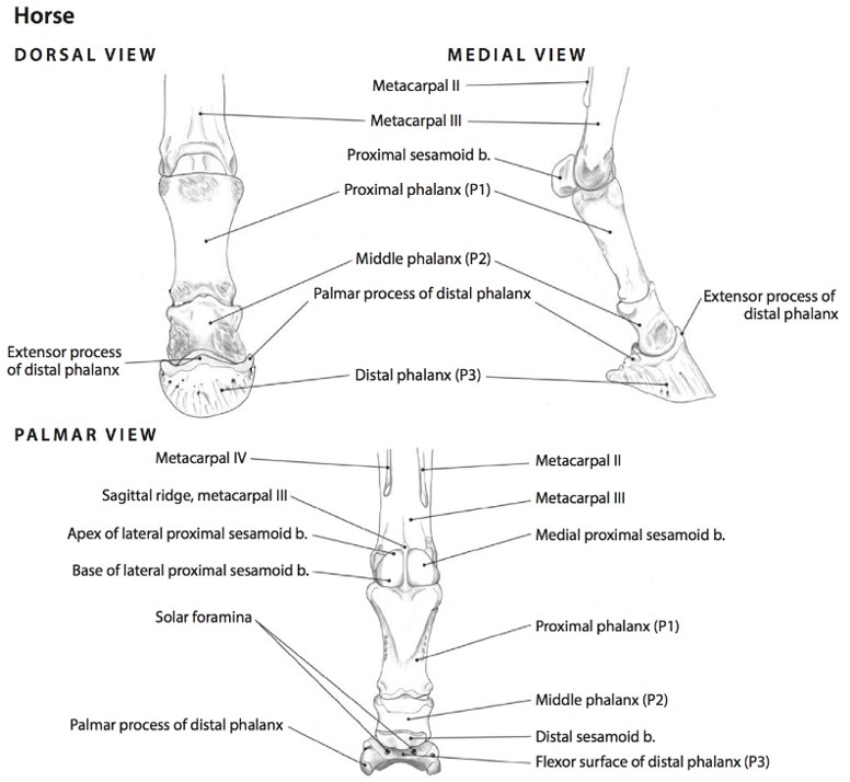
- Left Digit of the Horse, Dorsal, Medial, and Palmar Views. 6
-
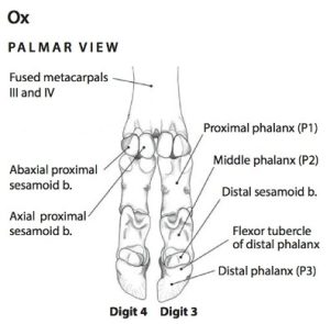
- Left Digits of the Ox, Palmar View. 6
-
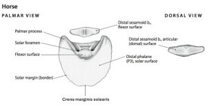
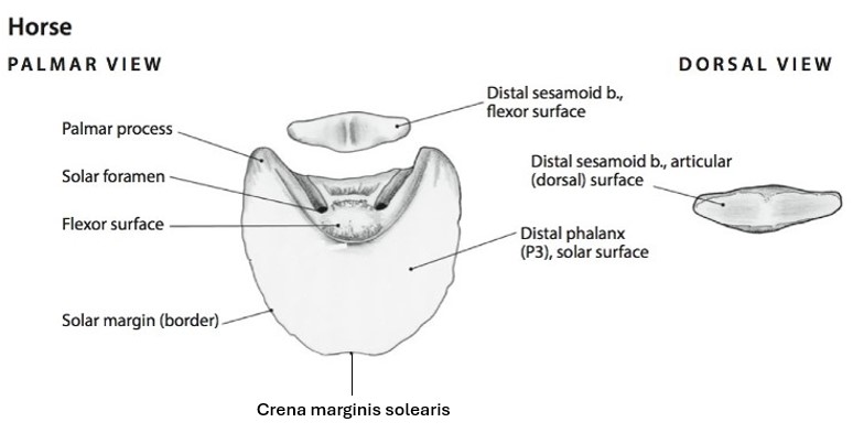
- Distal Phalanx and Distal Sesamoid (Navicular) bone of the Horse – Palmar and Dorsal View.6
-
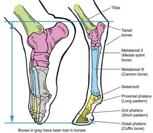
- Horse vs human foot. American Farriers Journal
-
- Ox tarsus
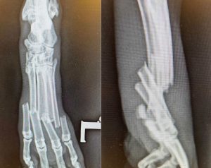
Clinical Relevance: Metatarsal Fractures in the Carnivore
You are presented with a two year old male castrated domestic short-haired cat with a pronounced swelling of his left hind foot. His owners state that a radiator cover had fallen on his foot. Now he is non-weight bearing and displays signs of moderate to severe pain upon manipulation of the affected paw. His physical examination is otherwise unremarkable. Radiographs of the cat’s foot are below. Would you recommend surgery? Why or why not (think back to the discussion on thoracic limb fractures in this region)?
Click here to see what this cat’s veterinarian did.
Joints & Ligaments
Pelvic ligaments and coxofemoral (coxal) joint and ligaments
The ischium and pubis of the right and left sides are joined on the median plane at the symphysis pelvis.
The sacroiliac joint is an articulation of stability rather than mobility. The right and left wings of the ilia articulate with the right and left wings of the sacrum. In the adult most of the apposed articular surfaces are united by fibrocartilage that surrounds a small area of articulating hyaline cartilage with synovial fluid. Dorsal and ventral sacroiliac ligaments reinforce the joint. Sacroiliac joint injections are performed in equine practice, particularly in sports medicine cases with lameness located to this site.
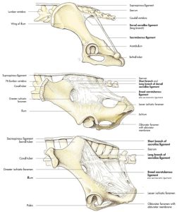
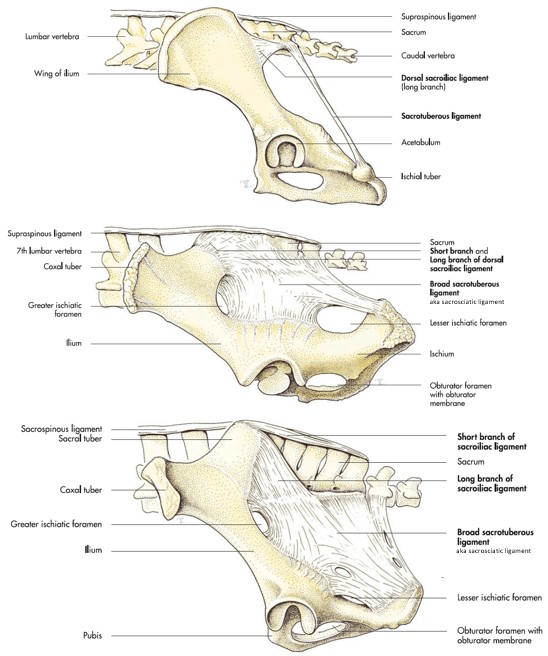
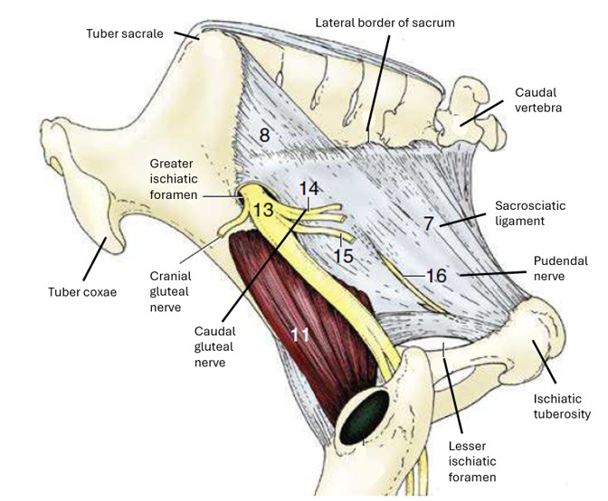
The sacrotuberous ligament in the carnivore runs from the transverse processes of the last sacral and first caudal vertebrae to the ischiatic tuberosity. It serves as an origin for several muscles that have been dissected. In the ungulates this ligament has expanded into a sheet and is referred to as the sacrosciatic (or broad sacrotuberous) ligament.
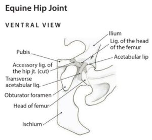
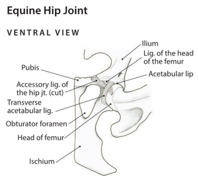
As mentioned, the hip joint is a ball-and-socket joint whose main movements are flexion and extension, although any direction is possible, to a degree. The joint capsule passes from the neck of the femur to a line peripheral to the acetabular lip. Also already mentioned, the ligament of the femoral head is a thick band of collagenous tissue that extends from the acetabular fossa to the fovea capitis, anchoring the femoral head into the acetabulum. See the horse addition to this ligament below.
The transverse acetabular ligament is a small band that extends from one side of the acetabular notch to the opposite side. It is located at the ventrocaudal aspect of the acetabulum and continues as the acetabular lip, which deepens the acetabulum by forming a fibrocartilaginous border around it.
-

- Ligaments of the dog pelvis, ventral view. 1
-
- Canine sacrotuberous ligament and bovine sacrosciatic ligament. 8
-
- Comparative pelvic ligaments. 7
-
- Horse hip. 6
-
- Ventral view of the horse hip. 14

-

- Lateral view of the sacrosciatic ligament of the horse. 8
-
- Dog hip ligaments 40

Coxal (hip) joint – horse specific feature.
Besides the standard ligament of the femoral head, uniquely, the horse has an additional accessory ligament of the femoral head passing from the femoral fovea capitis through the acetabular notch to the pectin pubis where it inserts in the prepubic tendon. This additional powerful ligament in the horse may be one explanation for the lower incidence of hip luxation in this species.
Observe: Review the cut ends of ligaments on the femoral head and within the acetabulum for the species studied. Separate identification of the two ligaments in the horse is not required but do be aware that two are present in that cut stump of ligament.
Clinical Relevance: Partial or Complete Coxofemoral Joint Luxation
- Coxofemoral luxation refers to the displacement of the head of the femur outside of the acetabulum, a relatively common occurrence in dogs and cats, while it is much less frequent in artiodactyls and is a very uncommon condition in the horse. At the time of acute injury, animals with this condition will develop a sudden onset, non-weight bearing lameness and will hold the leg at an angle depending on the direction of the luxation. For a complete luxation to occur, both the ligament of the head of the femur and the joint capsule must rupture. Thus it takes a significant blunt force, such as being hit by a car, for dislocation of a normal hip to occur. Due to the pull of the gluteal muscles on the proximal femur, the most common direction for the luxation to occur is craniodorsal to the acetabulum.
- Dislocation of the coxal (hip) joint is much less common in the horse, in part because of the accessory femoral head ligament.


Clinical relevance: Hip dysplasia
Hip dysplasia refers to the abnormal development or growth of the coxofemoral joint, usually occurring bilaterally. It is manifested by varying degrees of laxity of surrounding soft tissues, instability, malformation of the femoral head and acetabulum and resulting osteoarthrosis. Must be diagnosed using special radiographic technique, and animals who have hip dysplasia should not be bred, as it is an inherited trait. Most common cause of degenerative joint disease of the canine hip. Treatment may occur when patient is young to prevent degenerative changes. Curative treatment of older patient with already existing degenerative joint disease (DJD) is a total hip replacement.


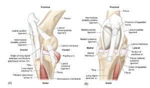
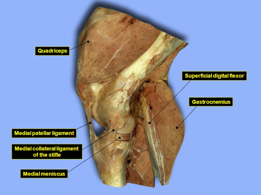
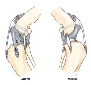
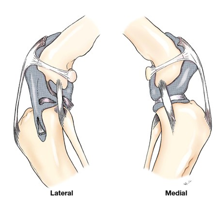
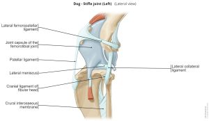
Stifle joint and ligaments
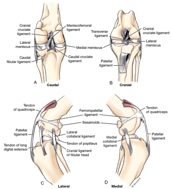
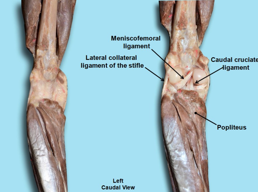
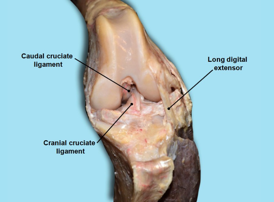
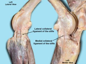
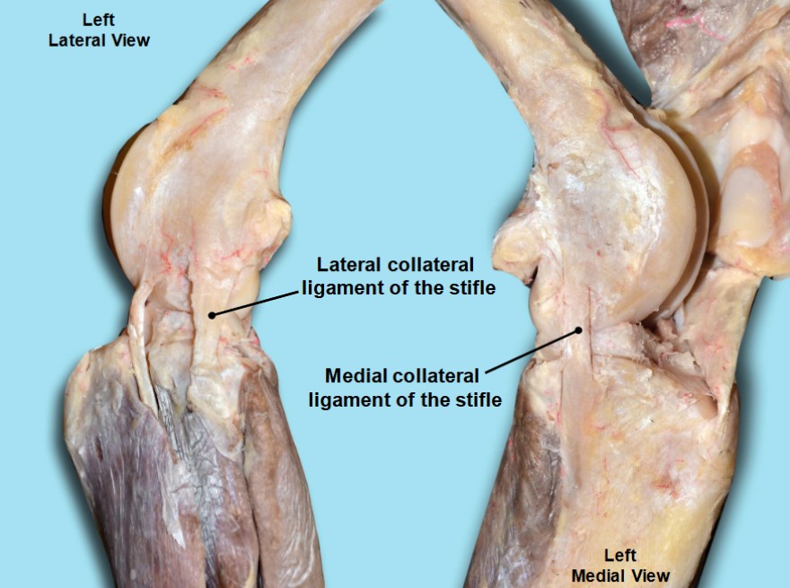
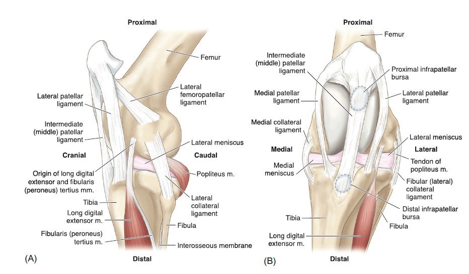
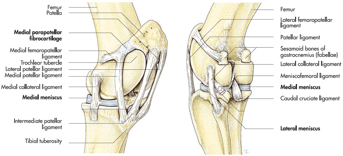
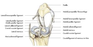
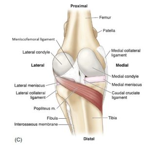
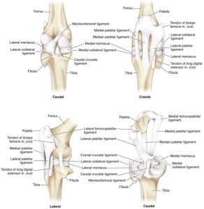
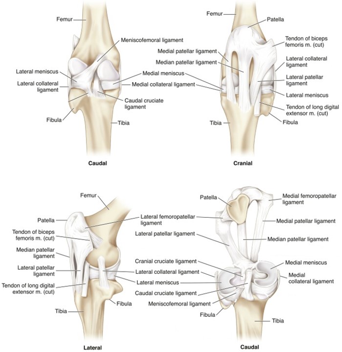
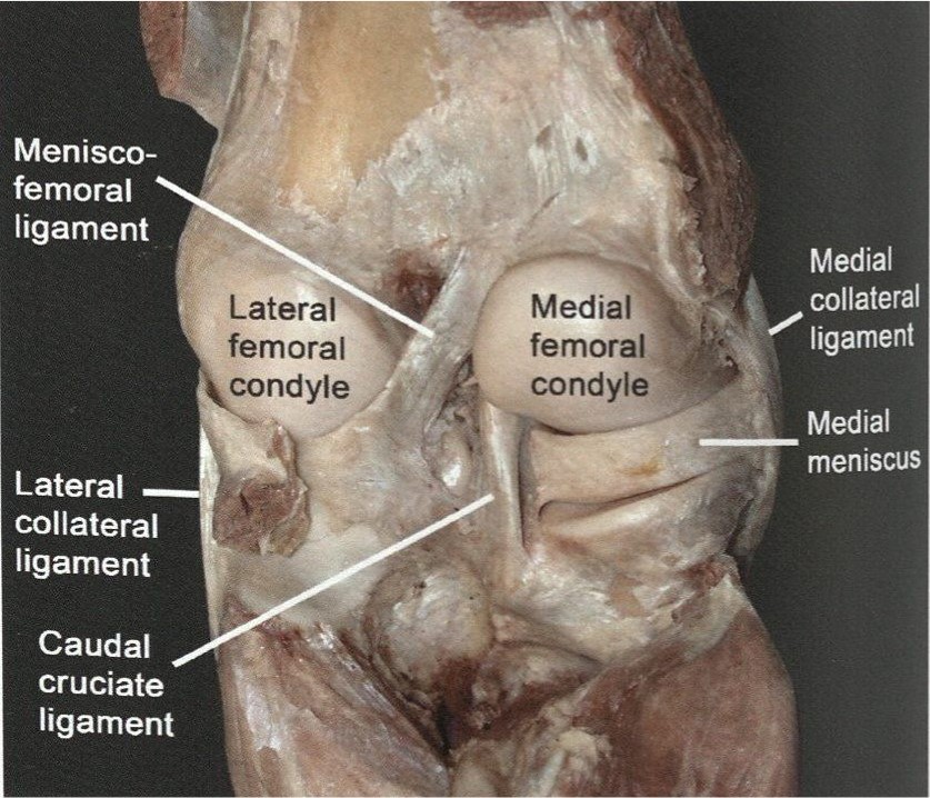
The stifle is a complex, condylar joint, with motion possible in 3 planes. Primarily it acts as a hinge joint (extension and flexion) in the craniocaudal plane, supported by extra- and intra-articular ligaments. The femur, patella (‘kneecap’ in human), and tibia contribute to the joint surfaces of the stifle, forming femoropatellar and femorotibial joints. Collateral, femoropatellar, and patellar ligaments are extra-articular ligaments and the cranial and caudal cruciate ligaments are intra-articular. The cruciate ligaments are named for their attachment on the tibia, e.g. the cranial cruciate ligament attaches to the cranial tibial plateau. There is a single patellar ligament in the carnivore, swine, and small ruminant and three patellar ligaments in the ox and horse, namely the lateral, intermediate and medial patellar ligament. Fat accumulates between the patellar ligament(s) and the deeper stifle joint capsule and is noted on radiographs as the infrapatellar fat body. Medial and lateral, C-shaped, cartilaginous menisci, lie between the femoral condyles and the tibial plateau, increasing joint congruency between these two bones. These menisci are attached by ligaments to each other and to the tibia and stifle. The meniscofemoral ligament connects the caudal part of the lateral meniscus to the femur.
In the carnivore, the medial and lateral femoropatellar ligaments are thin fascial bands extending between the patella and the sesamoids of the gastrocnemius muscle bellies. The gastrocnemius sesamoids are more commonly called the fabellae (singular, fabella) and form an important anchor for suture in stifle surgeries (e.g. stabilization after cruciate ligament rupture).
A little more on the all important cruciate ligaments. The cruciate ligaments limit craniocaudal motion of the femur and tibia. The cranial cruciate ligament keeps the tibia from sliding cranially beneath the femur when the limb bears weight. It also limits medial rotation of the tibia when the stifle is flexed. The caudal cruciate ligament prevents caudal movement of the tibia beneath the femur when the limb bears weight. See clinical relevance box below.
Lastly, the equine stifle has been designed to allow for a patellar locking mechanism that results in holding the stifle in extension with minimal muscle activity required (this helps the horse stand on the ‘locked’ leg for a bit, while resting the other pelvic limb). The mechanism involves an anatomical loop of structures being ‘hooked’ over the very large medial trochlear ridge of the stifle. The loop of structures includes the intermediate patellar ligament, the patella, the medially located parapatellar fibrocartilage (or cartilaginous process of the patella), and the medial patellar ligament. This locking mechanism will be revisited in detail in a later lab.
Carnivore stifle
-
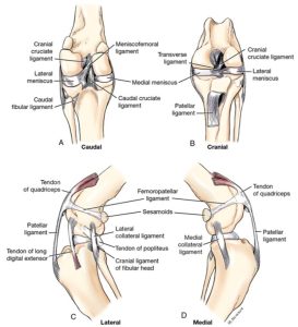
- A, Ligaments of the dog’s left stifle joint, caudal view. B, Cranial view. C, Lateral view. D, Medial view. 1
-

- Dog popliteus 40
-

- Dog cranial cruciate ligament 40
-
- Dog stifle ligaments 40
Ungulate stifle
-
- Bones of the equine stifle region. (A) Lateral view and (B) cranial view. 9

-
- Ligaments and menisci of the equine stifle region. (A) Lateral view, (B) cranial view, and (C) caudal view. 9
-

- Horse stifle, left medial aspect. 7
-
- Ligaments of the left horse stifle joint after removal of the femur, caudal aspect. 7

-
- Ligaments and menisci of the equine stifle region. (A) Lateral view, (B) cranial view, and (C) caudal view. 9

-
- Anatomy of the cow stifle joint. (A) Caudal view. (B) Cranial view. (C) Lateral view. (D) Caudal view with femur removed. 9
-

- Horse stifle. 16
-

- Horse medial stifle 40
Interactive review content:
Clinical Application – Cranial Cruciate Ligament Rupture
To diagnose rupture of the cranial cruciate ligament in the carnivore we perform mechanical tests of the integrity of the ligament. In the cranial drawer test the tibia is moved cranially while holding the femur firmly to prevent its movement. In the normal animal the tibia should not significantly displace/move cranially because the cranial cruciate when intact prevents this movement. When the cranial cruciate ligament is completely ruptured the tibia is able to noticeably slide forward under the femur causing a dramatic positive drawer test and confirming the diagnosis. Partial ligament ruptures are harder to diagnose because of the less displacement likely. The tibial thrust test is an alternative method of examining the function of the cranial cruciate ligament. Check out the videos below.
Drawer test for CCL rupture in a dog
Drawer test and tibial thrust test in a dog
WATCH: Presentation and Demonstration on the Cranial Drawer and Tibial Thrust Tests
Stifle joint synovial sacs
The stifle consists of femoropatellar and medial femorotibial and lateral femorotibial synovial sacs. These sacs are named for the bones they are between. In the carnivore and pig these 3 sacs communicate freely as one large joint sac, meaning synovial fluid and anything injected into one of the sac spaces will readily move between all three sacs. The ruminant typically has communication between the 3 synovial sacs. In the horse the medial femorotibial and femoropatellar sacs communicate most of the time and the lateral femorotibial sac is more commonly (but not always!) a closed space, not connected to the other sacs. Knowledge of the communications between synovial sacs has important bearing on joint injection sites for therapy and diagnostic analgesia during lameness exams (with particular reference to the horse lameness exam).
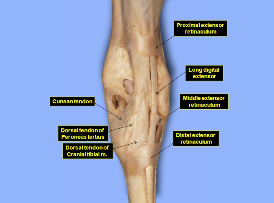
The lateral femorotibial sac continues distally through the extensor groove of the tibia as the tendon sheath for the tendon of origin of the long digital extensor m. (and fibularis tertius m.).
-
- Capsule of the dog’s left stifle joint. 1
-
- Dog stifle joint capsule. 24

-
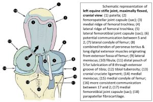
- Schematic representation of left equine stifle joint, maximally flexed, cranial view. 2
-
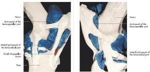
- Acrylic cast of the left stifle joint of a horse, medial and lateral aspect. 7

Tarsal joint and ligaments
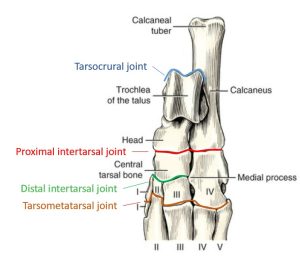
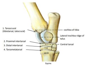
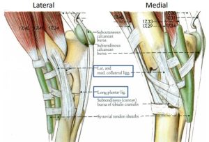
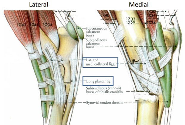
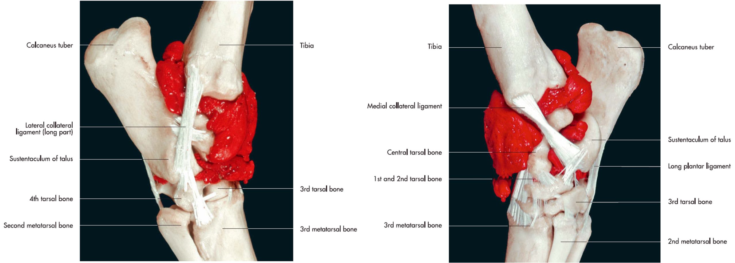
The tarsus is a composite of several articulations, supporting ligaments, and intervening joint sacs. Proximal to distal the joints are the tarsocrural, proximal intertarsal, distal intertarsal and tarsometatarsal joints. Greatest flexion and extension movement in the tarsus occurs at the tarsocrural joint. The remaining tarsal joints are considered low motion joints, i.e. they move very little, however they do handle the very high loads of weight bearing. Powerful and complex medial and lateral collateral ligaments cross the tarsus from the tibia (and fibula, as present per species) to the metatarsal bones, providing stability on each side of the tarsus. Numerous ligaments join the individual tarsal bones to each other. The long plantar ligament extends from the calcaneus to the 4th tarsal and 4th metatarsal bones, acting to anchor the calcaneus distally to counter the pull of the common calcanean tendon proximally. This ligament will be revisited in a later ungulate lab.
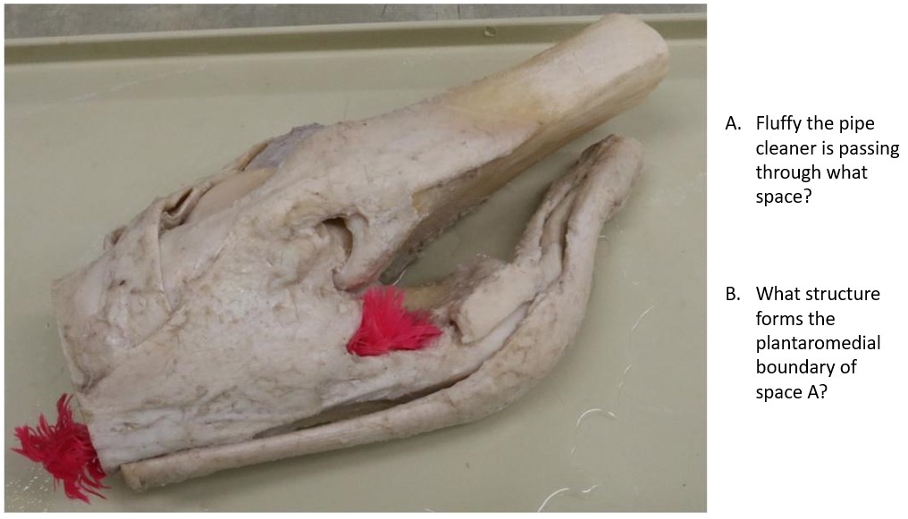
The flexor canal of the tarsus is the equivalent of the thoracic limb carpal canal. This space provides passage for tendons of the deep digital flexor to pass through, enroute to the digit. The canal is bound by the calcaneus laterally, the sustentaculum tali of the calcaneus dorsally, and the flexor retinaculum plantaromedially. A tarsal sheath wraps around the tendons to lubricate their path through the canal.
-
- Joints of the dog tarsus. 1

-
- Joints of the horse tarsus. 14

-
- Tarsal ligaments of the horse. 14
-
- Horse tarsus 40
Tarsal joint synovial sacs
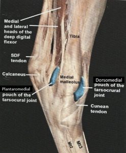
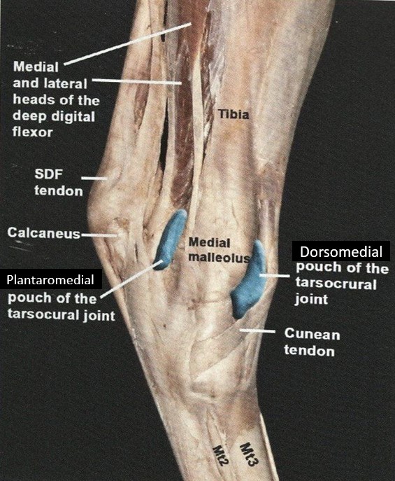
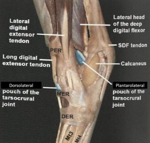
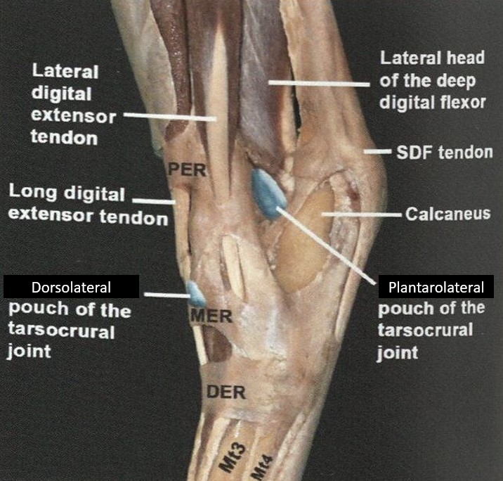
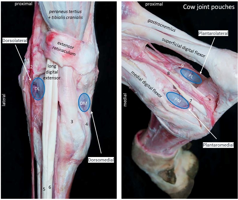
Along with the four major articulations of the tarsus there are four joint sacs. The tarsocrural joint sac is the largest and incorporates the articulations of the distal tibia (and fibula as present for the species) with that of the talus and calcaneus. Synovial fluid sampling is relatively easy from the spacious tarsocrural joint sac. Notable in the horse, the tarsocrural joint sac projects outward (superficially) in spaces between tendons, ligaments and bones, to form 4 pouches. These pouches are so named for their location on the tarsus – dorsomedial, dorsolateral, plantaromedial, and plantarolateral pouch. The dorsomedial pouch is invariably the largest and most accessible for joint access. The pouches become very apparent with excessive fluid distension of the tarsocrural joint sac. The other 3 joint sacs include the proximal and distal intertarsal sacs and the tarsometatarsal sac. With reference to clinically relevant anatomy, the tarsocrural and proximal intertarsal joint sacs communicate 100% of the time in the horse and ox (but not in the pig). The distal intertarsal and tarsometatarsal joint sacs have variable communication in the horse. Knowledge of these communications is important to planning joint therapies and diagnostic analgesia strategies during lameness exams.
-
- Horse hock. 16
-
- Horse hock. 16
-
- Articular spaces of the tarsal joint of the horse, lateral and medial views. 7
-
- Tarsus of horse, dorsal view. 2

-
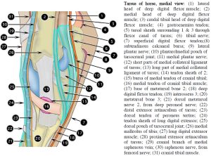
- Tarsus of horse, medial view. 2
-

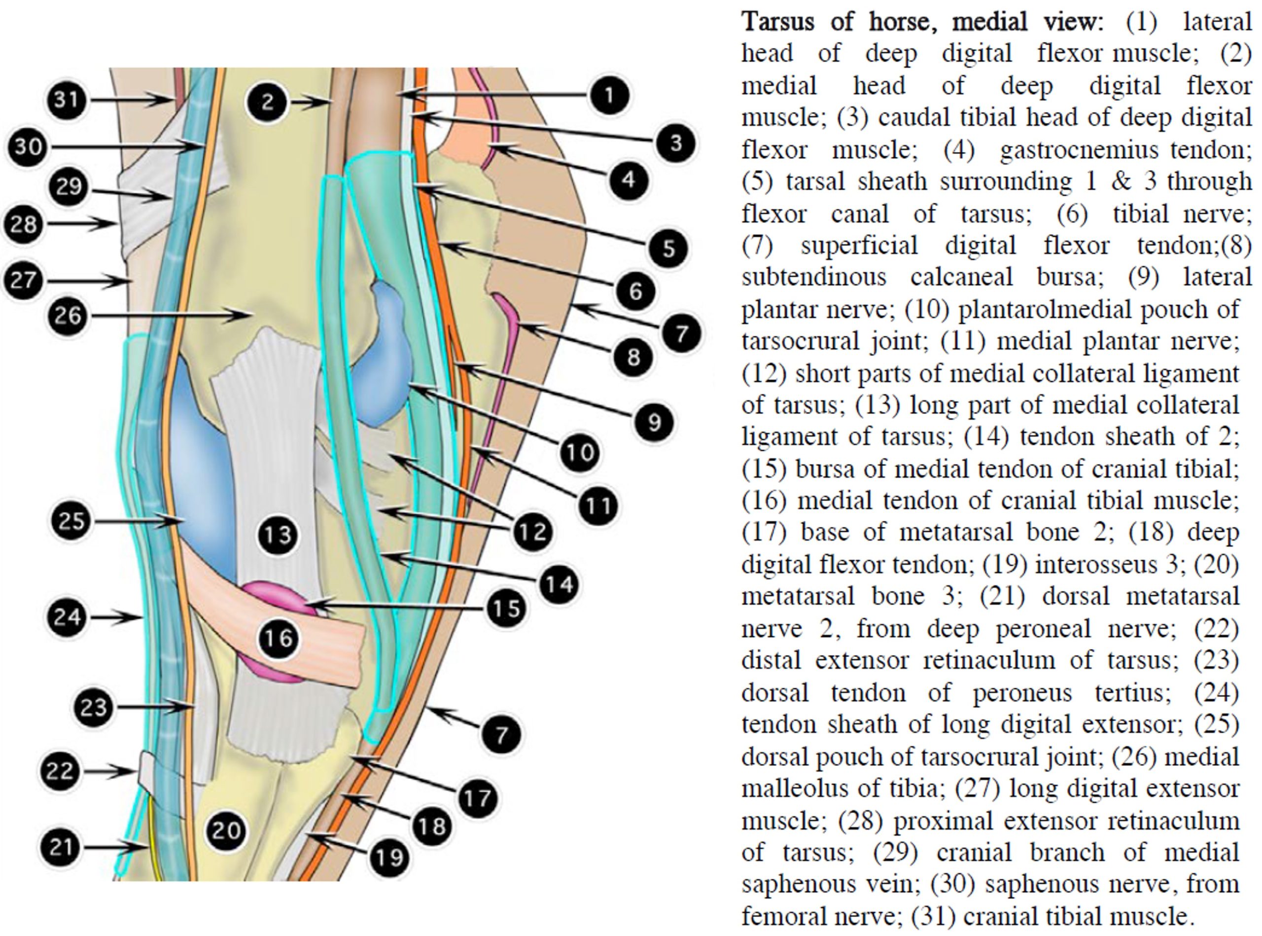
- Tarsus of horse, lateral view. 2
-
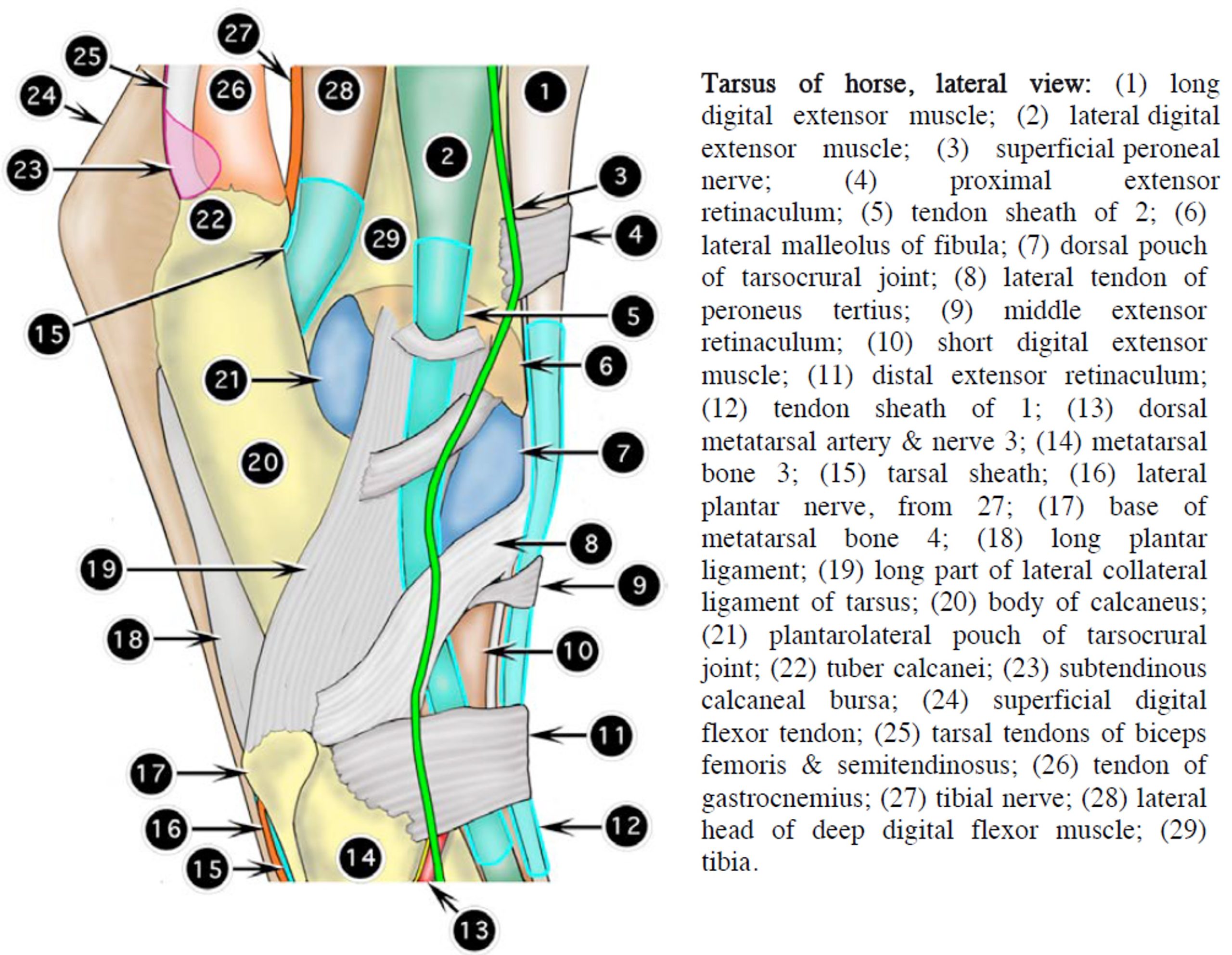
- Joint pouches of the right tarsocrural joint of an adult Holstein cow. H. Larde
Review Videos
Carnivore Pelvic Limb Osteology – 4 minutes
Horse and Cow Pelvic Limb Osteology – 24 minutes
Horse stifle and hock injections – 9 minutes
Terms
| Regions of the pelvic limb (proximal to distal) | Common Name | Species |
| Gluteal | Horse – croup
Ruminants & Pig – rump |
All |
| Thigh | All | |
| Crus | Gaskin | All |
| Pes | Limb distal to crus. i.e. includes tarsus, metatarsus and digit(s) | All |
| Tarsus | Hock | All |
| Metatarsus |
Ungulates – cannon/shank |
All |
| Digit | Toes. In ungulates, includes pastern and “foot” | All |
| Pastern | Ungulates | |
| “Foot” | Hoof | Ungulates |
| Joints of the pelvic limb (proximal to distal) | Common Name |
| Coxofemoral or coxal | Hip |
| Stifle | |
| Tarsus | Hock |
| Metatarsophalangeal | Ungulates – fetlock or ankle(s) |
| Proximal interphalangeal | Ungulates – pastern jnt(s) |
| Distal interphalangeal | Ungulates – coffin jnt(s) |
Osteology of Pelvis/Os coxae |
||
| Osteology | Features | Species Differences/comments |
| Ilium | Auricular surface – articulation with sacrum=sacroiliac joint. |
|
| Tuber sacrale | ||
| Tuber coxae | Ruminant = “hook bones” | |
| Greater ischiatic notch | ||
| Ischium | Ischiatic tuberosity | Ruminant = “pin bones” |
| Ischiatic arch | ||
| Ischiatic spine | ||
| Lesser ischiatic notch | ||
| Pubis | ||
| Acetabulum | Partly formed by acetabular bone, plus ilium, ischium, pubis | |
| Acetabular notch | ||
| Acetabular fossa | ||
| Obturator foramen | Bordered by pubis and ischium | |
| Pelvic symphysis | Midline fusion of left and right ossa coxae | |
Osteology of Thigh & Crus |
||
| Osteology | Features | Species Differences/comments |
| Femur | Head | |
| Fovea capitis | ||
| Neck | ||
| Body (shaft) | ||
| Greater trochanter | Horse – cranial & caudal parts | |
| Lesser trochanter | Attachment for iliopsoas m. | |
| Trochanteric fossa | ||
| Intertrochanteric crest | ||
| Third trochanter | Horse – superficial gluteal m. attachment | |
| Medial/lateral supracondylar tuberosities | Attachment for gastrocnemius mm. | |
| Supracondylar fossa | Ungulate – attachment for SDF m. | |
| Trochlear groove | ||
| Medial/lateral trochlear ridges of femur | ||
| Resting and gliding surfaces of femoral trochlea | Ungulate | |
| Medial/lateral condyles of femur | ||
| Medial/lateral epicondyles of femur | ||
| Extensor fossa of femur | Attachment of long digital extensor m., (and fibularis tertius m. in ungulates) | |
| Patella | Ungulate – apex (distal end), base (proximal end) | Horse – resting and gliding surfaces |
| Sesamoids of gastrocnemius mm. = Fabellae | [L] faba = bean; the fabellae are bean-shaped sesamoids | Carnivore |
| Sesamoid of popliteus m. | Carnivore | |
| Tibia | Body (shaft) | |
| Tibial tuberosity | ||
| Medial malleolus | All | |
| Lateral malleolus | Horse, because fibula fused to tibia. | |
| Distal intermediate ridge | Horse – this is a specific clinical/radiographic imaging term for the cochlea of the tibia where OCD occurs. | |
| Fibula | Lateral malleolus | Carnivore, ruminant, pig |
Osteology of Tarsus |
||
| Osteology | Features | Species Differences |
| Calcaneus | Tuber calcanei (calcaneal tuberosity) | |
| Sustentaculum tali | ||
| Talus | Trochlea – with medial and lateral ridges of trochlea | Artiodactyls have a proximal and distal trochlea on the talus |
| Tarsal bones – Central, I, II, III, IV | Variation in number present and fusion between tarsal bones, be able to identify in carnivore, horse, ruminant. | |
Osteology of Metatarsus and Digit |
||
| Osteology | Features | Species Differences |
| Metatarsal bone I | Carnivore only (variable) | |
| Metatarsal bone II | Horse – button of splint bone at distal end | Horse – MT2 is the medial splint bone |
| Metatarsal bone III | Horse – sagittal ridge of MC3 | Ruminant – MT3 + MT4 fused |
| Metatarsal bone IV | Horse – button of splint bone at distal end | Horse – MT4 is the lateral splint bone; Ruminant – MT3+MT4 fused |
| Metatarsal bone V | Carnivore only | |
| Metatarsal sesamoid bone | Ruminant | |
| Proximal sesamoid bones | Base, apex, axial and abaxial surfaces | Ungulate only |
| Distal sesamoid bone = Navicular bone | distal border, proximal border, flexor and articular surfaces, sagittal ridge of navicular bone | Horse only |
| Digits (I)II-V – species variation | Consist of proximal, middle and distal phalanges; axial and abaxial side to each digit (or medial and lateral sides in horse since only one digit) | Carnivores – 4 digits typical, ie digits 2-5
Ruminant and Pig – 4 digits (3 and 4 are weight bearing; 2 and 5 = dewclaws) Horse – 1 digit (3rd digit, bears all weight of limb!) |
| Proximal phalanx | Ungulate – Pp, P1, long pastern bone | |
| Middle phalanx | Ungulate – Pm, P2, short pastern bone | |
| Distal phalanx | Ungulate – Pd, P3, coffin or pedal bone | |
| Extensor process | All (attachment of CDE) | |
| Flexor tubercle of Pd | Carnivore, Ruminant | |
| Solar border (margin) | Horse | |
| Crena marginis solearis | Horse | |
| Medial and lateral plantar processes (‘wings’) of Pd | Horse | |
| Solar surface with smooth area and flexor surface | Horse (DDFT attaches to flexor surface of solar surface) | |
Joints and Ligaments of the Pelvic Limb |
|
| Features | Species Differences |
| Sacroiliac joint | |
| Sacrotuberous ligament | Dog |
| Sacrosciatic ligament | Ungulate |
| Coxal joint |
|
| Ligament of the femoral head | Cut end can be identified (in horse combined with accessory ligament) |
| Accessory ligament of the femoral head | Horse only (no need to identify separately to regular ligament) |
| Acetabular lip | Carnivore – cartilaginous extension of rim of acetabulum |
| Transverse acetabular ligament | Carnivore – bridges acetabular notch |
| Stifle (genual) joint | |
| Patellar ligament(s) | Horse & Ox – medial, intermediate/middle, lateral |
| Carnivore, small ruminant, pig – single ligament | |
| Femoropatellar ligament | Medial and lateral |
| Collateral ligament of stifle (may also refer to as femorotibial ligament) | Medial and lateral |
| Cruciate ligament | Cranial and caudal |
| Meniscus | Medial and lateral |
| Meniscofemoral ligament | |
| Femoropatellar joint | |
| Medial femorotibial joint | |
| Lateral femorotibial joint | |
| Parapatellar fibrocartilage (cartilaginous process of patella) | Horse only, see cross section specimens |
| Tarsus – Tarsus and distal not studied in pig | |
| Flexor canal of tarsus | |
| Collateral ligament of tarsus | Medial and lateral |
| Long plantar ligament | Horse only |
| Tarsocrural joint | Articular surface |
| Tarsocrural joint pouches | Horse – dorsolateral, dorsomedial, plantarolateral, plantaromedial |
| Proximal intertarsal joint | |
| Distal intertarsal joint | |
| Tarsometatarsal joint | |
| Metatarsophalangeal and interphalangeal joints |
The detailed arthrology and ligaments is similar to the thoracic limb and will not be revisited here. It suffices to be able to name the bones and the joints of this region. |