
MSK LAB 21A – Pelvic Limb Ungulate Highlights
Learning Objectives
- Identify the structures of joints, including the stifle and tarsus in detail; connect structures to the clinically applied anatomy as emphasized.
- Identify the anatomy of the tarsal canal
- Identify the tendons and ligaments of the metatarsal region
- Identify bursae and tendon sheaths as noted.
- Connect structures to the clinically applied anatomy as emphasized.
- List, identify and explain the key anatomy and mechanism of the pelvic limb passive stay apparatus in the horse.
Lab instructions:
Dissection teams are to dissect an equine thoracic limb, as provided on the day of the lab. Supplement dissection learning with liberal study and review of the limb models available in lab, wet and dry. The limbs can be tagged and stored at the conclusion of the lab for subsequent review. In addition, external anatomy of the hoof capsule (see LAB 23A) may be reviewed on these limbs.
This lab will start with important anatomy at the ungulate stifle joint and this content is to be learned on lab prosections and models. Following stifle anatomy, the lab guide is split into a horse section (which has dissection instructions) and a brief ruminant section (content learnt on prosections and models). It is ok to start with the equine limb dissection and return to the stifle anatomy of the horse and ruminant, as specimens are available for review.
Observe: Firstly, take a moment to review and identify the regions and joints of the ungulate pelvic limb.
Stifle (genual) joint – horse and ruminant
The stifle joint consists of the femur, tibia, and patella, three joint compartments, and various ligaments and menisci. Attachments of muscles are also incorporated. The fibula does not contribute directly to the stifle joint.
Observe: Refer to articulated skeletons to review the bony structures of the stifle (stifle ligaments from LAB 16A) and to recognize the location of the femoropatellar joint, the medial femorotibial joint and the lateral femorotibial joint.
Refer to the equine and ruminant prosected cadavers, models and wet and dry specimens to view and study the anatomy of the stifle.
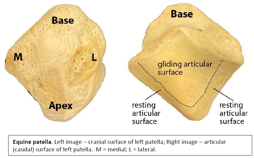
Examine the articular surfaces of the patella and femoral trochlea. Note the significant size difference between the medial and lateral femoral trochlear ridges, in the horse and ox. The much larger medial ridge has an important role in the patellar locking mechanism, which is of specific interest in the horse (see below).
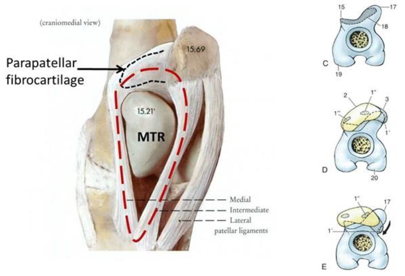
Identify the trochlear groove of the femur and the resting and gliding surfaces of the patella and femoral trochlea. The patella has a medial cartilaginous extension, the parapatellar fibrocartilage (aka the cartilaginous process of the patella). Identify this structure, it has a role in the patella locking mechanism (see below).
Recall that the patella is attached to the tibial tuberosity by a single patellar ligament in the carnivore. The ox and horse have three (3) patellar ligaments, the pig and small ruminants have one (1). The three ligaments in the ox and horse are termed the medial, intermediate (or middle) and lateral patellar ligament.
In addition the patella is attached to the femur via medial and lateral femoropatellar ligaments. The stifle joint is stabilized by very strong medial and lateral collateral ligaments of the stifle, and by cranial and caudal cruciate ligaments. A medial meniscus and lateral meniscus are slotted between the femoral condyles and tibial plateau, to increase joint congruence. The lateral meniscus is bound to the femur by the caudally located meniscofemoral ligament.
-
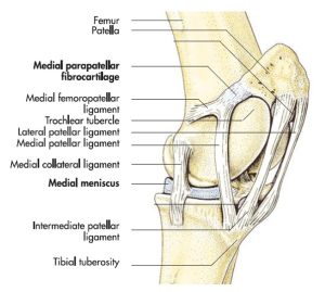
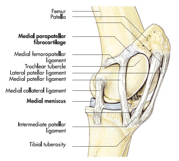
- Horse stifle, left medial aspect. 7
-
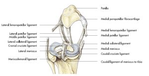
- Ligaments of the left horse stifle joint after removal of the femur, caudal aspect. 7

-
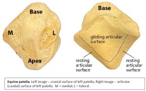
- Horse patella, resting and gliding surfaces. Gerard, M., 2020
-
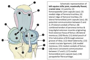
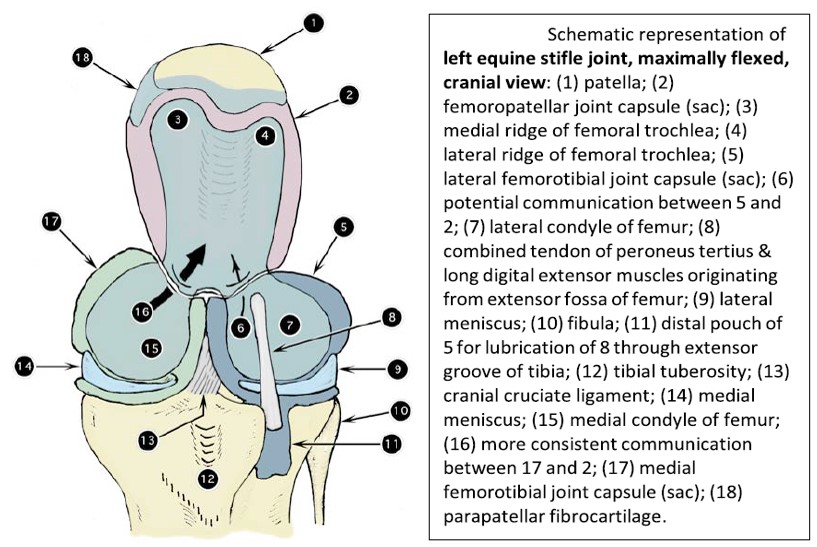
- Schematic representation of left equine stifle joint, maximally flexed, cranial view. 2
-

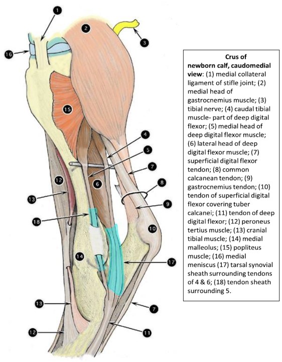
- Crus of the newborn calf; caudomedial view. 2
-
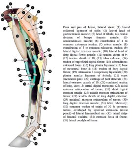
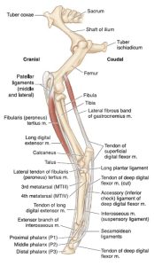
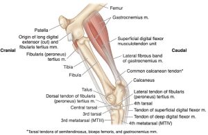
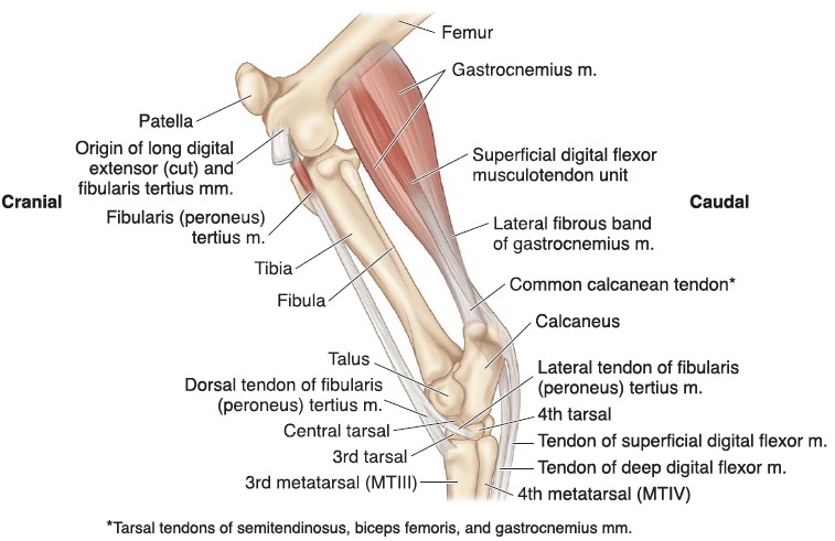
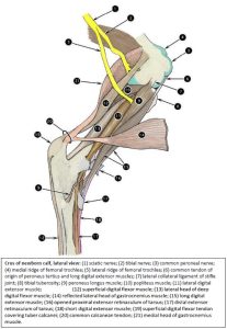
- Crus and pes of the horse, lateral view. 2
-
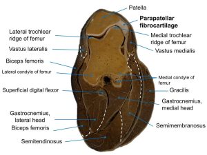
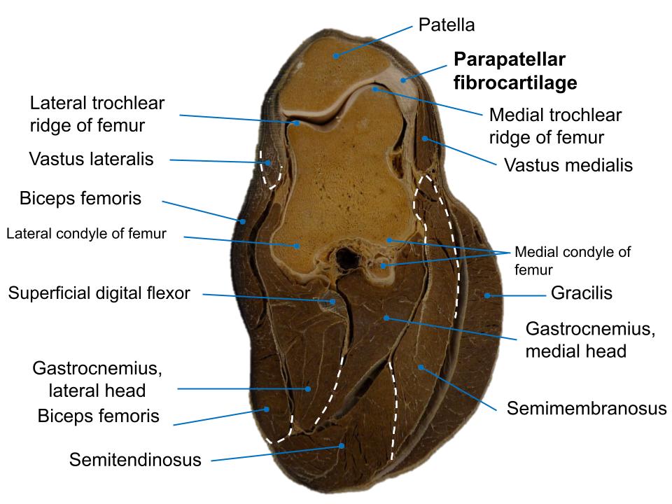
- Horse cross section at the level of the stifle, showing the parapatellar fibrocartilage.
Clinical relevance: Q6
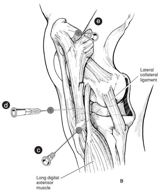
The stifle is frequently accessed with a needle to get a synovial fluid sample or to make an intraarticular injection (lameness exam or treatment). The stifle is a common site for causes of lameness, and in the horse lesions include: osteochondrosis dessicans (OCD) of the lateral femoral trochlear ridge, subchondral bone cyst of the medial femoral condyle, patellar luxation and upward fixation of the patella, and meniscal injuries.
Q6: What stifle joint sac (compartment) has been entered by the needle C, as shown in the image?
A: lateral femorotibial joint
B: medial femorotibial joint
C: femoropatellar joint
Patella locking mechanism – horse
Extending from the medial aspect of the patella is the hook-shaped parapatellar fibrocartilage (aka cartilaginous extension of the patella) that is continuous with the medial patellar ligament. During flexion and extension of the joint, the large gliding surfaces of the patella and trochlea are in contact. Narrow resting surfaces located at the distal aspect of the patella and its fibrocartilage, and the proximal aspect of the medial ridge of the femoral trochlea, engage when the patella is pulled proximally and rotated medially to ‘lock’ over the medial ridge in a resting position. This is the normal patellar locking mechanism of the horse. You may have noticed that when horses are standing around, taking it easy, they tend to stand with all their weight on one pelvic limb, with the other one relaxed and sort of half-cocked, resting on the toe of the hoof. When they do this, they lock the patella of the supporting limb over the medial ridge of the femoral trochlea (i.e., they “put it in park”). Fixation of the stifle joint by the patellar locking mechanism is the key to converting the pelvic limb into a ‘passive stay’ for support of the body weight (more of this passive stay apparatus is revealed as we continue distal dissection).
-
- Horse stifle, left medial aspect. 7
-
- Ligaments of the left horse stifle joint after removal of the femur, caudal aspect. 7
-

- Horse patellar locking mechanism. 7
Clinical relevance:
Patellar locking mechanism in horse is part of the passive stay apparatus of the pelvic limb. When it is stuck in ‘lock’, it is called ‘upward fixation of the patella’ (“locking stifles”).
Structures of tarsus – horse
At this point, dissection of the provided limb specimen is to be performed. Learn the following equine limb anatomy from directed dissection activities and reference to prosections and models (wet, dry, plastinated).
Dissect: Make a skin incision on the medial aspect of the horse limb, extending to the epidermal hoof margin (the junction of skin with the epidermal hoof is called the coronet). Reflect the skin completely from the limb and discard it. Superficial vessels and nerves can be removed with the skin. Our focus is primarily on tendons, ligaments, bursa, and synovial structures.
Excise crural fascia, from around the limb. Dissect muscle margins to the level of their tendons. Muscle identification is facilitated by tracing known tendons at the tarsus and metatarsus, proximally to the muscle belly. Once tendons become familiar trace them back to identify the muscles. Refer to prosected models to assist with identification.
Observe: We will focus on the tarsus for a bit. This complex joint and related structures is a frequent site of examination during lameness work ups and subsequent treatment procedures. Refamiliarize yourself with the articulations and bones of the tarsus and their prominent landmarks. Compare the horse and bovine tarsus to note the difference in tarsal bones and their fusions. For a review of the anatomy of synovial structures, refer back to the foundations content. Study prosections of the tarsus as well as the cadavers.
-
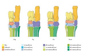
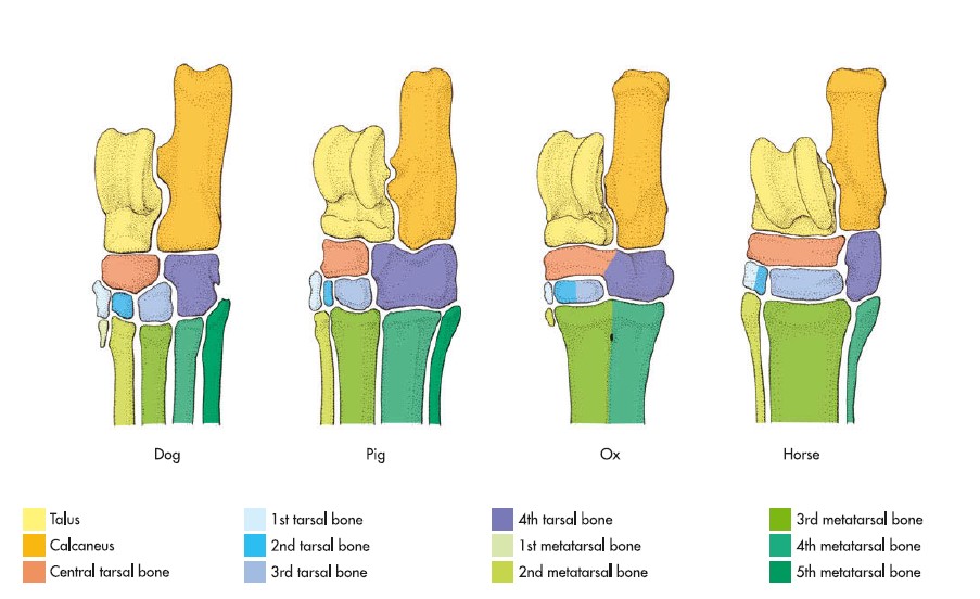
- Skeleton of the tarsus in the domestic mammals. 7
-
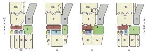
-
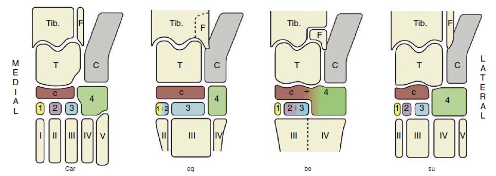
The bones of the tarsal skeleton in the carnivores (Car), horse (eq), cattle (bo), and pig (su), schematic. Roman numerals identify the metatarsal bones, Arabic numerals the distal tarsal bones. Tib., Tibia; F, fibula; T, talus; C, calcaneus;
c, central tarsal bone. 8
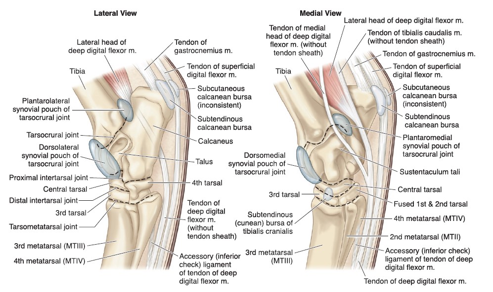
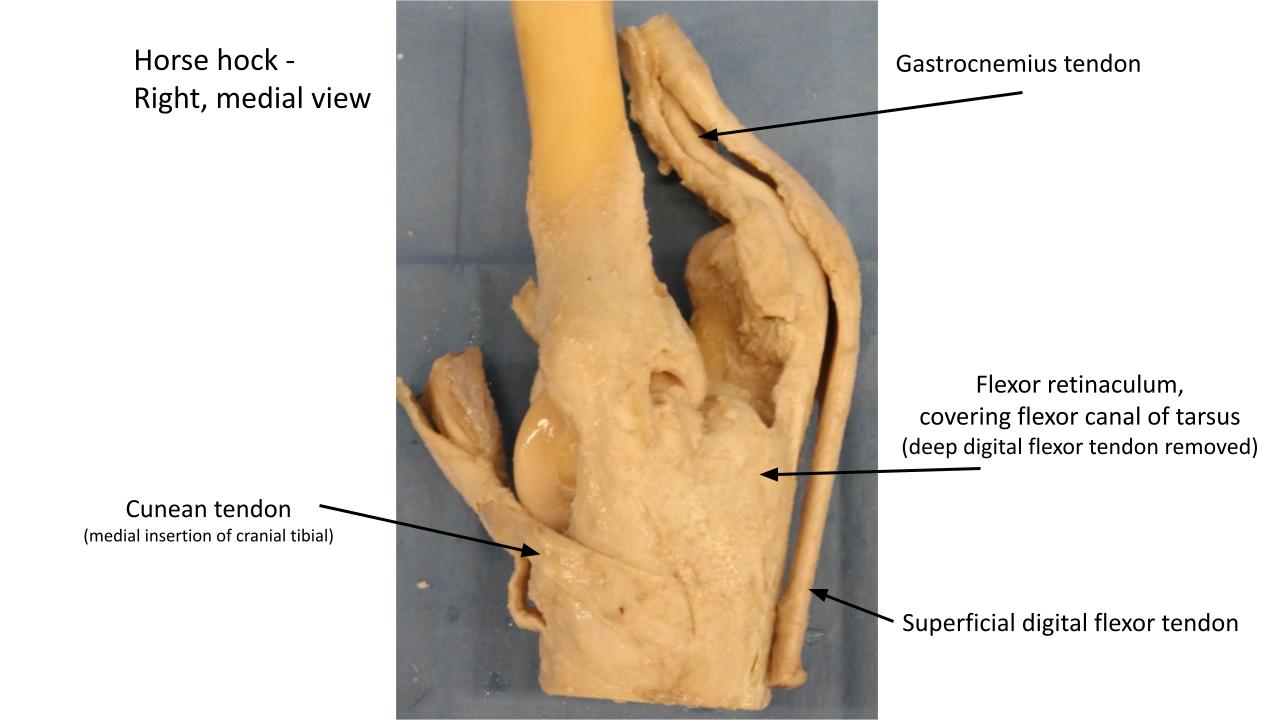
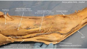
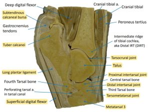
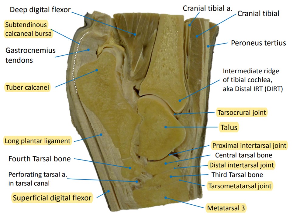
Starting our tarsus dissection on the plantar aspect of the joint, the flexor canal of the tarsus is a space for tendons to pass through, heading to the digit. It is bound dorsally by the sustentaculum tali of the calcaneus, laterally by the calcaneus, and plantaromedially, by the fibrous structure, the flexor retinaculum. A tarsal sheath envelops the tendons. There are calcaneal bursae at the tarsus, cushioning tendons as they pass by or insert in the region. The subtendinous calcaneal bursa lies between the SDF tendon and the calcaneal tuberosity, i.e. at the site the tendon forms a cap over the bony prominence.
Dissect: On the limb specimen (now devoid of skin and crural fascia), follow the tendons of the transected heads of the deep digital flexor m. from the crus distally. Transect the flexor retinaculum, from its proximal to distal margin. This fibrous tissue is very dense and thick – it takes a few good scalpel incisions to cut through it. Once the flexor retinaculum is transected the combined tendons of the lateral head and caudal tibial head of the deep digital flexor can be identified passing through the flexor canal of the tarsus, enveloped in the tarsal sheath. Note the tendon of the medial head of the DDF does not pass through the flexor canal.
Follow the path of the SDF tendon over the tuber calcanei and into the metatarsus. Cut the medial attachment of the superficial digital flexor tendon to the calcaneus and reflect it off the tuber calcanei to reveal the underlying subtendinous calcaneal bursa. Appendix on synovial structures.
-
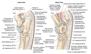
- Joints and bursae of the equine tarsus. 9
-
- Crus and pes of the horse, lateral view. 2
-
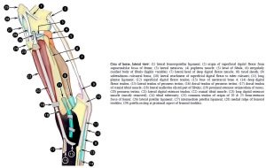
- Crus of the horse, lateral view. 2

-
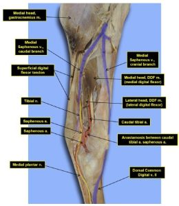
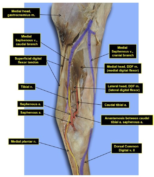
- Medial crus and tarsus of the horse. 16
-
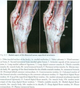
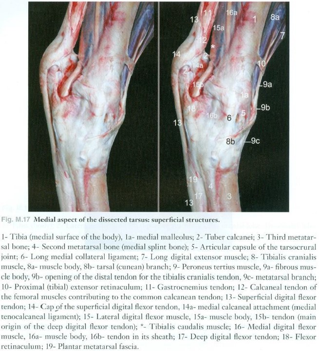
- Medial aspect of the dissected horse tarsus; superficial structures. 17
-

- Horse tarsus
-
- Horse crus
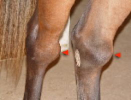
Clinical relevance: “Thoroughpin” and calcaneal bursitis.
“Thoroughpin” (tarsal tendosynovitis) and calcaneal bursitis in horses.
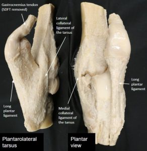
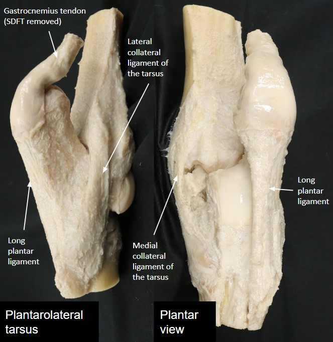
The tarsus is supported and stabilized by strong, complex medial and lateral collateral ligaments. The long plantar ligament binds the calcaneus to the distal tarsus and metatarsus, counteracting the proximal pull of the common calcaneal tendon.
Observe and dissect: Study wet and dry prosections of the tarsus and refer to figures to identify the collateral ligaments and long plantar ligament of the equine tarsus. Identification of these ligaments on your dissected specimen is also possible.
-
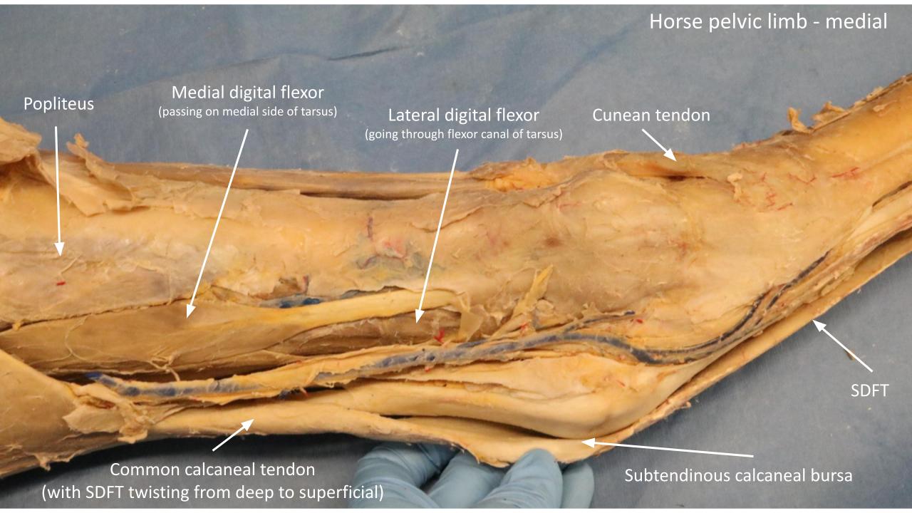
- Tarsus of horse, dorsal view. 2

-

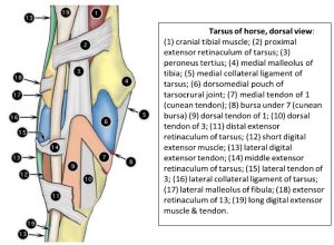
- Tarsus of horse, lateral view. 2
-
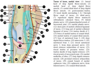
- Tarsus of horse, medial view. 2
-
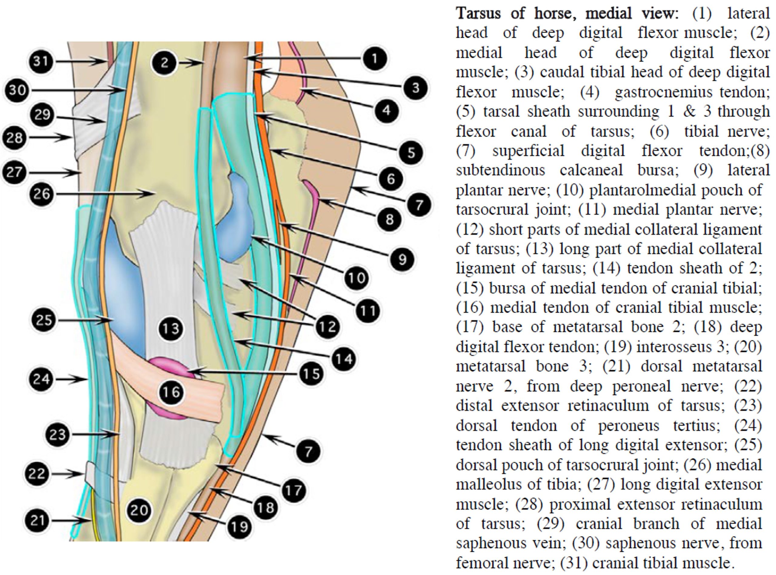
- Equine tarsus. (A) Lateral view of the tarsus depicting the muscles, tendons, ligaments, synovial sheaths, and bursae. (B) Medial viewof the same region. 9

-

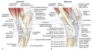
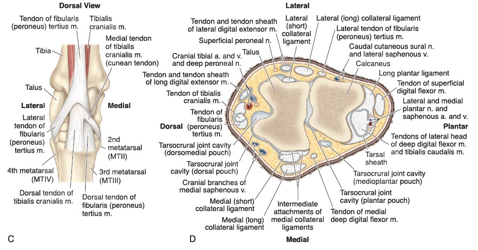
- (C) Dorsal view of the tarsus depicting the arrangement of the tibialis cranialis and fibularis (peroneus) tersius muscles. (D) Cross-sectionof the tarsus at the level of the talus and calcaneus including the vessels and nerves. Notice the 3 heads of the deep digital flexor muscle: lateral (largest) andmedial heads of the deep digital flexor muscle and tibialis caudalis muscle. 9
-
- Horse tarsus
-
- Horse tarsus
Clinical relevance: “Curb”
Ligament inflammation (= desmitis) from strain or tear; long plantar (ligament) desmitis = “curb”.
Moving to the cranial crus and dorsal tarsus, the tendons of the cranial tibial m., fibularis tertius m., long digital extensor m. and lateral digital extensor m. all cross the tarsus. The extensor muscle tendons continue to the digit and we will follow those shortly. Recall from LAB 20A that the tendons on the craniolateral crus of the horse are bound down to the limb by three (3) horizontal bands of thickened fascia, the extensor retinacula (pl.), as they cross over the tarsal region.
Dissect: Identify the craniolateral crus muscles and their tendons and follow them as they pass over the tarsus. Transect the extensor retinacula to facilitate tracing of the tendons. Synovial sheaths surround the tendons and may also be opened to examine the tendons.
-
- Tarsus of horse, dorsal view. 2
-
- Tarsus of horse, lateral view. 2
-
- Equine tarsus. (A) Lateral view of the tarsus depicting the muscles, tendons, ligaments, synovial sheaths, and bursae. (B) Medial viewof the same region. 9
At the distal attachments of the cranial tibial and fibularis tertius mm. both of these muscles have two tendons of insertion that are closely related. The medial tendon of the cranial tibial m. is referred to as the cunean tendon and it passes over the cunean bursa before it inserts on the fused first and second tarsal bones.
Dissect: Clean the margins to isolate the distal tendon of attachments of the cranial tibial and fibularis tertius mm. Observe how the cranial tibial tendons become apparent as they pass between the bifurcating fibularis tertius. Follow the medial tendon of the cranial tibial muscle (i.e. the cunean tendon) to its insertion point. Transect this tendon at its midpoint and elevate it towards its attachment medially. Carefully observe for the low profile cunean bursa being opened when elevating the tendon.
-
- Tarsus of horse, dorsal view. 2
-
- Equine tarsus. (A) Lateral view of the tarsus depicting the muscles, tendons, ligaments, synovial sheaths, and bursae. (B) Medial viewof the same region. 9
-
- (C) Dorsal view of the tarsus depicting the arrangement of the tibialis cranialis and fibularis (peroneus) tersius muscles. (D) Cross-sectionof the tarsus at the level of the talus and calcaneus including the vessels and nerves. Notice the 3 heads of the deep digital flexor muscle: lateral (largest) andmedial heads of the deep digital flexor muscle and tibialis caudalis muscle. 9
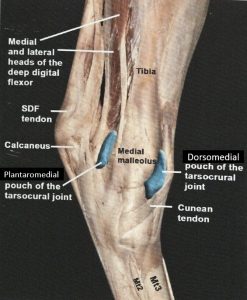
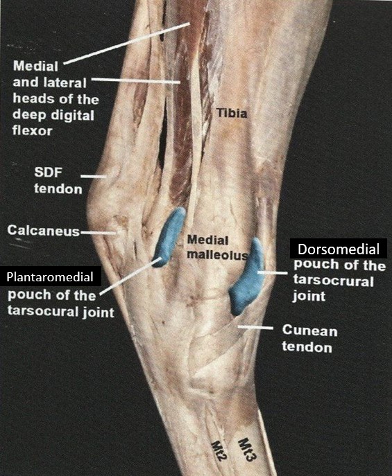
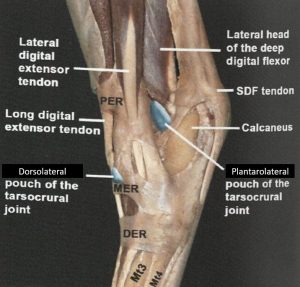
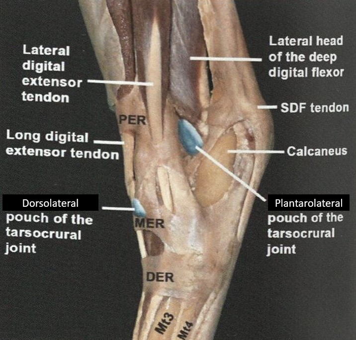
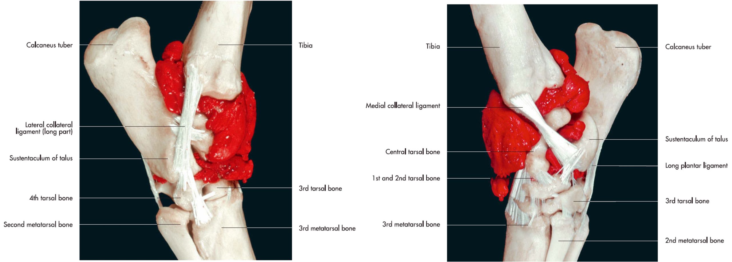
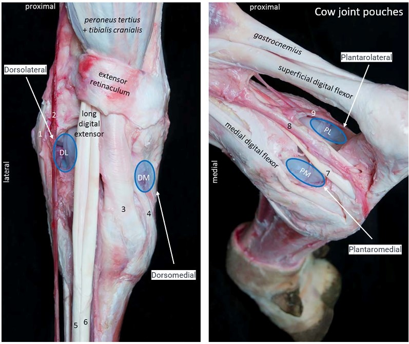
Okay, before we dissect more distally, lets consider the tarsocrural joint in detail. This is the most proximal joint of the four joints that make up the tarsus. The tarsocrural joint in the horse is lined by one extensive synovial joint sac that is subdivided into four (4) pouches: the dorsomedial, dorsolateral, plantaromedial and plantarolateral tarsocrural joints pouches. The pouches represent clinically accessible regions of the joint sac, located between bones, tendons and ligaments. The pouches are quite obvious when there is synovial effusion present (increased volume of synovial fluid) causing excessive filling of the joint sac and bulging of the sac between bones, tendons and ligaments, at the pouch locations.
Dissect: On the dissection specimen, palpate the locations of the 4 tarsocrural joint pouches. Incise and open the dorsomedial pouch of the tarsocrural joint and note synovial fluid and the articular surface of the joint. You may also identify synovial villi lining the synovial membrane of the joint (review material regarding synovial anatomy is located in the thoracic limb content).
-
- Horse hock. 16
-
- Horse hock. 16
-
- Articular spaces of the tarsal joint of the horse, lateral and medial views. 7
-
- Tarsus of horse, dorsal view. 2
-
- Tarsus of horse, medial view. 2
-
- Tarsus of horse, lateral view. 2
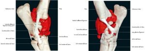
-
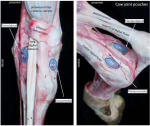
- Joint pouches of the right tarsocrural joint of an adult Holstein cow. H. Larde
Clinical relevance: “Bog spavin”
Tarsal hydrarthrosis/effusion (“bog spavin”), arthrocentesis, surgical access via arthroscopy to tarsocrural joint (making sure to avoid the cranial branch of the medial saphenous v. passing over the dorsomedial pouch).
Metatarsus – horse
The next step is to continue following the tendons extending distally through to the digit. Dorsally, the lateral digital extensor tendon combines with the long digital extensor tendon, usually in the proximal half of the metatarsus, such that only the long digital extensor tendon travels into the digit. On the plantar aspect, the flexor tendons pass into the digit as they do in the thoracic limb. The suspensory ligament is also of a similar construct as was dissected in the thoracic limb. In the pelvic limb, only an accessory (check) ligament of the DDF may be present, if at all. A small band of fibrous tissue passing from the plantar proximal metatarsal region to the DDF tendon represents the accessory ligament of the DDF – it may be vestigial (small, underdeveloped) or absent. There is no accessory (check) ligament of the SDF in the pelvic limb.
Dissect: Remove superficial fascia to isolate and dissect the tendons and ligaments of the dorsal and plantar metatarsus, as far distally as the fetlock (metatarsophalangeal joint). Separate between structures, removing fascia surrounding tendons to isolate their borders for identification. Identify: tendons of Long digital extensor, Lateral digital extensor, Superficial digital flexor, Deep digital flexor; Accessory ligament of DDF (if present); Suspensory ligament (aka, third interosseous m.)

Clinical relevance:
Transection of lateral digital extensor tendon in proximal dorsolateral metatarsus to treat stringhalt; SDF and DDF tendonitis (“bowed tendons”); suspensory desmitis; lacerations of SDFT, DDFT and suspensory ligament; role of DDFT and its accessory ligament, and the suspensory ligament, in the PL passive stay apparatus.
Digit – horse
The pelvic limb digit anatomy is essentially identical to that already studied in the thoracic limb and it will not be considered further in this section. However, if time allows and you wish to explore further, please feel free to dissect ligaments and tendons in the digit area. Refer back to this lab for assistance with identification.
Hoof (foot) anatomy will be studied in LAB 23A and these limbs can also be used in that lab to identify external anatomy of the hoof capsule.
Passive Stay Apparatus – Horse
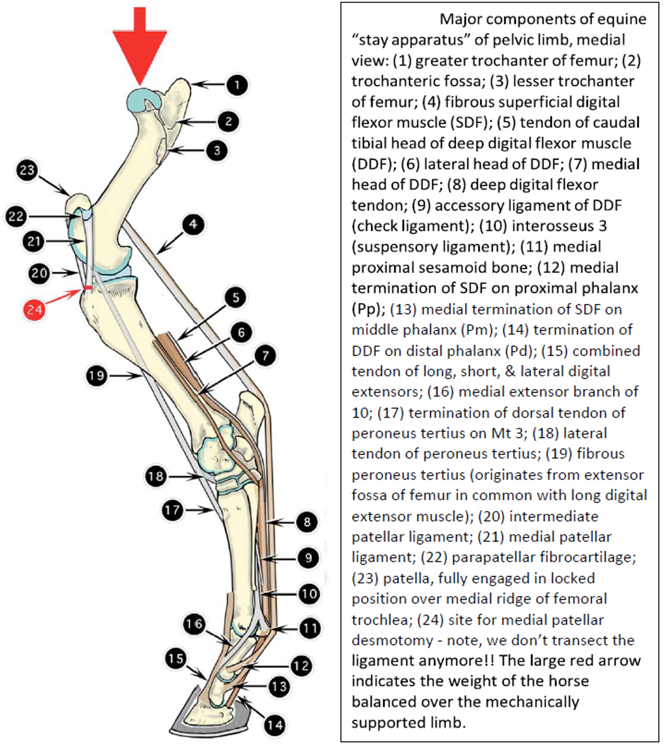
Horses spend much of their adult life standing, and have a mechanism to rest one of their hindlimbs while the other one is “locked” or “placed in park” and is carrying the bulk of the weight in the hind end. This mechanism is referred to as the Pelvic Limb (Passive) Stay Apparatus. The limb that is bearing most of the weight is ‘locked’ anatomically to decrease the need for muscle activity to keep the limb extended and the horse standing! The ‘passive’ in the term refers to being able to keep the limb in extension with minimal active muscle energy required. So the horse can weight bear on this limb for an extended period, giving the opposite limb a good rest.
The horse has a set of fibrous support structures that convert the limb into a passive, weight-bearing stilt (stay). These structures make up the stay apparatus. Being the “efficiency experts” that they are, horses only have to lock the patella over the medial ridge of the femoral trochlea, and what do you know, they’ve “put the limb in park.” If the stifle can’t flex, neither can the tarsus (due to the reciprocal apparatus). The lower joints are passively supported by the fibrous SDF, the DDF with its fibrous accessory ligament (variably developed), and the fibro-osseous sling, known as the suspensory apparatus, formed by the 3rd interosseus muscle (= suspensory ligament), the proximal sesamoid bones, and the distal sesamoid ligaments (straight, oblique, cruciate, short). But, aren’t we forgetting the highly movable hip joint? No problem. It’s not going anywhere, as long as the horse’s weight is balanced over and pushing straight down on the limb. In order for the head of the femur to move downward (as in hip flexion on a weight bearing limb), the medial trochlear ridge must be free to rotate upward. This is prevented by the patellar lock – the femur is hooked under the patella like a crowbar under the edge of a building. Unless the horse shifts backward or forward, the hip joint doesn’t need to be fixed in any way; it’s a balancing act.
-
- Lateral view of the stay apparatus of the horse pelvic limb. 9

-
- Lateral view of the reciprocal apparatus of the horse pelvic limb. 9
-

- Major componets of equine stay apparatus of pelvic limb, medial view. 2
Clinical relevance:
Upward fixation of the patella (UFP) or “locked stifles” and conservative and surgical correction (see clinical video in stifle section above); rupture of fibularis tertius (what does it look like? see previous lab); laceration of SDFT, DDFT, or breakdown of the suspensory apparatus (how do these injuries affect locomotion?).
Highlights of the pelvic limb – ruminant
The stifle of the ruminant has been considered at the start of this lab. Notably, small ruminants have one patellar ligament, while the large ruminant (ox in our course) has 3 patellar ligaments (similar to the horse).
The ruminant tarsus has unique bone anatomy that should be reviewed. The flexor canal of the tarsus and the tendons and ligaments of the plantar tarsus are similar to the horse. Moving to the cranial crus and dorsal tarsus, the tendons of the cranial tibial m., fibularis tertius m., fibularis longus m., long digital extensor m. (with medial and lateral bellies in the ruminant), and lateral digital extensor m. all cross the tarsus. The extensor muscle tendons continue to the digit. Recall from LAB 20A the tendons on the craniolateral crus in the ruminant are bound down to the limb by two (2) horizontal bands of thickened fascia, the extensor retinacula (pl.), as they cross over the tarsal region.
Recall the metatarsus in the ruminant consists skeletally of the fused 3rd and 4th metatarsal bones. Dorsally, the long digital extensor tendon will have a medial and lateral tendon extending from the respective muscle bellies. The medial tendon attaches to the medial digit (digit 3). The lateral tendon bifurcates at the metatarsophalangeal joint to send a branch to the medial and lateral digits (digits 3 and 4). The lateral digital extensor tendon passes down independently to attach to the lateral digit only. Therefore each digit receives two extensor tendon attachments. On the plantar aspect of the metatarsus the flexor tendons pass into the digit as they do in the thoracic limb. There are no accessory ligaments of the flexor tendons present in the pelvic limb of the ruminant. The suspensory ligament (fused 3rd and 4th interosseous mm.) is also of a similar construct as was dissected in the thoracic limb.
The pelvic limb digit anatomy is essentially identical to that already studied in the thoracic limb and it will not be considered further in this section.
Observe: study and review ruminant anatomy on prosections and models.
-
- Crus of the newborn calf; caudomedial view. 2
-

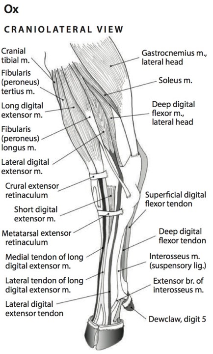
- Muscles of the distal pelvic limb of the ox, craniolateral view. 6
-
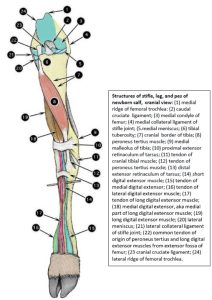
- Structures of stifle, leg, and pes of newborn calf; cranial view. 2

-
- Crus of the newborn calf; lateral view. 2

Review Videos
Horse proximal muscles – 35 minutes (whole limb)
Calf pelvic limb muscles – 24 minutes (whole limb)
Goat pelvic limb muscles – watch until 18 minutes (whole limb)
Pig pelvic limb muscles – watch until 16 minutes
Horse passive stay apparatus – 23 minutes
Horse intraarticular injections – 22 minutes
Terms chart
NOTE – All terms below except those highlighted have been covered in previous labs. There are only two new terms introduced in this chart.
| Primary structure |
Feature | Horse and Ruminant (Ox), unless noted |
| Stifle | Medial and lateral trochlear ridges, femur | |
| Medial and lateral condyles, femur | ||
| Trochlear groove, femur | ||
| Patella | ||
| Resting and gliding surfaces of patella | horse | |
| Resting and gliding surfaces of trochlea, femur | horse | |
| Parapatellar fibrocartilage | horse | |
| Medial and lateral collateral ligaments, stifle | ||
| Medial and lateral femoropatellar ligaments | ||
| Medial, intermediate, lateral patellar ligaments | ||
| Cranial and caudal cruciate ligaments | ||
| Meniscofemoral ligament | ||
| Medial and lateral meniscus | ||
| Femoropatellar joint | ||
| Medial femorotibial joint | ||
| Lateral femorotibial joint | ||
| Patella locking mechanism – 4 structures involved | horse | |
| Tarsus | Tarsocrural joint | |
| Dorsomedial, dorsolateral, plantaromedial, plantarolateral pouches of tarsocrural joint | horse | |
| Proximal intertarsal joint | ||
| Distal intertarsal joint | ||
| Tarsometatarsal joint | ||
| Talus – medial and lateral trochlear ridges | ||
| Calcaneus – tuber calcanei, sustentaculum tali | ||
| Tarsal bones – central, 2nd, 3rd, 4th | species specific fusion | |
| Flexor canal of tarsus and its boundaries | ||
| Flexor retinaculum | ||
| Subtendinous calcaneal bursa | ||
| Medial and lateral collateral ligaments | ||
| Long plantar ligament | ||
| Tendons of caudal crus muscles at tarsus | ||
| Tendons of cranial crus muscles at tarsus | ||
| Extensor retinaculum | ||
| Cunean tendon | horse | |
| Cunean bursa | horse | |
| Metatarsus | Tendons of digital flexors and extensors | |
| Suspensory ligament | ||
| Accessory ligament of DDFT – as present | horse | |
| Pelvic Limb Passive Stay Apparatus – horse |
Patellar locking mechanism | horse |
| Reciprocal apparatus – fibularis tertius and SDFT | horse | |
| Suspensory apparatus | horse | |
| SDFT, DDFT (+/- accessory lig.) | horse | |