MSK LAB 5A – Thoracic Limb Antebrachial muscles
Learning Objectives
- Identify the mm. of the antebrachium.
- Understand and know the attachments of the antebrachial mm.
- Understand and know the actions of the antebrachial mm.
- Categorize the antebrachial mm. into muscle action groups
- Understand the function of the dorsal (elastic) ligaments in the cat.
Lab instructions:
For this lab, teams will focus on dissecting the muscles of the antebrachium, in canine and feline cadavers, continuing on the left limb. This carnivore understanding can then be applied to the prosected large animal cadavers to identify the same muscles (and any species specific variations) as directed. A link to the muscle/structure terms chart for this lab is provided below.
Thoracic Limb: Antebrachium Muscle Chart
Cranial and lateral (i.e. craniolateral) muscles of the antebrachium (forearm):
- Brachioradialis (vestigial or not present in dog, consistently present in cat)
- Extensor carpi radialis
- Common digital extensor
- Lateral digital extensor
- Ulnaris lateralis
- Abductor digit I (pollicis) longus (aka extensor carpi obliquus)
- Supinator
Observe: Re-examine the foot pads in the cadaver on the right and/or left thoracic limb. The small pad that protrudes palmar to the carpus is the carpal pad. The largest in the paw, the metacarpal pad is on the palmar side of the metacarpophalangeal joints and is triangular. The digital pads are ovoid and flattened. Each is located palmar to the distal interphalangeal joint.
Dissect: Examine the left thoracic limb from the elbow, distally. Remove the strong fibrous antebrachial fascia. Incise it with a scalpel and then lift it up, reflecting it off the underlying muscles and cutting the tissue away completely. After doing this, the identification and cleaning of individual antebrachial muscles will be possible by finding the fascial seams between the muscle bellies and separating them along those seams.
Clean and observe the muscles as described below.
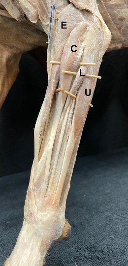
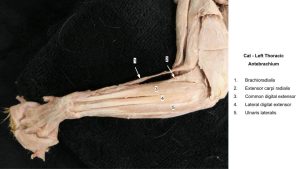
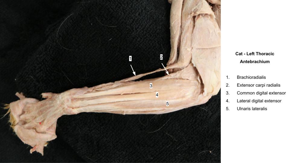
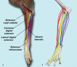
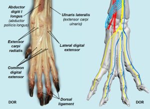
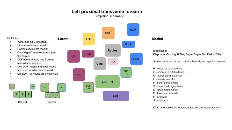
The cranial and lateral antebrachial muscles are, from cranial to caudal, the extensor carpi radialis, supinator, common digital extensor, lateral digital extensor, ulnaris lateralis, and abductor digiti I (pollicis) longus (aka extensor carpi obliquus). Most of these muscles arise from the lateral epicondyle of the humerus. A thin strap of muscle (if present!), the brachioradialis, is also included in the craniolateral antebrachial muscle group and will be the first described. We commonly abbreviate these craniolateral muscles as “ECLU” to remember the order – do you have another mnemonic?! Dr. Stewart likes “Elephants Can Lay U Flat; Super Duper Flat. Period, Silly” to cover the entirety of the antebrachial muscles.
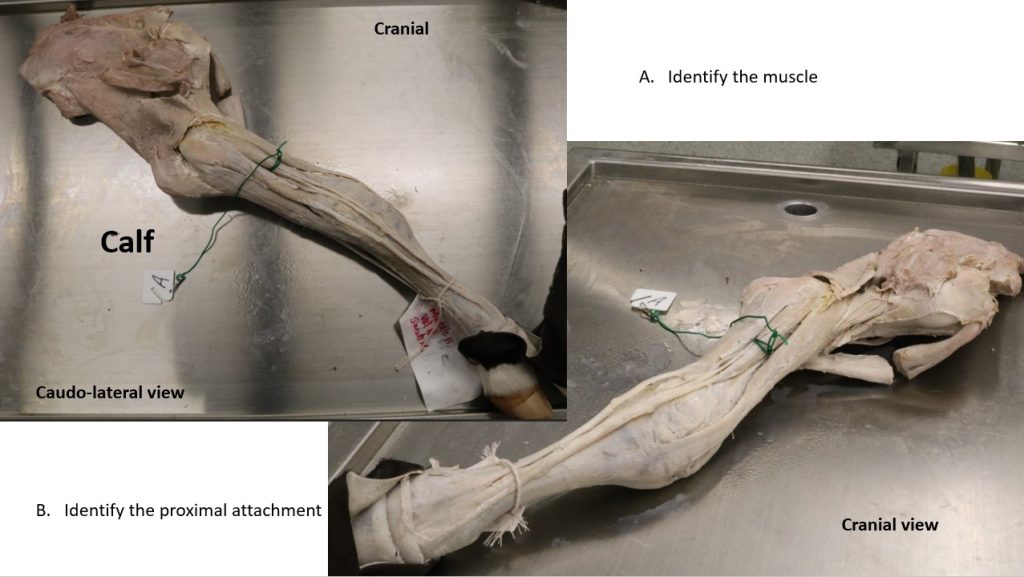
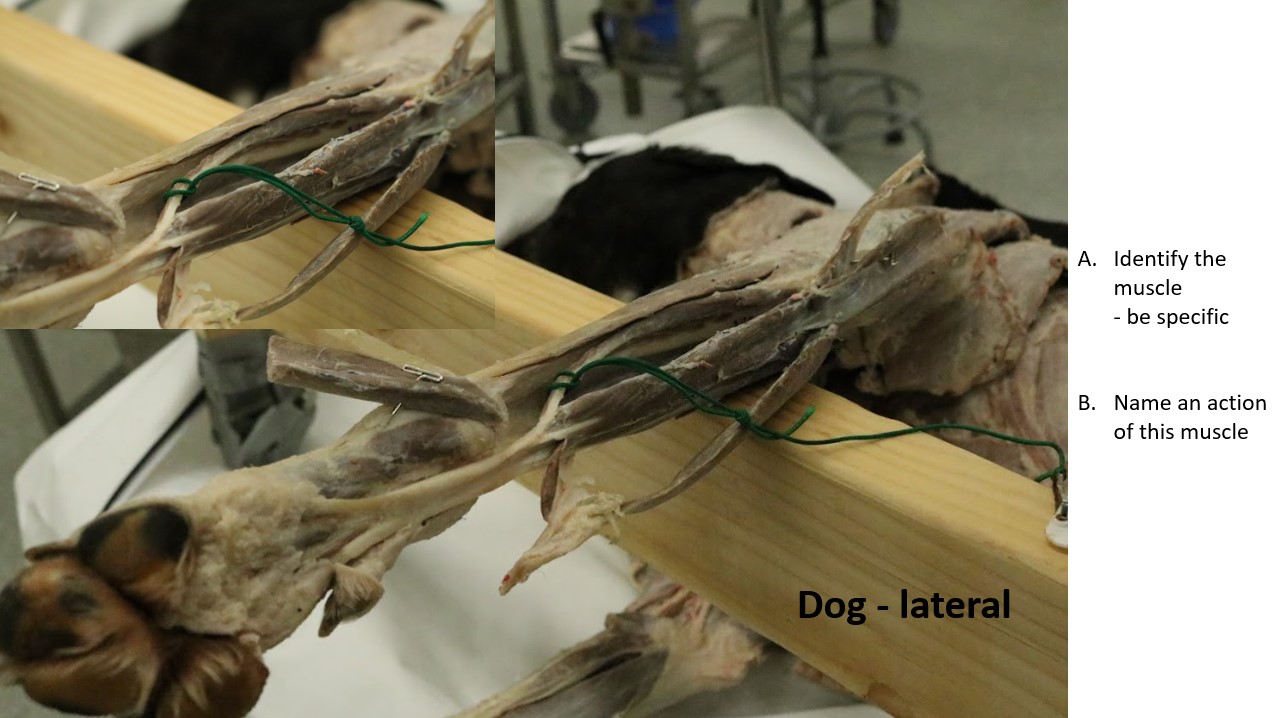
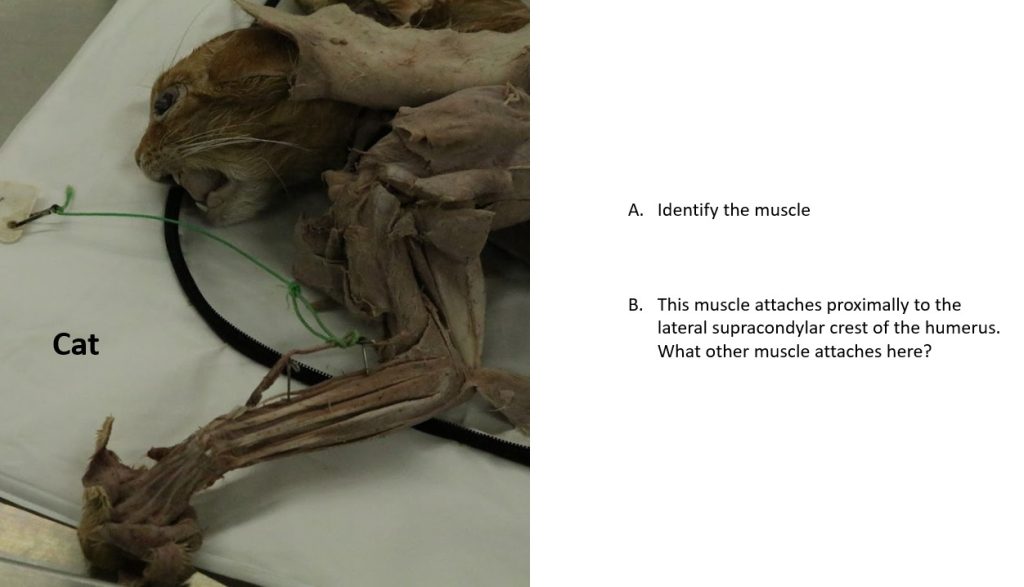
Brachioradialis m.
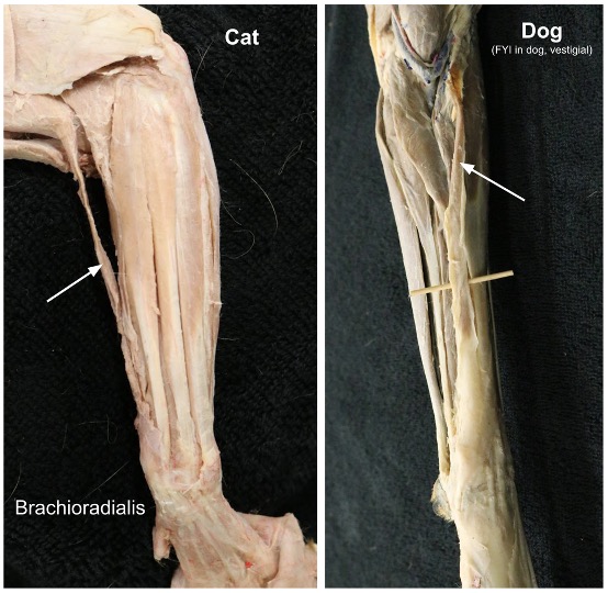
The brachioradialis functions as a supinator of the antebrachium. It is a slender, inconstant muscle in the dog but comparatively well developed and consistently present in the cat (why might this be so?). The brachioradialis arises from the lateral supracondylar crest of the humerus, adjacent to the extensor carpi radialis, and passes distally and medially to insert on the distal fourth of the radius. The muscle is frequently removed with the skin when not being specifically preserved.
PROXIMAL ATTACHMENT: Lateral, distal humerus
DISTAL ATTACHMENT: Distal radius
ACTION: Supination
Observe: Ideally observe this muscle in at least a feline cadaver.
-

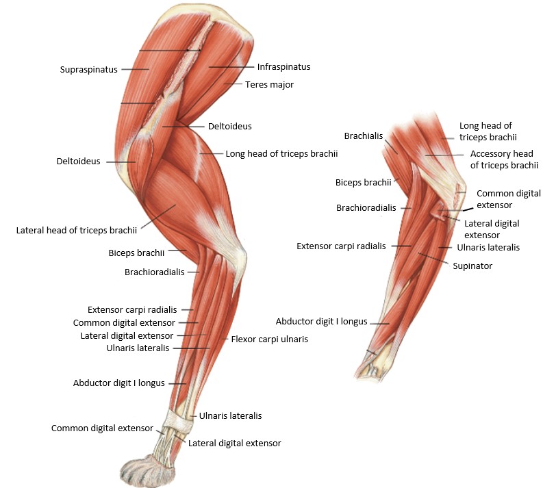
- Intrinsic muscles of the cat thoracic limb. 4
-

- Brachioradialis m.
-
- Cat brachioradialis. 40

Extensor Carpi Radialis m.
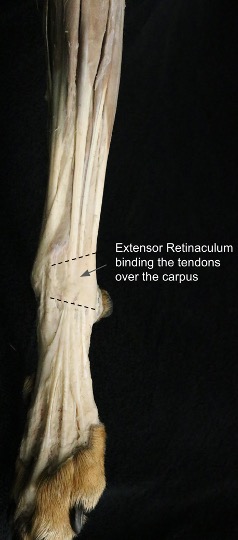
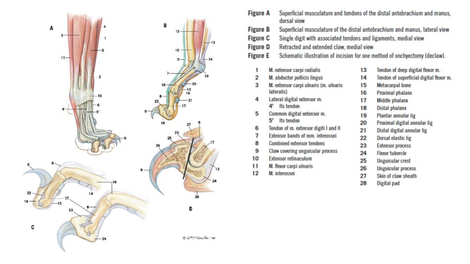
The extensor carpi radialis is the largest of the craniolateral antebrachial muscles. It lies on the cranial surface of the radius throughout most of its course and is easily palpated in the live dog. The tendon looks single but is distinctly double throughout its distal third. [Two tendons are present because the extensor carpi radialis m. has two muscle bellies, fused in the canine and remain more separable in the feline. So, unlike the dog, individual muscles bellies to the ECR m. may be readily noted in the cat, each belly continuing as one of the two tendons.] These closely associated tendons run first under the tendon of the abductor digiti I longus, then in the middle groove of the radius farther distally and on over the carpus. They are bound and held in place on the dorsal surface of the carpus by the extensor retinaculum. The extensor retinaculum is a transversely oriented condensation of carpal fascia that aids in holding all the tendons that cross the dorsum of the carpus in their grooves. Between bundles of tendons the extensor retinaculum dips down to blend with the fibrous dorsal part of the joint capsule.
PROXIMAL ATTACHMENT: Lateral supracondylar crest of the humerus
DISTAL ATTACHMENT: Dorsal base of metacarpals (note: base of metacarpal bone = its proximal end)
ACTION: Extend carpus
Dissect: Review prosections to visualize the extensor retinaculum and its proximal and distal margins. On your cadaver, transect the extensor retinaculum in multiple locations and reflect the tissue so the extensor tendons can be observed.
-
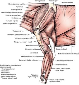
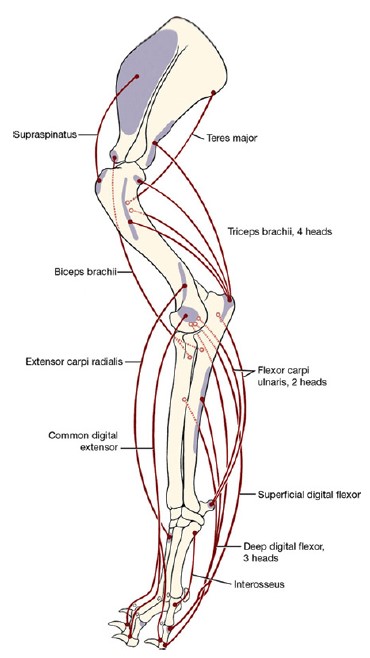
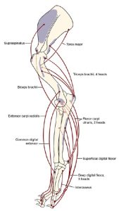
- Deeper muscles of the left scapula, brachium, and antebrachium, lateral view. 1

-
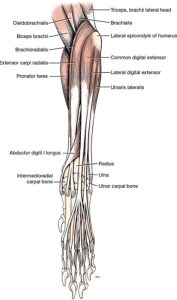
- Muscles of left canine antebrachium, cranial view.1

-
- Intrinsic muscles of the cat thoracic limb. 4
-
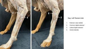
- Dog antebrachium

-
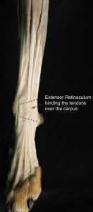
- Dog extensor retinaculum
-
- Cat antebrachium
Common Digital Extensor m.
The common digital extensor is shaped like, and lies caudal to, the extensor carpi radialis, on the lateral side of the forearm. The four individual tendons that leave the muscle are closely combined where they cross the cranial surface of the abductor digiti I longus. As they cross the carpus; they are held in the lateral groove of the radius by the extensor retinaculum. Distal to the retinaculum the four tendons diverge, and each goes to the distal phalanx of the four main digits. Likely in the dog, a dorsal sesamoid bone may be present in the tendons of the common digital extensor as they cross the metacarpophalangeal joint.
PROXIMAL ATTACHMENT: Lateral epicondyle of the humerus
DISTAL ATTACHMENTS: Extensor process of the distal phalanges
ACTION: Extend digits and carpus
Dissect: Dissect the tendons of the common digital extensor to the level of the mid-metacarpal region. Your team can dissect more distally as wished, otherwise refer to models and plastinated specimens in the lab to further examine the tendon insertions on the digits.
-
- Deeper muscles of the left scapula, brachium, and antebrachium, lateral view. 1
-
- Muscles of left canine antebrachium, cranial view.1
-
- Intrinsic muscles of the cat thoracic limb. 4
-
- Dog antebrachium
-
- Cat antebrachium
-
- Dog craniolateral antebrachium. 40

Observe: Notice that in the cat, the distal interphalangeal joint (the joint between the middle and distal phalanges) is in a marked degree of hyperextension (overextension). This is brought about by a pair of dorsal elastic ligaments at each distal interphalangeal joint. One ligament lies on each side of the common digital extensor tendon inserting on the distal phalanx. The ligaments attach from the distal phalanx to the middle phalanx. Acting like rubber bands the ligaments allow for passive claw retraction with the distal phalanx resting (claw sheathed) lateral to the respective middle phalanx. When the cat unsheathes its claws, the deep digital flexor m. must contract to overpower the dorsal elastic ligaments (i.e. stretch out the rubber bands). Dogs have dorsal elastic ligaments too, with partial claw retraction possible, but clearly not to the degree that cats can achieve.
Lateral Digital Extensor m.
The lateral digital extensor is about half the size of the common digital extensor. It lies between the common digital extensor and the ulnaris lateralis. Its tendon begins at the middle of the forearm, passes under the extensor retinaculum in a groove between the radius and ulna, and immediately splits into three branches (four in the cat). The main part of each tendon attaches to the extensor process of the distal phalanx of digits III, IV, and V in the dog, in common with the common digital extensor tendon. In the cat, a tendon attaches to digit II as well.
PROXIMAL ATTACHMENT: Lateral epicondyle of the humerus
DISTAL ATTACHMENTS: Dorsal base (i.e. proximal end) of phalanges, extensor processes of distal phalanges
ACTION: Extend digits and carpus
Dissect: Clean the lateral digital extensor and its tendons to the level of the mid-metacarpal region. Your team can dissect more distally as wished, otherwise refer to models and plastinated specimens in the lab to further examine the tendon insertions on the digits.
-
- Deeper muscles of the left scapula, brachium, and antebrachium, lateral view. 1
-
- Muscles of left canine antebrachium, cranial view.1
-
- Intrinsic muscles of the cat thoracic limb. 4
-
- Dog antebrachium
-
- Cat antebrachium
-
- Dog craniolateral antebrachium. 40
Ulnaris Lateralis m.
The ulnaris lateralis is wider than the lateral digital extensor and lies caudal to it. It is bounded deeply by the ulna, and the flexor group of muscles, which lie caudal and medial to it. Because of its attachments it acts as a flexor on the carpus, and abducts the manus. However the muscle is innervated by the radial n., the same nerve that supplies all the extensors of the carpus and digits. A detail to keep straight as your knowledge grows.
PROXIMAL ATTACHMENT: Lateral epicondyle of the humerus
DISTAL ATTACHMENT: Base of metacarpal V and accessory carpal bone
ACTION: Flex carpus; abduct manus at carpus
Dissect: Clean the muscle and observe its tendons of insertions.
-
- Deeper muscles of the left scapula, brachium, and antebrachium, lateral view. 1
-
- Muscles of left canine antebrachium, cranial view.1
-
- Intrinsic muscles of the cat thoracic limb. 4
-
- Dog antebrachium
-
- Cat antebrachium
-
- Dog craniolateral antebrachium. 40
Abductor Digiti I Longus m.
The abductor digiti I longus (aka the abductor pollicis longus or extensor carpi obliquus) lies primarily in the groove between the radius and ulna and is triangular.
PROXIMAL ATTACHMENT: Craniolateral ulna
DISTAL ATTACHMENT: Base of metacarpal I
ACTION: Abduct first digit and extend carpus
Dissect: Retract the digital extensors so that the bulk of the muscle is visible for identification. Track its tendon passing obliquely across the distal antebrachium towards the medial proximal metacarpus. There is a sesamoid bone in its tendon where it crosses the medial surface of the carpus (seen on radiographs!).
-
- Muscles of left canine antebrachium, cranial view.1
-
- Intrinsic muscles of the cat thoracic limb. 4
-
- Dog abductor digiti I longus. 40

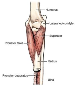
Supinator m.
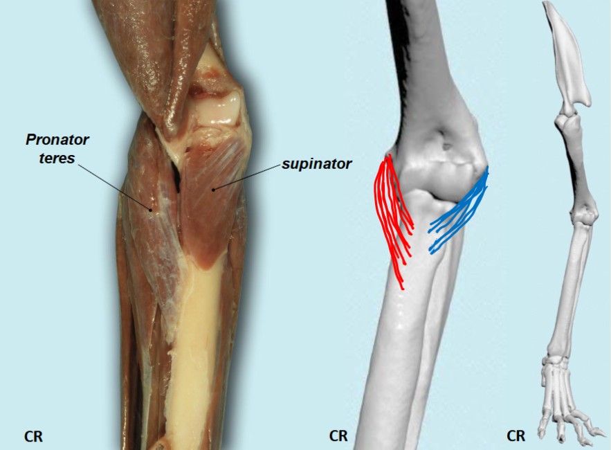
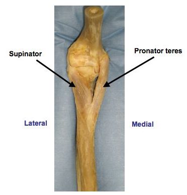
The supinator is short, broad, and flat and obliquely placed across the lateral side of the flexor (also the cranial) surface of the elbow joint. The supinator lies principally on the proximal fourth of the radius. There is a sesamoid bone in this muscle where it crosses the head of the radius. The cat has a greater ability to supinate the paw (recall it has a well developed brachioradialis m.!); this aids in climbing and defense.
PROXIMAL ATTACHMENT: Lateral epicondyle of the humerus
DISTAL ATTACHMENT: Cranial, proximal radius
ACTION: Supination of distal limb (secondary – flex elbow joint)
Observe: The supinator is covered superficially by the extensor carpi radialis and common digital extensor muscles. These will need to be retracted (pulled to the side) to see the supinator. Also look at prosected models for a clear view of the muscle along with its partner the pronator teres.
-
- Intrinsic muscles of the cat thoracic limb. 4
-

- Dog pronator and supinator. 40
-

- Dog pronator & supinator mm.
Caudal and medial (i.e. caudomedial) muscles of the antebrachium (forearm):
- Pronator teres
- Flexor carpi radialis
- Superficial digital flexor
- Deep digital flexor
- Flexor carpi ulnaris
- Pronator quadratus
Dissect: If not already done, excise the thick, deep antebrachial fascia on the caudal and medial aspects of the antebrachium to reveal and facilitate separation and cleaning of the underlying muscles.
The muscles in this group include, starting from the craniomedial side of the radius and passing caudally, the pronator teres, the flexor carpi radialis, the deep digital flexor, the superficial digital flexor, and the flexor carpi ulnaris. The pronator quadratus m. is deep and lies directly between the ulna and radius, attached to the shaft of those bones. While the craniolateral forearm muscles are typically straightforward to remember, adding in the caudomedial muscles may get confusing. Here is a simplified schematic of the forearm muscles with learning tips.
Pronator Teres m.
The pronator teres extends obliquely across the medial side of the flexor (also the cranial) surface of the elbow joint. It is round in transverse section at its origin and flat at its insertion. It lies between the extensor carpi radialis cranially and the flexor carpi radialis caudally.
PROXIMAL ATTACHMENT: Medial epicondyle of the humerus
DISTAL ATTACHMENT: Medial border of the proximal mid radius
ACTION: Pronation of the distal limb (secondary – flex elbow joint)
Dissect: Retract adjacent muscles to see its origin and insertion. Also look at prosected models for a clear view of the muscle along with its partner the supinator.
-
- Muscles of left canine antebrachium, cranial view.1
-
- Intrinsic muscles of the cat thoracic limb. 4
-
- Dog pronator & supinator mm.
Flexor Carpi Radialis m.
The flexor carpi radialis lies between the pronator teres cranially and the superficial digital flexor caudally. It covers part of the deep digital flexor. The flexor carpi radialis has a thick, fusiform belly, which, partly embedded in the deep digital flexor, extends only to the middle of the radius. There it gives rise to a flat tendon that is augmented by fibers leaving the medial border of the radius.
PROXIMAL ATTACHMENT: Medial epicondyle of the humerus and medial radius
DISTAL ATTACHMENT: Palmar base of metacarpals
ACTION: Flex carpus
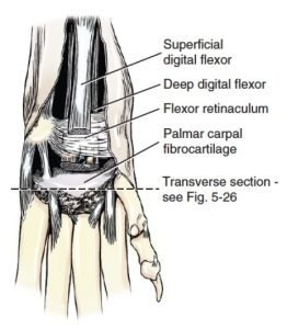
Dissect: Clean the tendon to the point where it passes through the carpal canal, deep to a thick layer of fibrous tissue on the palmar side of the carpus, called the flexor retinaculum (considered in more detail below). The flexor retinaculum serves the same purpose as the extensor retinaculum, except it binds the flexor tendons in place. The flexor retinaculum has a superficial and deep layer to it – some tendons are bound by the superficial layer only.
-
- Canine antebrachial muscles, caudal aspect. 1
-

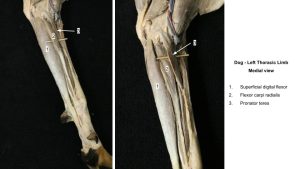
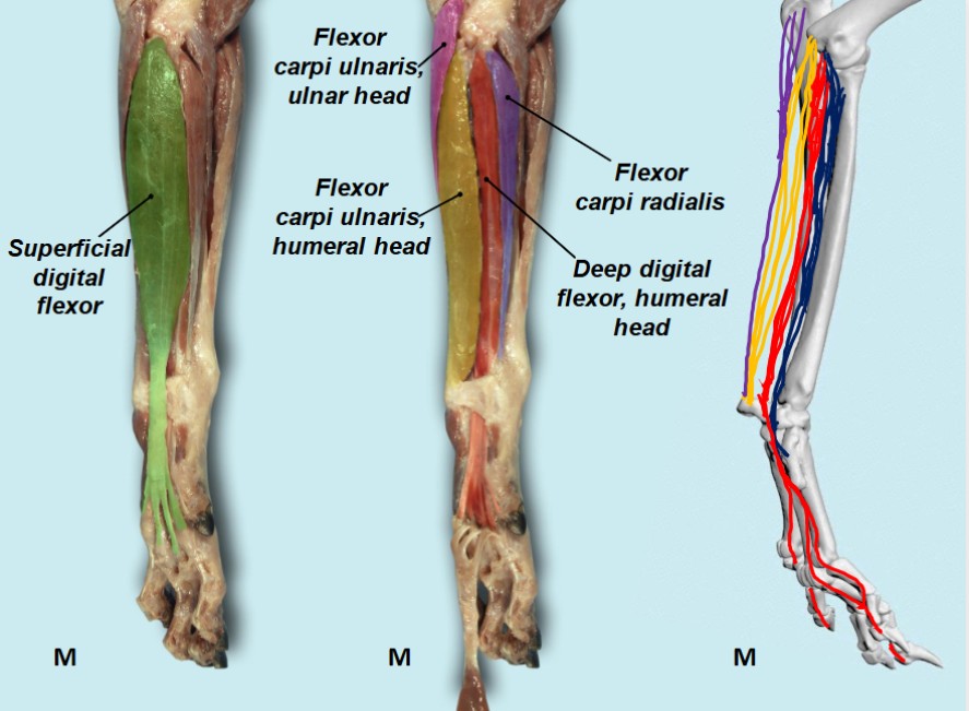
- Intrinsic muscles of the cat thoracic limb, medial view. 4
-
- Dog antebrachium

-

- Dog caudomedial antebrachium. 40
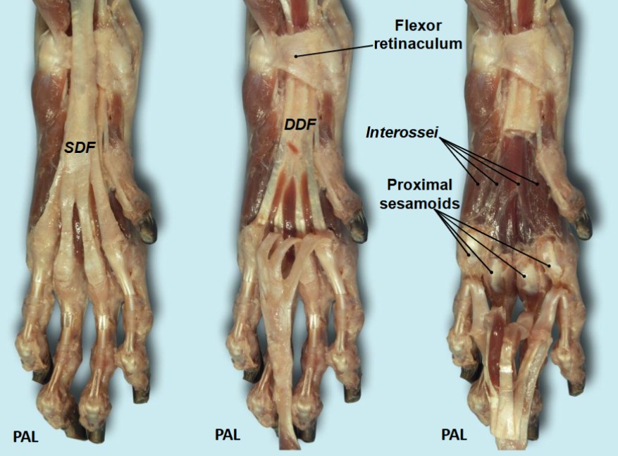
Flexor Retinaculum
-
- Tendons and Ligaments of left canine carpus, palmar aspect. 1

-
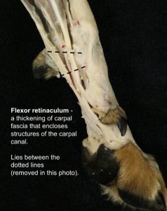
- Dog flexor retinaculum

-
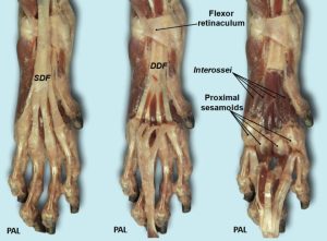
- Dog flexor retinaculum. 40
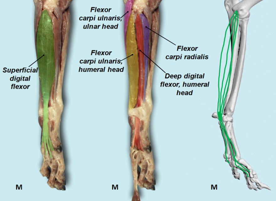
Superficial Digital Flexor m.
The superficial digital flexor lies on the caudomedial side of the forearm. It covers the deep digital flexor and is fleshy almost to the carpus. Its tendon is at first single, then crosses the palmar (flexor) surface of the carpus medial to the accessory carpal bone in the carpal canal, where it is covered by the superficial part of the flexor retinaculum and finally divides into four tendons of nearly equal size. These insert on the palmar surfaces of the base of the middle phalanges of the four principal digits (II-IV). At the metacarpophalangeal joint, each tendon forms a collar (called the manica flexoria), around the deep digital flexor tendon that passes through it. The manica flexoria will be studied in detail in the ungulate digit.
PROXIMAL ATTACHMENT: Medial epicondyle of the humerus
DISTAL ATTACHMENTS: Palmar base of middle phalanges
ACTION: Flex proximal interphalangeal joints, (Secondary – flex carpus and metacarpophalangeal joints)
FYI – The superficial and deep digital flexor tendons are held firmly in place at the metacarpophalangeal joint by the palmar annular ligament and by digital annular ligaments at the digit level. The superficial flexor tendon and the deep flexor tendon are in a common synovial membrane, the digital synovial sheath. (Note, all this anatomy will be explored in depth in ungulate cadavers – it is clinically relevant anatomy during evaluation and treatment of distal limb conditions, particularly in the horse).
Dissect: When dissecting the superficial and deep digital flexor mm. it is very helpful to first isolate their tendons distally where they pass through the carpal canal and then to track those tendons proximally to lead to the associated muscle bellies of these muscles and distally to their divergence onto digits. Transect the superficial part of the flexor retinaculum at its medial margin and isolate the tendon unit of the superficial digital flexor at the carpus. Follow this tendon proximally to confirm the muscle belly of the superficial digital flexor, follow the tendon distally to the diverging 4 individual tendons as far as the distal metacarpal region.
-
- Canine antebrachial muscles, caudal aspect. 1
-
- Intrinsic muscles of the cat thoracic limb, medial view. 4
-
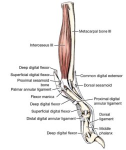
- Canine Third digit, medial view. 1

-

- Dog superficial digital flexor. 40
-
- Dog antebrachium
-
- Cat antebrachium

-
- Dog superficial digital flexor

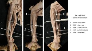
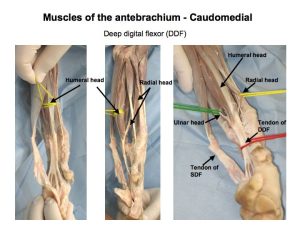
Deep Digital Flexor m.
The deep digital flexor has three heads of origin, of dissimilar size in the dog, and relatively equal size in the cat, which arise from the humerus, radius, and ulna. Their bellies lie on the caudal surfaces of the radius and ulna.
In the dog, the ulnar head is small and arises from the caudal border of the ulna. The radial head is the smallest and arises from the medial border of the radius. The humeral head is much larger. In the cat – all three heads are of roughly equal diameter. The tendons of all three heads fuse at the carpus level to form a single tendon. This tendon is held in place in the carpal canal by the thick, deep part of the flexor retinaculum. The carpal canal is formed by the accessory carpal bone laterally, the palmar carpal ligament dorsally (covering the palmar surfaces of the carpal bones), and the flexor retinaculum on the palmaromedial surface.
Distal to the carpus, the deep digital flexor tendon divides into five branches. Each branch goes to the palmar surface of the base of the distal phalanx of its respective digit.
PROXIMAL ATTACHMENT: Medial epicondyle of the humerus (humeral head), caudal ulna (ulnar head), medial radius (radial head)
DISTAL ATTACHMENTS: Flexor tubercle of distal phalanges
ACTION: Flex digits, flex carpus
Dissect: To definitively identify the heads of this muscle it is very helpful to isolate the combined tendon unit of the 3 heads, at the level of the carpus, and then to trace the tendon a short distance proximally to its separation into the 3 individual heads. To expose the combined tendon passing within the carpal canal, first, cut the deep part of the flexor retinaculum medially and reflect it. The tendon is now accessible. It may be helpful to transect the tendon branch distally from digit I in order to visualize the remaining 4 tendon branches going to digits II-V. To visualize the muscle, retract the flexor carpi ulnaris and superficial digital flexor mm. in the middle of the antebrachium. The large muscle mass remaining on the caudal antebrachium is the deep digital flexor m.
In the dog, the humeral head of this muscle is much larger than the other two heads and it has 3 ‘sub’ bellies itself (try not to separate these three sub-bellies to avoid confusion, but if they are, use their proximal attachment to appreciate they are all part of the humeral head).
In the cat, all three heads of the deep digital flexor are roughly the same diameter (they need a well-developed deep digital flexor for climbing). The humeral head has 3 sub-bellies, as in the dog. Take care not to confuse these 3 sub-bellies as the main heads of the deep digital flexor m.
-
- Canine antebrachial muscles, caudal aspect. 1
-
- Intrinsic muscles of the cat thoracic limb, medial view. 4
-
- Canine Third digit, medial view. 1
-
- Dog antebrachium

-
- Cat antebrachium
Flexor Carpi Ulnaris m.
The flexor carpi ulnaris consists of two muscle heads that are distinct throughout their length. The ulnar head arises from the caudal border of the proximal end of the ulna (the olecranon). It is thin and wide proximally but narrow distally. It lies between the ulnaris lateralis and superficial digital flexor. The humeral head is large and fleshy and lies cranial to the ulnar head, except distally, where its tendon lies caudal to it.
PROXIMAL ATTACHMENTS: Olecranon (ulnar head) and medial epicondyle of the humerus (humeral head)
DISTAL ATTACHMENTS: Accessory carpal bone
ACTION: Flex carpus
Dissect: Dissect the insertion of this muscle on the accessory carpal bone and clean its origins. There is no need to separate the ulnar and humeral heads – consider them as the one muscle.
-
- Canine antebrachial muscles, caudal aspect. 1
-
- Intrinsic muscles of the cat thoracic limb, medial view. 4
-
- Dog caudomedial antebrachium. 40
-
- Cat antebrachium
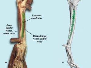
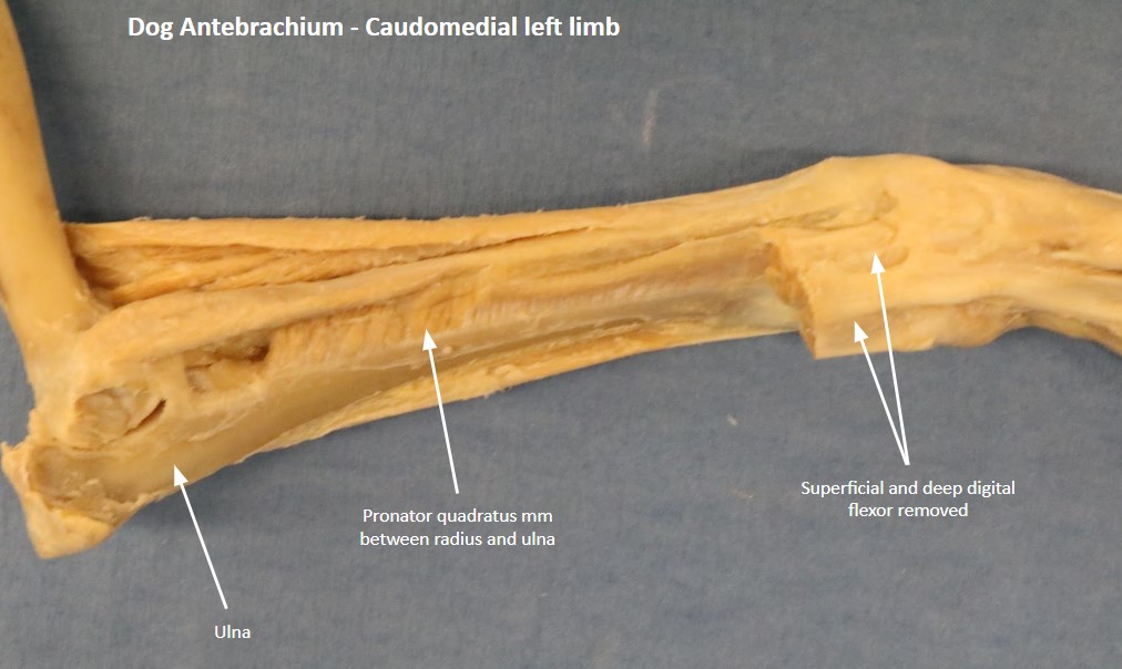
Pronator Quadratus m.
The pronator quadratus occupies the space between the radius and ulna. The fibers of this muscle run transversely between the ulna and radius.
ATTACHMENTS: Apposed surfaces of the radius and ulna
ACTION: Pronation of the distal limb.
Dissect: Retract the overlying flexor muscles and observe the pronator quadratus m. Holding the carpus and digits in slight flexion helps with retraction by taking tension off the flexor muscles. Also observe the pronator quadratus m. on prosections.
-
- Deep muscles of the dog antebrachium. 1

-
- Dog pronator quadratus. 40

-

- Dog antebrachium
Ungulate Comparative Anatomy
INTRINSIC TL MUSCLES of the antebrachium – horse and ruminant
Note: we are not concerned with learning antebrachial muscles of the pig – when you need to know them, look it up.
Intrinsic muscles of the antebrachium to identify on ungulates |
|
| Craniolateral muscles of the antebrachium |
|
| Caudomedial muscles of the antebrachium |
|
-
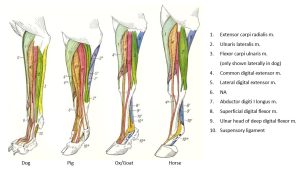
- Comparative craniolateral muscles of the antebrachium. 23

-

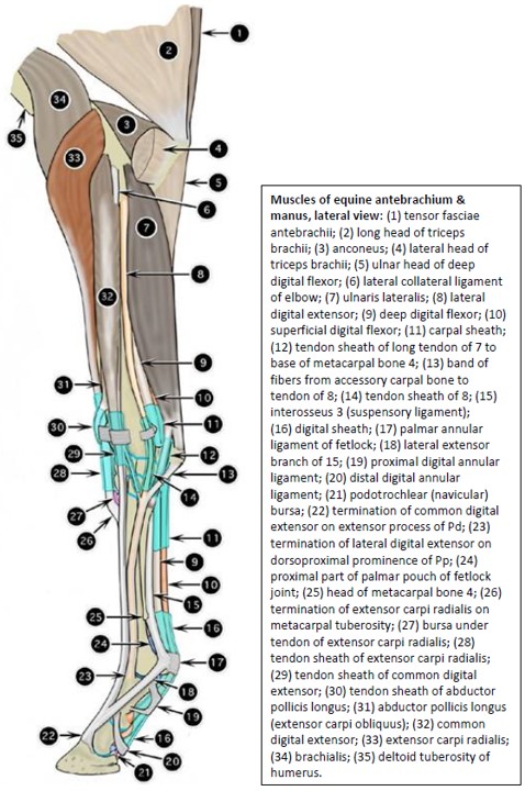
- Muscles of equine antebrachium & manus, lateral view 2
-
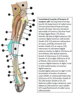
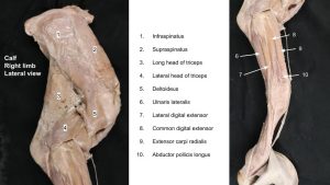
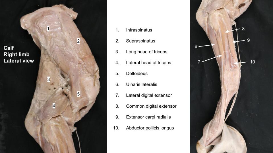
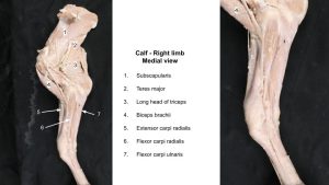
- Craniolateral muscles of forearm of newborn calf 2

-
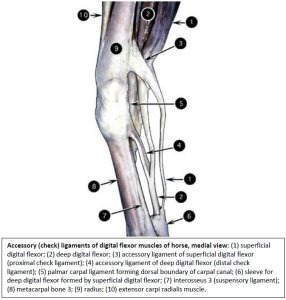
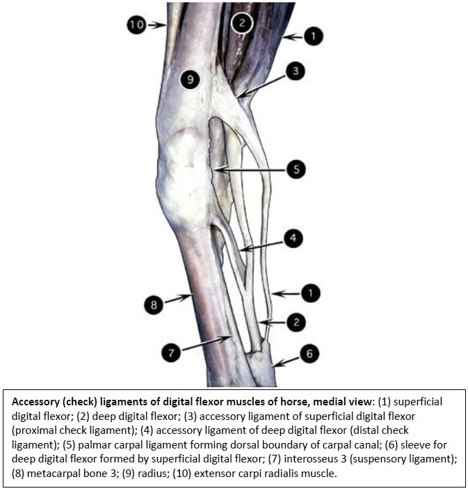
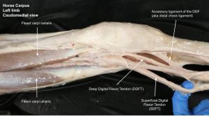
- Accessory (check) ligaments of digital flexor muscles of horse, medial view 2
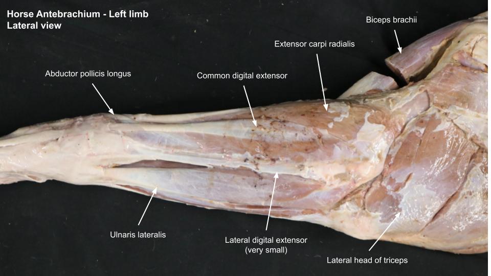
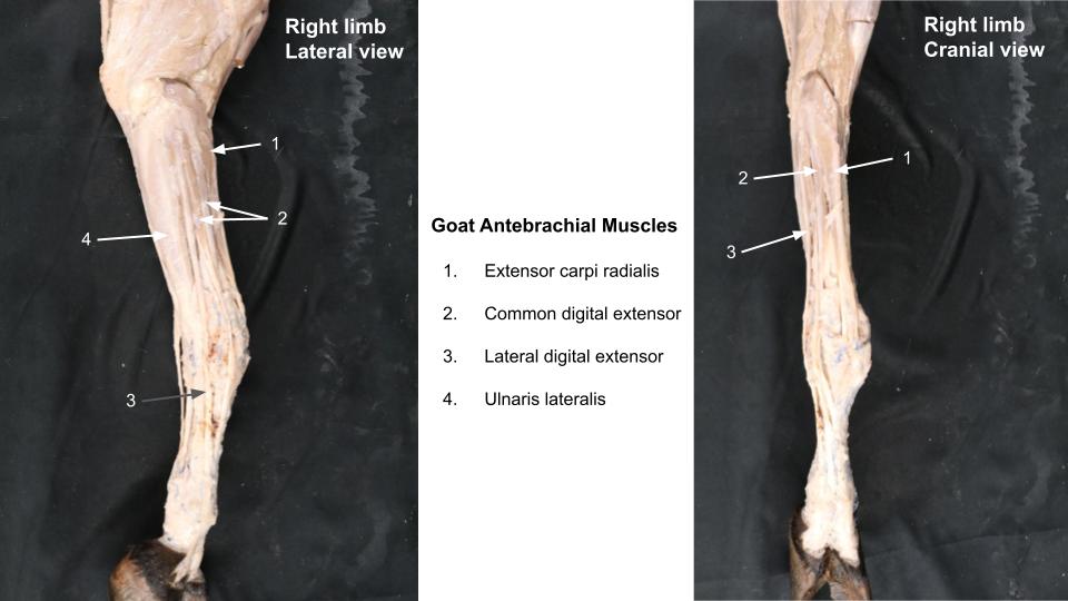
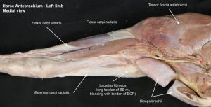
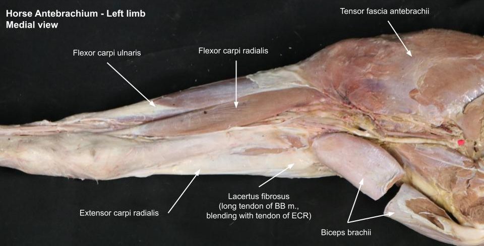
Craniolateral muscles – horse and ruminant
Observe: Review the prosected ungulate cadavers (horse and ruminant) to identify the comparative antebrachial anatomy as directed. It is helpful to isolate and then follow the tendons in the metacarpal region proximally, to match them with their muscle bellies. Learn the proximal and distal attachments of these muscles and their actions. Refer to the linked muscle chart.
In the horse the presence of the lacertus fibrosus attaching into the extensor carpi radialis m. should be noted. The common digital extensor m. has one tendon in the horse and two in the ruminant (three in the pig). Tendons passing over the carpus are bound down by the extensor retinaculum of the carpus and are enveloped by synovial sheaths. The lateral digital extensor m. is very small in the horse (there is no lateral digit in the modern domestic horse!)
Observe: Follow the tendon of the lateral digital extensor from the metacarpus proximally to identify the muscle belly. Note the tendon is wider in the metacarpus than proximal to the carpus, because at the carpus it receives fibers from the common digital extensor and a band of fibers passing from the accessory carpal bone. Note also that the lateral digital extensor tendon passes deep to the lateral collateral ligament of the carpus and runs palmar to the center of rotation for flexion of the carpus i.e. it may act more as a carpal ‘antihyperextensor’ (and a digit extensor, as expected).
The ulnaris lateralis m. lies more palmar than the lateral digital extensor and it acts as a carpal flexor. The ulnaris lateralis inserts on the accessory carpal bone and the base (i.e the proximal end) of MC4 (clinically, the proximal end of metacarpals 2 and 4 in the horse, the “splint bones”, are referred to as the ‘head’ of the splint bone). The extensor carpi obliquus m. (abductor digiti 1 [or pollicis] longus) passes diagonally across the cranial antebrachium. Synovial sheaths (tendon sheaths) envelop the tendons of muscles as they pass over the carpus. We will appreciate these tendon sheaths in the upcoming ungulate limb dissection lab.
Clinical relevance
Synovitis of the extensor tendon sheaths; trauma to these muscles can cause loss of carpal and digital extension leading to a “knuckling over” gait.
Observe: In the horse – retract/reflect the proximal stump of the transected ulnaris lateralis m. toward the elbow joint. Lying deep to the tendon of origin of this muscle is the ulnaris lateralis bursa, a bursa which may communicate with the elbow joint.
Clinical relevance
Infection of the ulnaris lateralis bursa may extend into elbow joint.
-
- Muscles of equine antebrachium & manus, lateral view 2
-
- Craniolateral muscles of forearm of newborn calf 2
-
- Accessory (check) ligaments of digital flexor muscles of horse, medial view 2
-
- Horse antebrachium

-

- Horse antebrachium
-
- Calf antebrachium
-
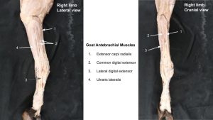
- Goat antebrachium
Caudomedial muscles – horse and ruminant
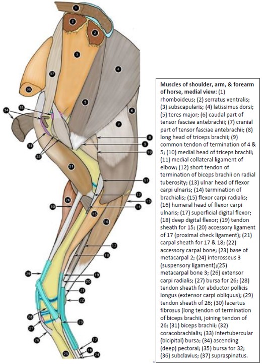
Observe: As was done for the craniolateral muscles, review the prosected ungulate cadavers (horse and ruminant) to identify the comparative antebrachial anatomy as directed. It is helpful to isolate and then follow the tendons in the metacarpal region proximally, to match them with their muscle bellies. Learn the proximal and distal attachments of these muscles and their actions. Refer to the linked muscle chart.
On the caudomedial side of the antebrachium note that a triangular area of the distal radius is subcutaneous; no muscles or tendons cross this area, and the deep fascia of the forearm blends with the periosteum of the radius. This heavy protective and supportive fascia was removed to observe the caudomedial muscles.
Observe: Identify the pronator teres (present in ruminant only, as a fibrous band) the flexor carpi radialis m., and the flexor carpi ulnaris m. (with humeral and ulna heads.)
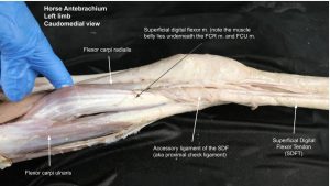
Observe: To aid in identifying the superficial and deep digital flexor muscles, in particular, identify their dissected tendons in the mid metacarpus first of all and then track them proximally into the muscle bellies. Note the flexor retinaculum of the carpus, that was incised, to follow these flexor tendons passing through the carpal canal.
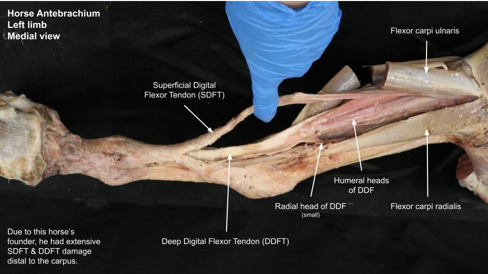
In the horse prosection – elevate and retract the superficial digital flexor tendon, just proximal to the carpus, and viewing from the medial aspect, note that the SDF tendon (SDFT) is attached to the distomedial aspect of the radius by a short fibrous band, the accessory ligament of the superficial digital flexor. This “ligament” represents the completely fibrous radial head of the muscle and forms a direct attachment of the tendon to the caudodistomedial aspect of the radius. It is frequently referred to as the proximal (or superior or radial) check ligament.
Note, in other species of domestic mammals, the SDF tendon terminates on the middle phalanx only. In the horse, its additional termination on the proximal phalanx is unique. By attaching both proximal and distal to the proximal interphalangeal joint (PIPJ), the SDF tendon in the horse assists in preventing the joint from flexing when the limb is bearing weight and as a result, there is virtually no movement in the joint during the support phase of the stride.
Clinical relevance:
-

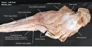
- Muscles of the shoulder, arm and forearm of the horse. Medial view. 2
-

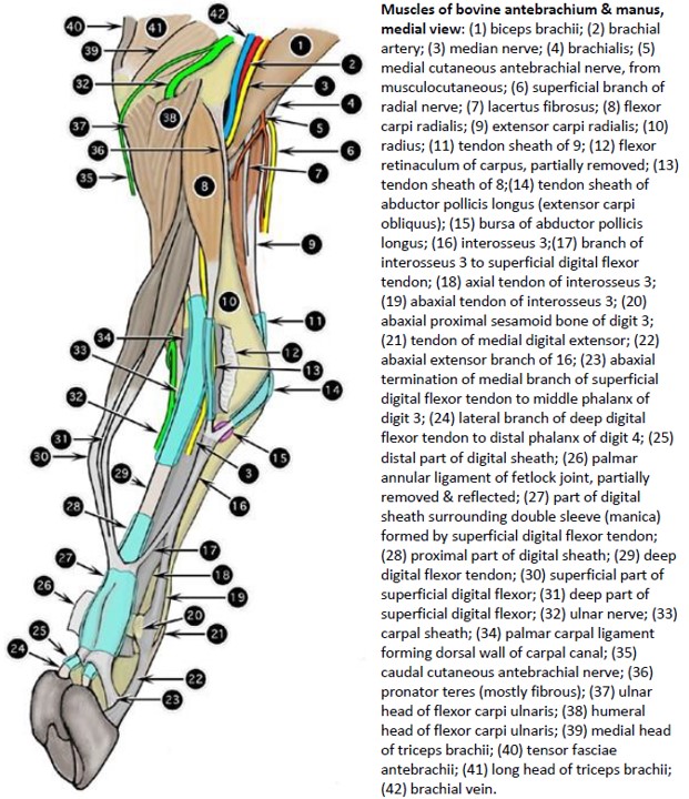
- Muscles of bovine antebrachium & manus, medial view 2
-
- Accessory (check) ligaments of digital flexor muscles of horse, medial view 2
-
- Horse antebrachium
-
- Calf antebrachium

-
- Horse antebrachium

Observe: Now locate the tendon of the deep digital flexor (DDFT) in the metacarpus. Follow this heavy tendon proximally through the carpal canal to observe that it is formed by three heads (humeral, radial and ulnar). Returning distally to the proximal metacarpal region, observe that the DDFT has a direct fibrous attachment that originates from the junction of the carpus and metacarpus. This is the accessory ligament of the deep digital flexor, which is frequently referred to as the distal (or inferior or carpal) check ligament.
Clinical relevance:
-
- Muscles of the shoulder, arm and forearm of the horse. Medial view. 2
-
- Muscles of bovine antebrachium & manus, medial view 2
-
- Accessory (check) ligaments of digital flexor muscles of horse, medial view 2
-

- Horse antebrachium
-
- Horse antebrachium

Observe: In the ruminant prosection – by pulling the superficial tendon of the superficial digital flexor palmarly in the metacarpus, a smaller tendon should be found to join it just above the fetlock. This is the tendon of the deep part of the superficial digital flexor. Follow this smaller tendon proximally through the carpal canal to identify this additional head of the superficial digital flexor. The flexor retinaculum of the carpus has been incised to view the tendons. In artiodactyls, this deep part of the SDF is intimately attached to the deep digital flexor tendon by one or more adhesions, which represent the interflexorii muscles.
Now locate the tendon of the deep digital flexor in the metacarpus. Follow this heavy tendon proximally through the carpal canal to observe that it is formed by three heads (humeral, radial and ulnar).
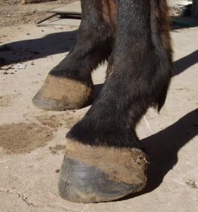
Clinical application: Q3
| Diagnose the condition.
What soft tissue structure would a veterinarian considering transecting first of all, to treat this condition?
(Answer) |
 |
Review Videos
Muscle Chart/Terms List
| Muscle Action Groups | |
| Extensors of carpus and digits |
|
| Flexors of carpus and digits |
|
| Supinators |
|
| Pronators |
|
|
Muscle/structure |
Attachments/comments |
Action/comments |
| EXTENSOR CARPI RADIALIS M. |
|
Extend carpus |
| COMMON DIGITAL EXTENSOR M. |
|
Extend digits and carpus |
| LATERAL DIGITAL EXTENSOR M. |
|
Extend digits and carpus |
| ABDUCTOR DIGITI I LONGUS M.(ABDUCTOR POLLICIS LONGUS) |
|
Abduct first digit (as exists) and extend carpus |
| Extensor retinaculum | Fibrous band of thickened fascia at level of dorsal carpus | Binds down extensor tendons |
| ULNARIS LATERALIS M. |
|
Flex carpus; abduct manus at carpus |
| Ulnaris lateralis bursa | Synovial ‘cushion’ deep to proximal tendon of origin | Horse |
| FLEXOR CARPI RADIALIS M. |
|
Flex carpus |
| SUPERFICIAL DIGITAL FLEXOR M. |
|
Flex proximal interphalangeal joints; flex carpus and metacarpophalangeal joints) |
| Accessory ligament of SDF | aka proximal or superior check ligament | Horse |
| DEEP DIGITAL FLEXOR M.
Humeral, Ulnar, Radial heads |
|
Flex digits and carpus |
| Accessory ligament of DDF | aka distal or inferior check ligament | Horse |
| Flexor retinaculum | Fibrous band with superficial and deep parts on the palmaromedial carpus | Binds digital flexors within carpal canal |
| FLEXOR CARPI ULNARIS M. (Humeral and Ulnar heads) |
|
Flex carpus |
| BRACHIORADIALIS M
(Cat Only; Vestigial, but frequently seen, in Dog) |
|
Supination |
| SUPINATOR M. |
|
Supination of distal limb (and flex elbow) |
| PRONATOR TERES M. |
|
Pronation of the distal limb (and flex elbow) |
| PRONATOR QUADRATUS M. |
|
Pronation of the distal limb |
| Synovial tendon sheaths | Surround tendons of muscles passing over joints | Also to view in next lab |
-
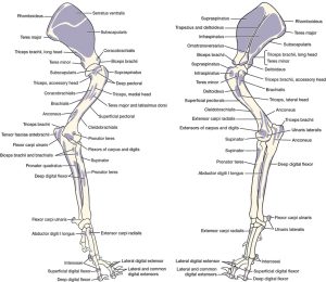
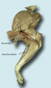
- Left thoracic limb skeleton with muscle attachments (medial and lateral views).1
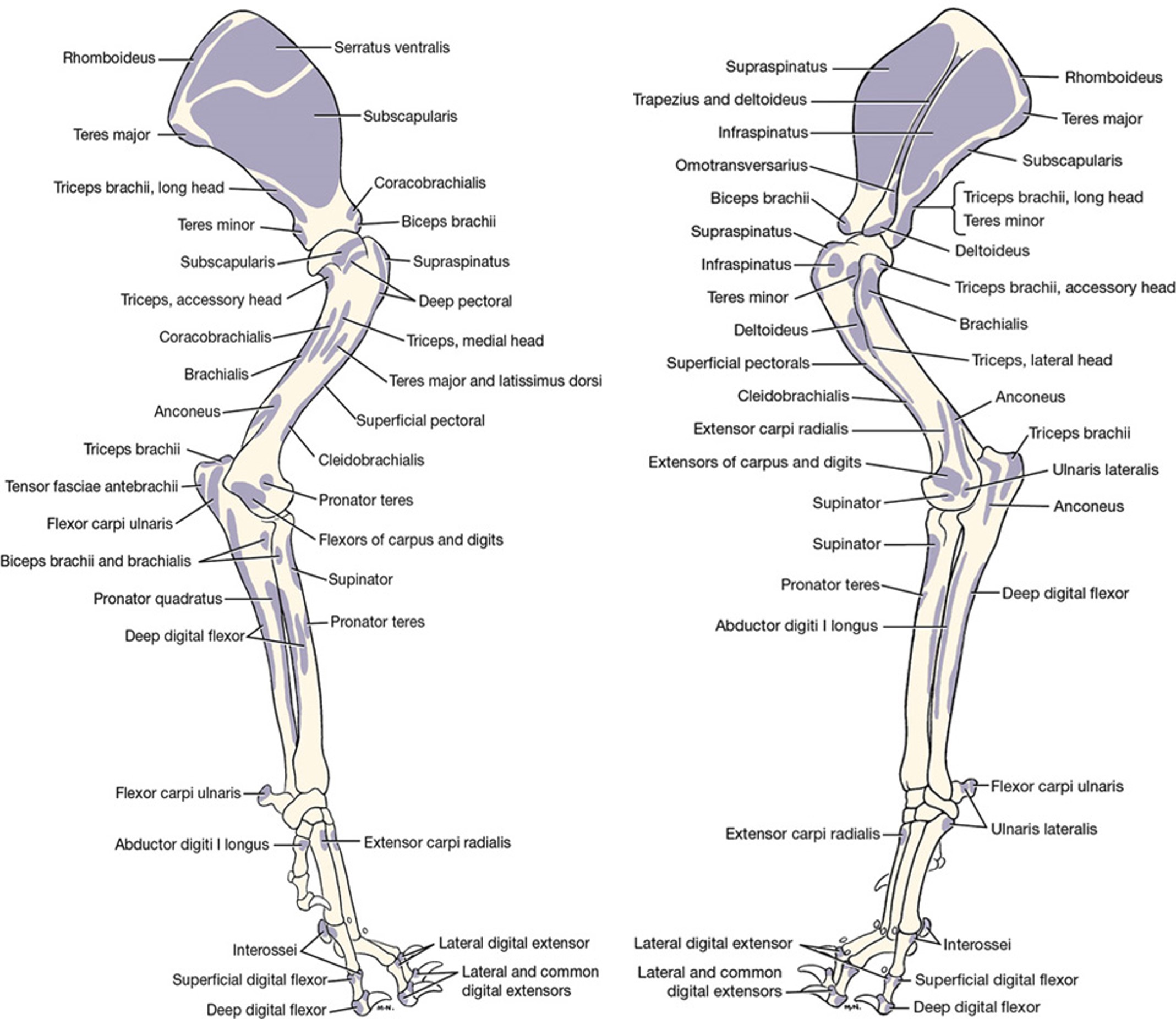
-
- Major extensors and flexors of the canine left forelimb.1