
MSK LAB 9A – Skull and Vertebrae: Osteology and Arthrology
Learning Objectives
- Identify skull bones, joints, and specific features as listed and differentiate between canine, feline, porcine, caprine, bovine, and equine skulls.
- Identify the bones of the hyoid apparatus.
- Recall the vertebral column formula for species studied (carnivore, horse, ruminant, pig)
- Identify cervical, thoracic, lumbar, sacral, and caudal vertebrae and their major features.
- Identify processes and foramina of the vertebrae.
- Understand the vertebral column as a unit and the structures and openings formed by adjacent vertebrae.
- Identify the joints and certain ligaments of the vertebral column.
- Describe the supraspinous ligament and its location and clinical relevance.
- Describe the yellow ligament and its location and clinical relevance
- Describe the intercapital ligament and its location.
Lab Overview and Instructions:
The axial skeleton consists of all the bones that are not part of the limbs (which form the appendicular skeleton). We can divide it into the skull, the hyoid apparatus, the vertebral column (with its regional subdivisions), the bones of the thoracic cavity wall (ribs, sternebrae), and also include the os penis. The bones of the pelvis, although directly associated with back and axial skeleton anatomy, are considered part of the pelvic limb (appendicular skeleton) and will be covered in that unit.
Observe: Use skeletal models, plastinated models and other specimens to cover the comparative species. Refer to linked diagrams and the tabulated terms to guide your learning.
Skull introduction
The skull is a conglomeration of approximately 29 bones, depending on how a ‘separate’ bone is defined. There are many, many features of the skull and for this section and unit we are focused on an understanding of the bones that combine to form the skull. Most holes in the skull (foramina, fissures, canals) will be considered in the Nervous System and Cardiovascular System, in association with the nerves and vessels that pass through those features. Similarly, the air-filled sinuses (paranasal sinuses) found within certain bones will be considered in detail in the Respiratory System.
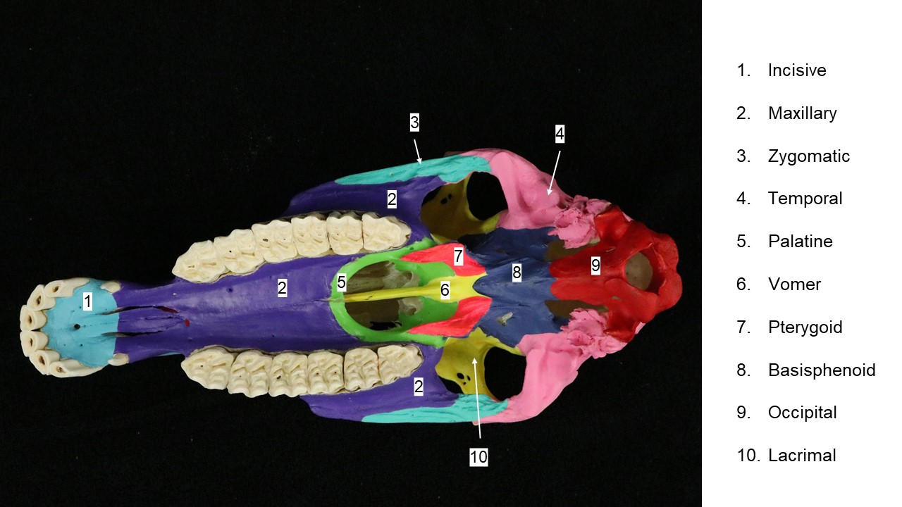
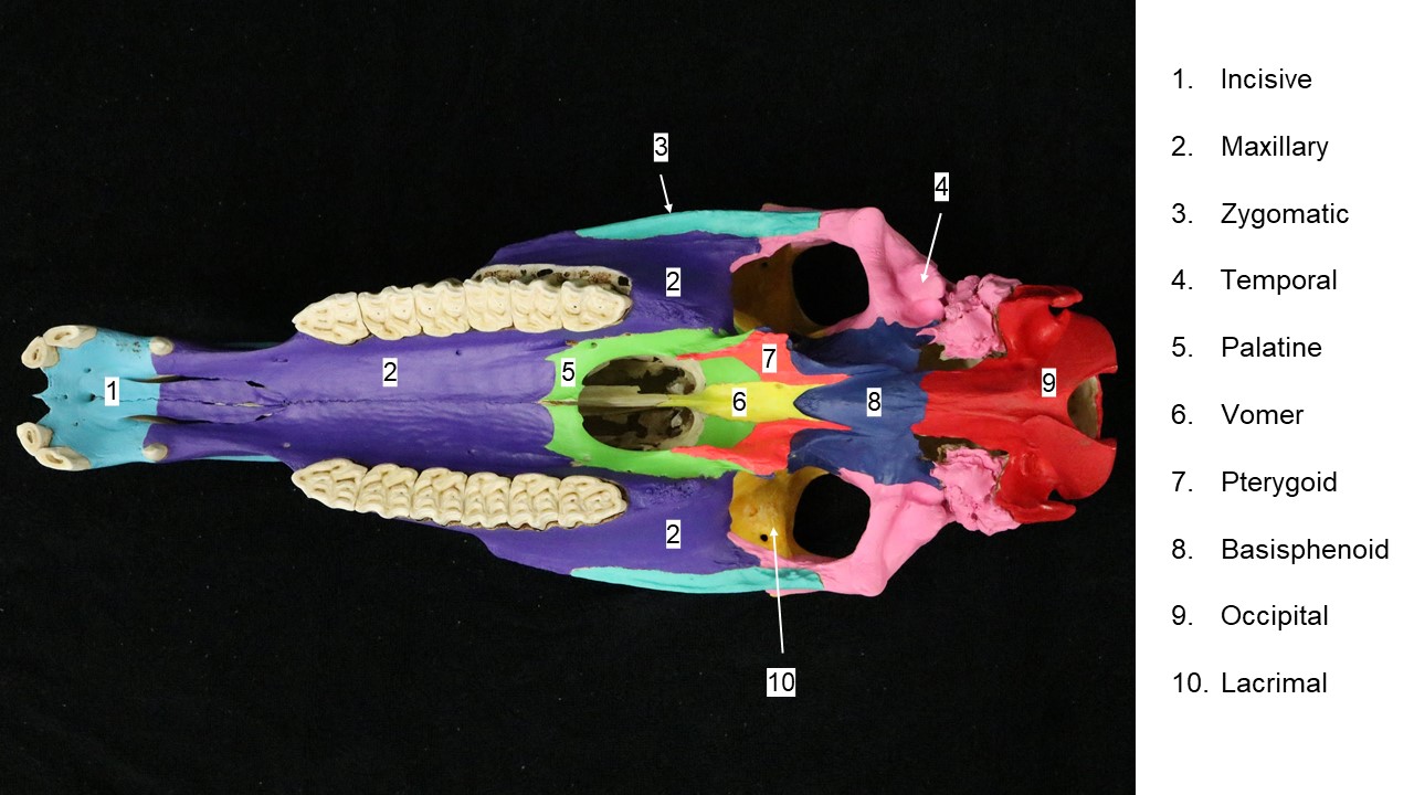
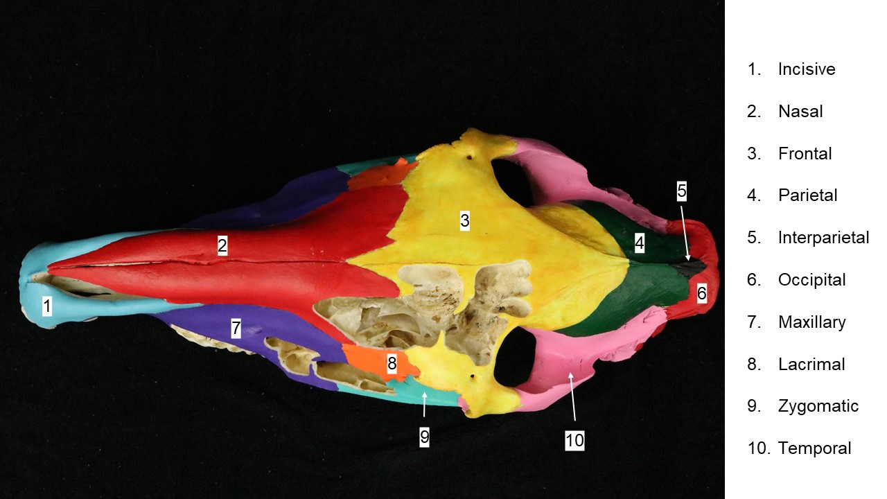
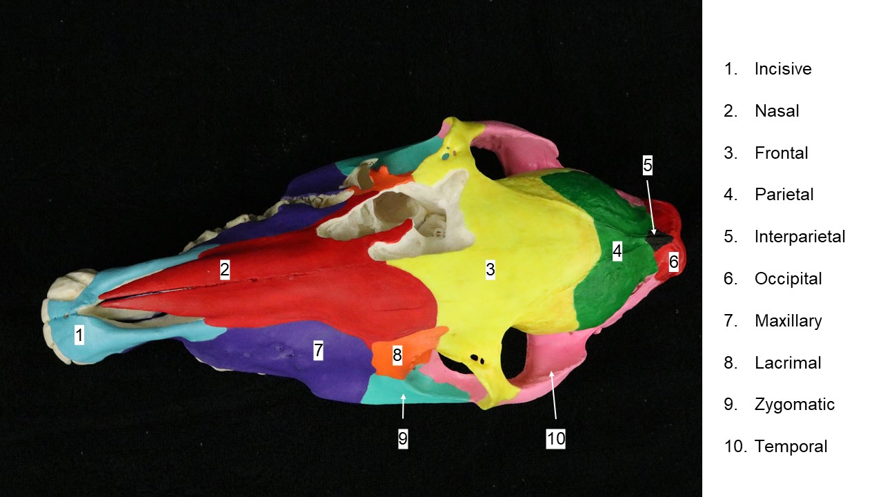
So, a place to start is to pick up a skull and to use the images below to identify the bones. Any species can be selected as a starting point and once names and locations of bones are familiar this knowledge can be applied to the skulls of other species. There can be slight variation between anatomy textbooks in the labeling of bones of the skull, in particular the bones of the sphenoid complex on the ventral aspect of the skull – use the terms list to guide your learning.
Carnivore skull
-
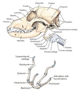
- Lateral view of dog skull with hyoid and laryngeal cartilages. 1

-
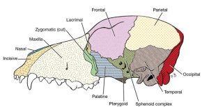
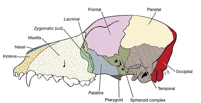
- Bones of the dog skull, lateral. 1
-
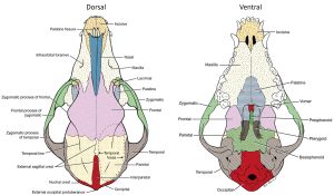
- Bones of the dog skull. 1
-
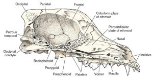
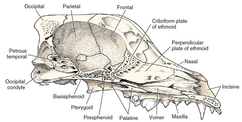
- Bones of the skull of the dog, medial aspect of sagittal section. 1
-

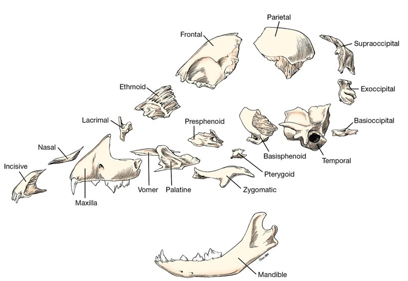
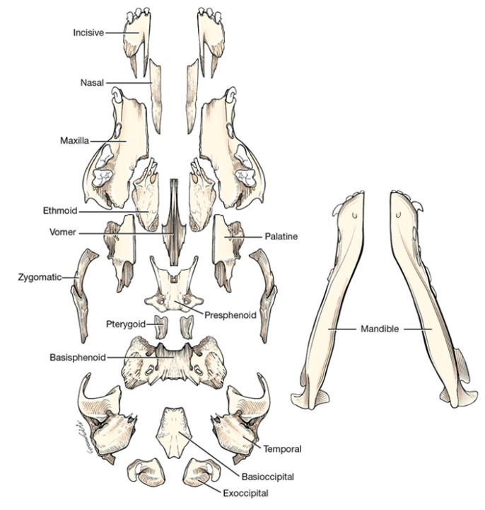
- Disarticulated puppy skull, left lateral view. 1
-

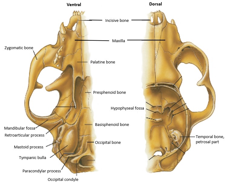
- Disarticulated puppy skull, ventral view. 1
-
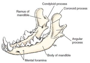
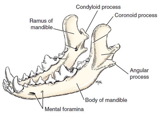
- Left and right mandibles, dorsal lateral aspect.1
-
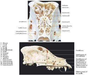
- Top: Transverse section of the nasal cavity of a dog with the cribriform plate of the ethmoid bone. Bottom: Medial sagittal section of the skull of a dog. 7
-
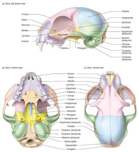
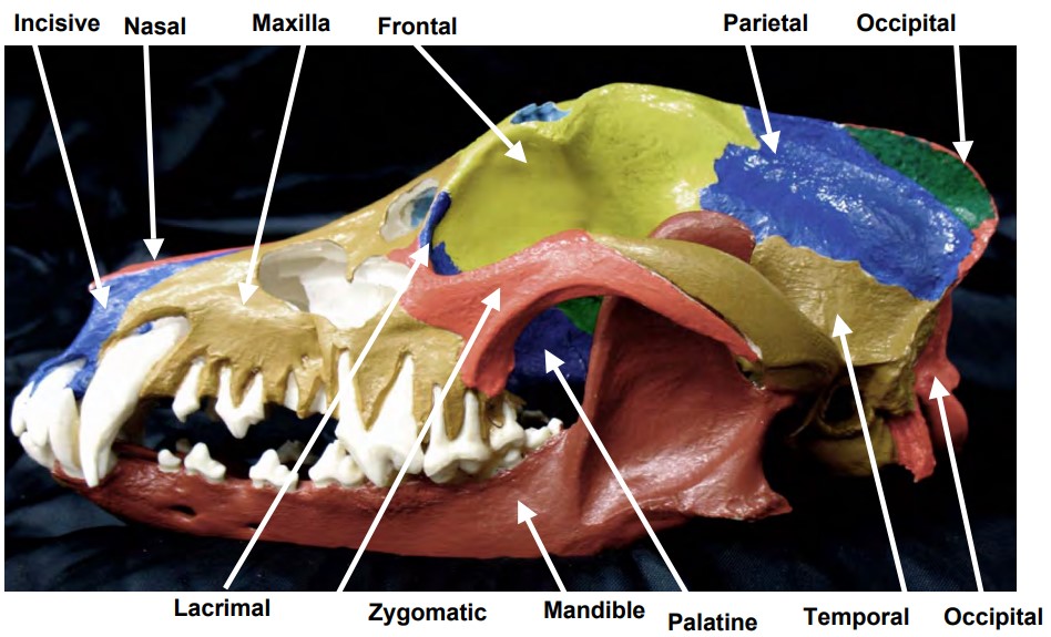
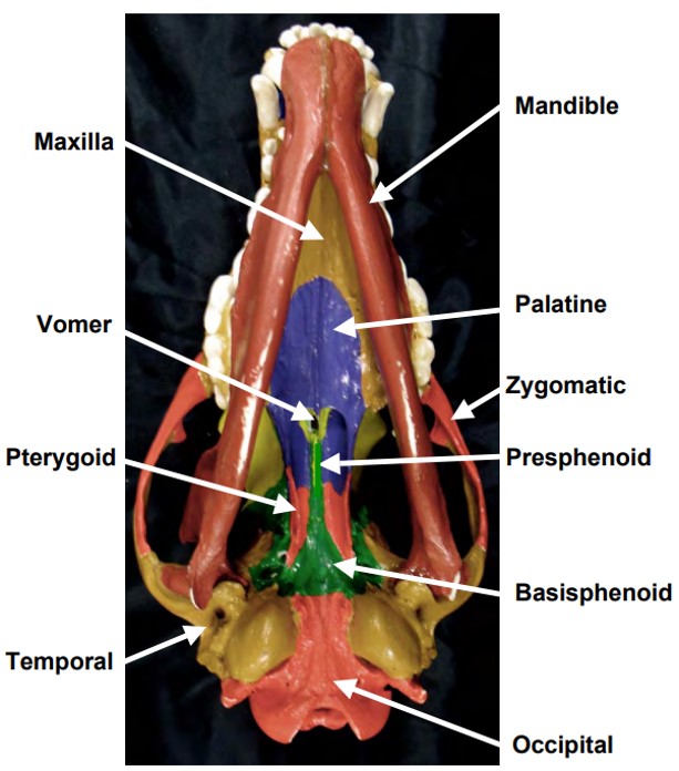
- Skull of the cat in (a) left lateral, (b) ventral, and (c) dorsal views, with bones color-coded. 5

-

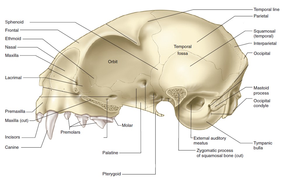
- Skull of the cat in left lateral view, with zygomatic arch removed.5
-
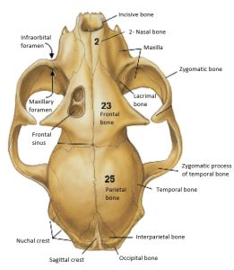
- Cat skull, dorsal view. 4

-

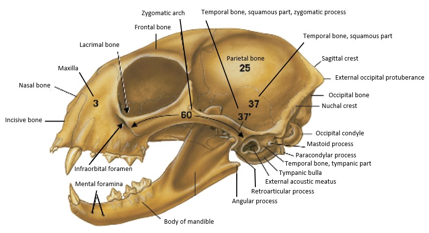
- Cat skull with mandible, left lateral view. 4
-
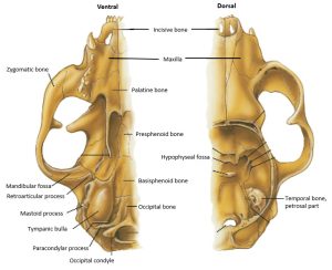
- Cat skull -ventral, and dorsal with cranial vault opened. 4
Dog skull bones:
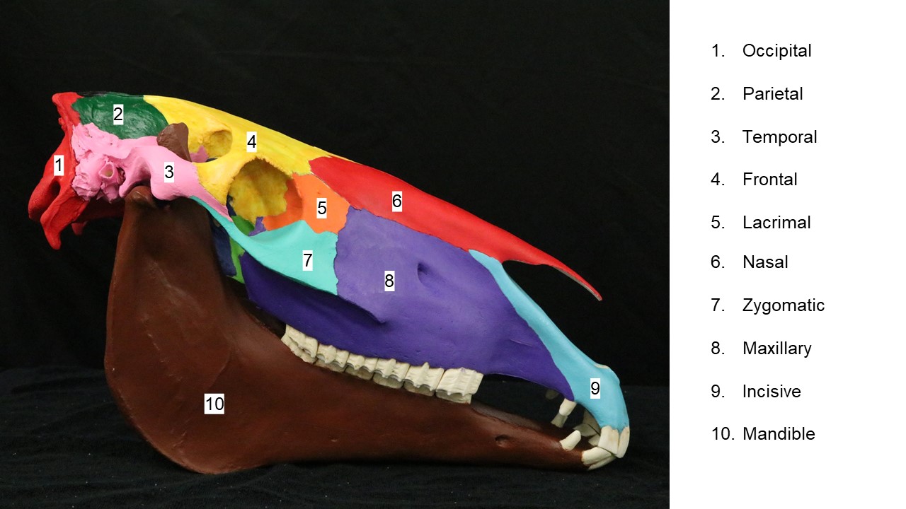
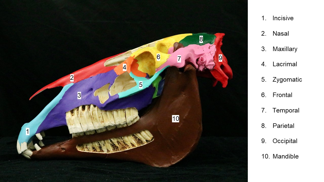
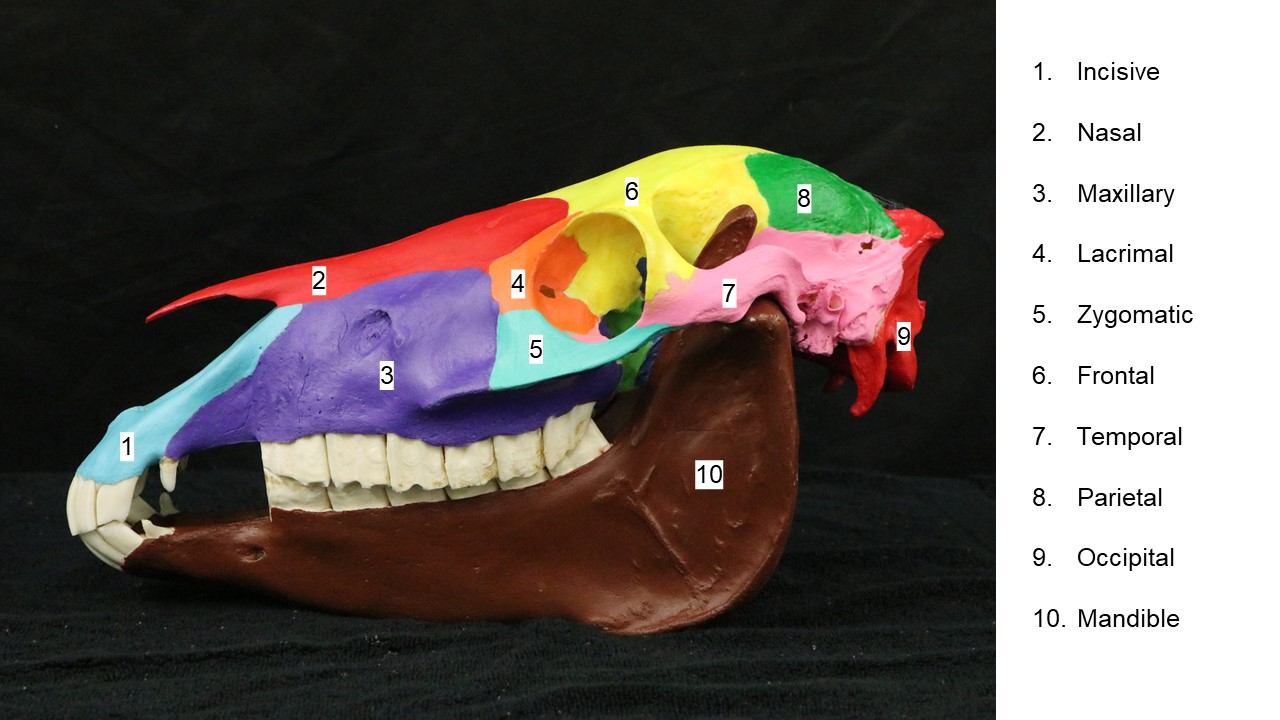
Horse Skull
-
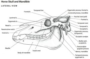
- Horse skull and mandible, lateral view. 6

-
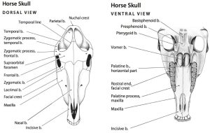
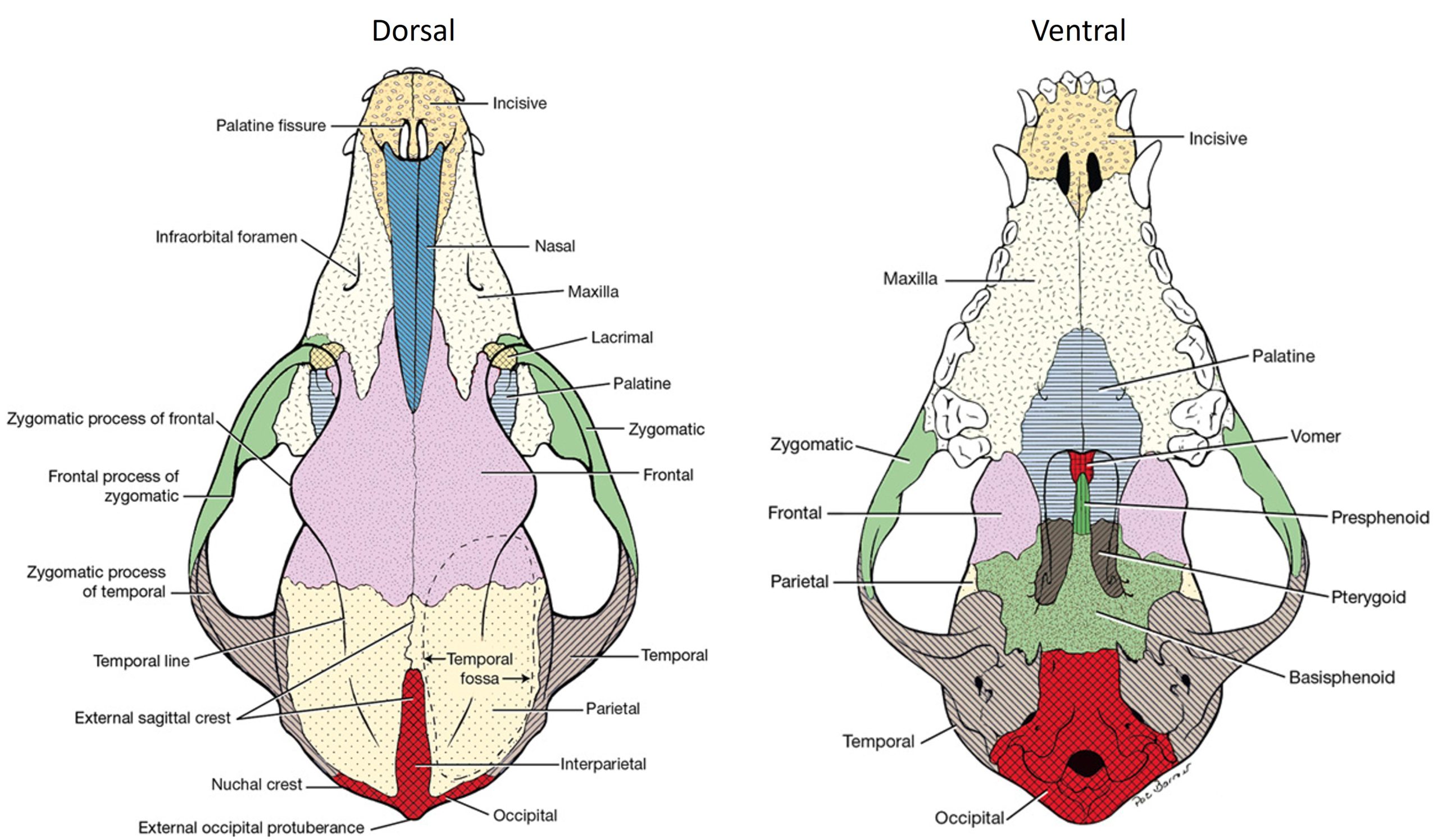
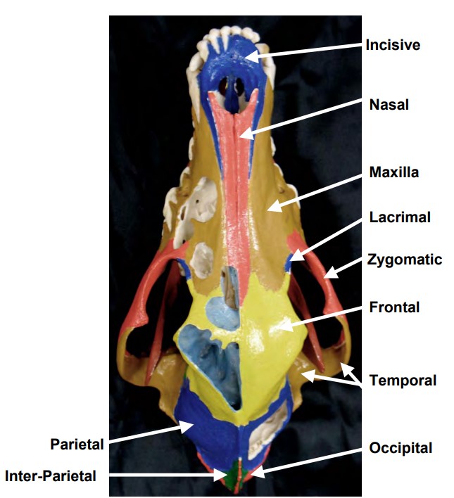
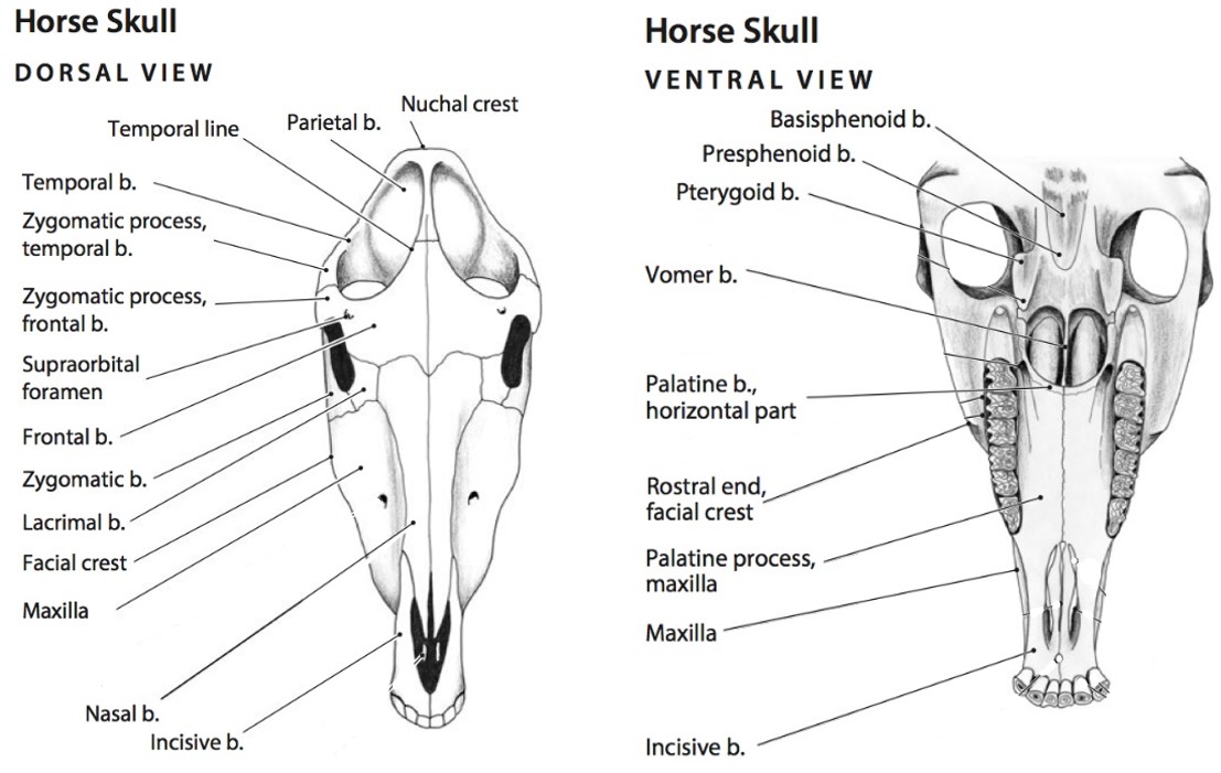
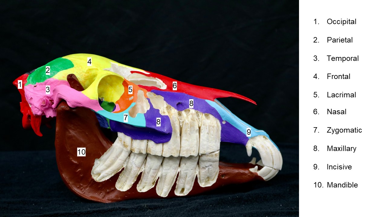
- Horse skull, dorsal and ventral views. 6
Horse skull bones:
Ruminant Skull
-
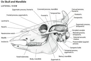
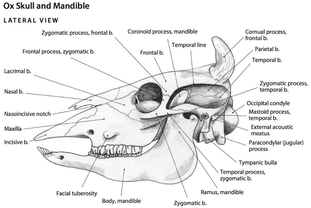
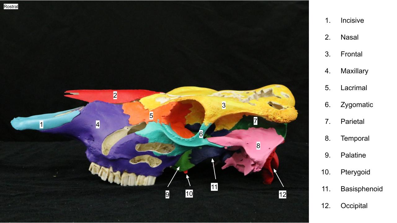
- Ox skull, lateral view. 6
-
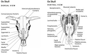
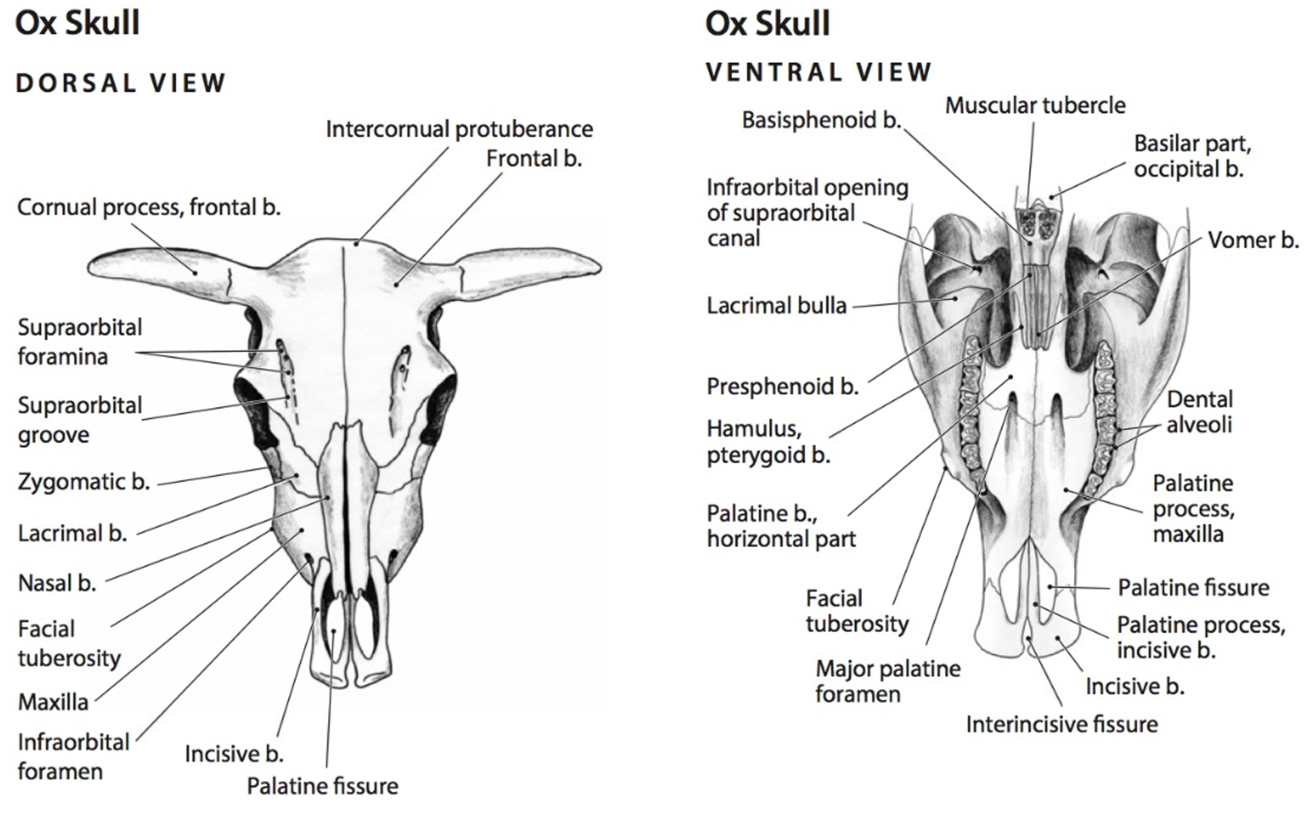
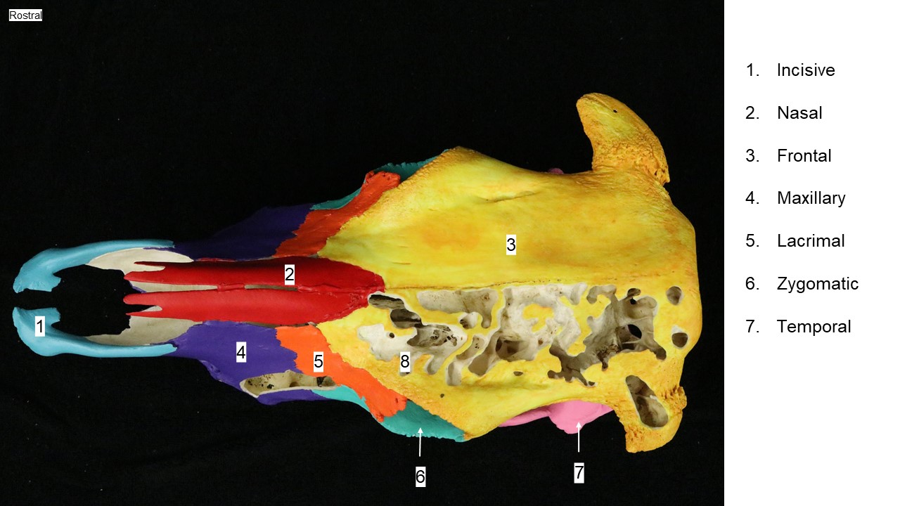
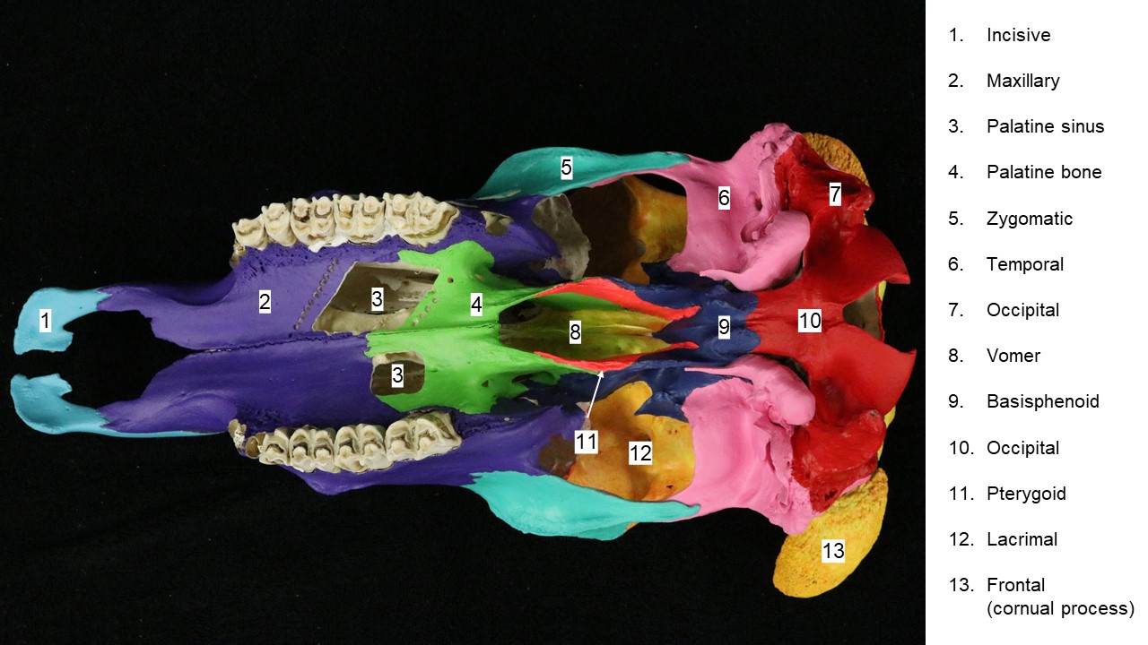
- Ox skull, dorsal and ventral views. 6
-
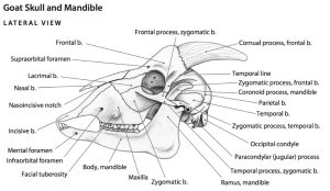
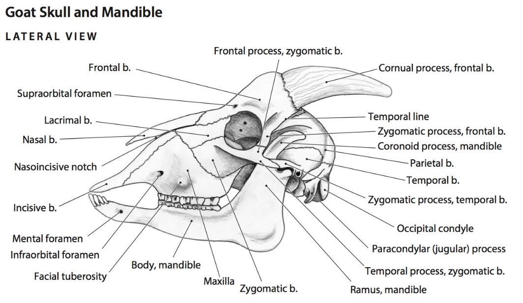
- Goat skull, lateral view. 6
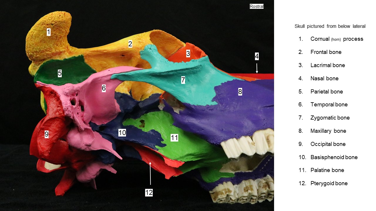
Ox skull bones:
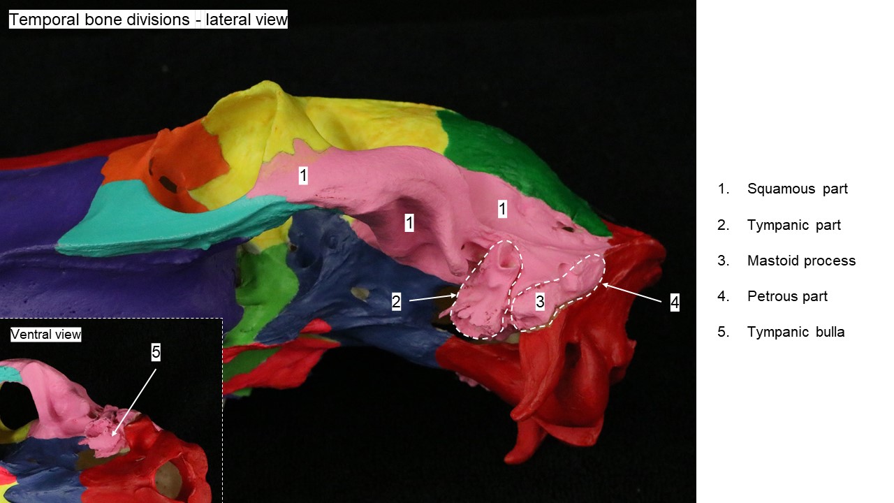
-
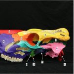
- Ox skull
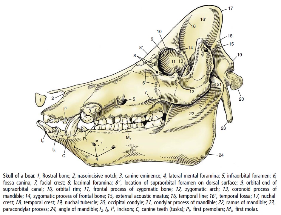
Pig skull
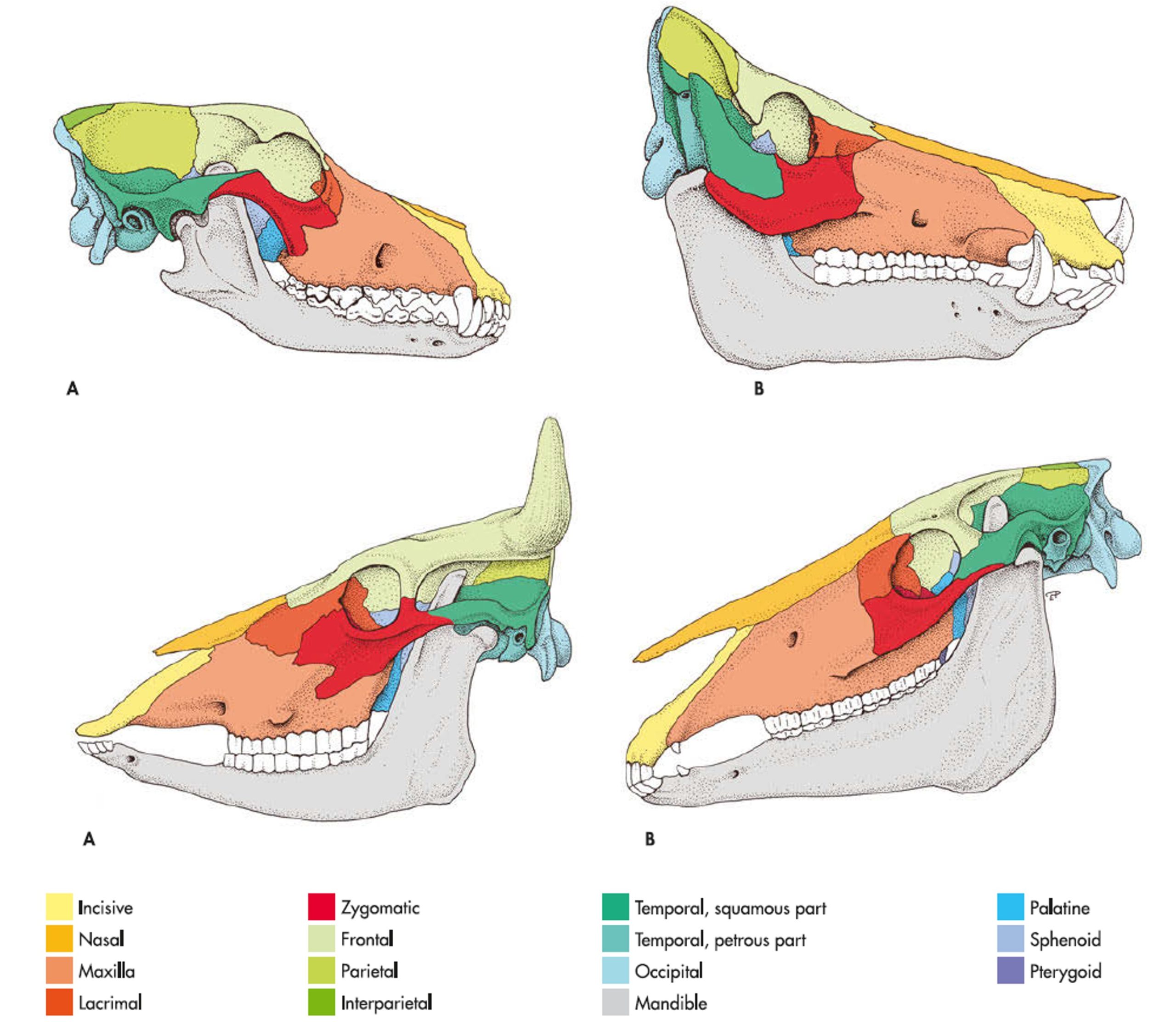
Comparative skulls
-
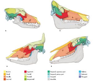
- Bones of the skulls of the dog, pig, ox and horse. 7
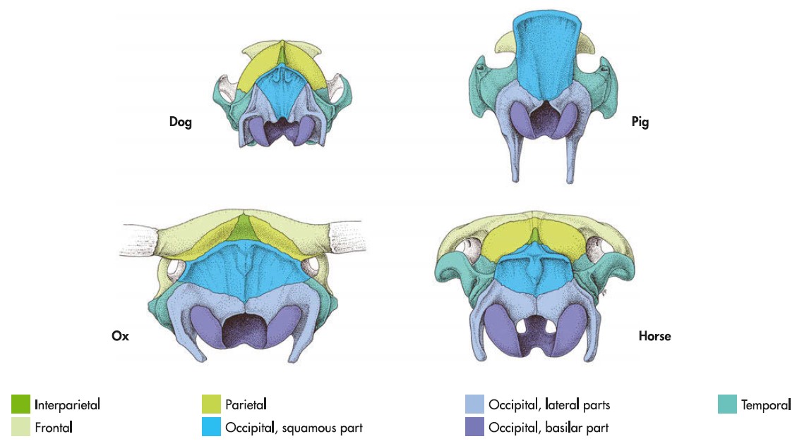
-

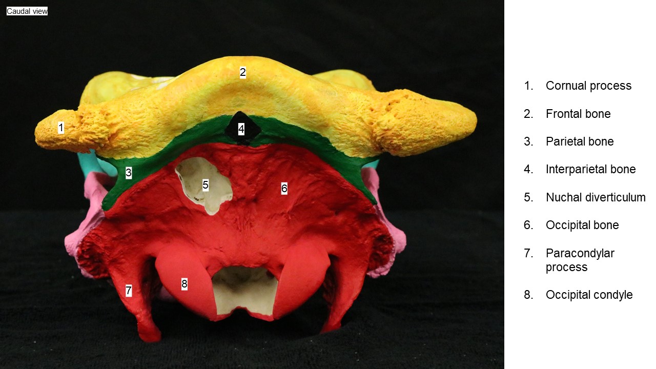
- Nuchal aspect of the canine, porcine, bovine and equine skull. 8
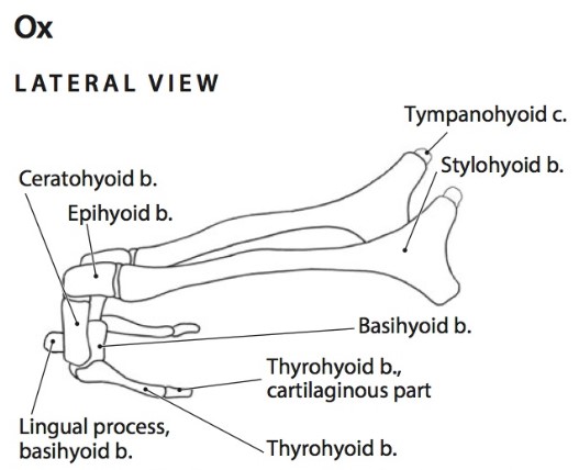
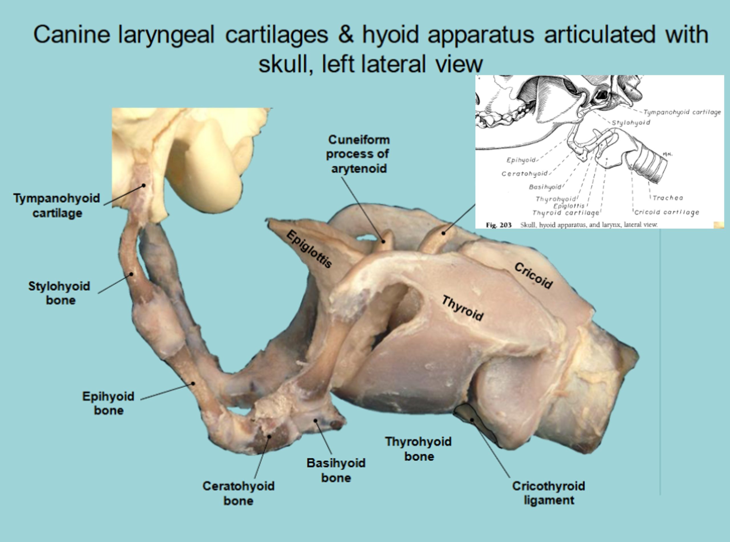
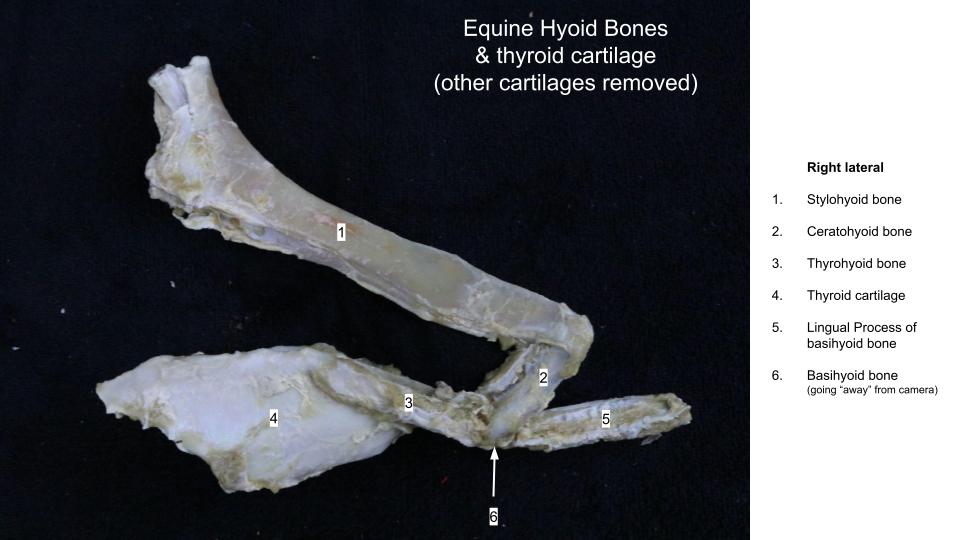
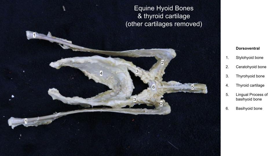
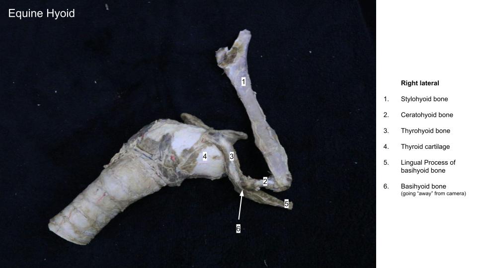
Hyoid apparatus
This is a construct of bones that attach the root of the tongue and larynx to the base of the skull. ‘Hyoid’ is from the root words ‘hyo’-[Gr] related to U, the letter Upsilon, and ‘-eidos’ [Gr] = shape or form. Therefore the hyoid is the U-shaped set of bones. The hyoid apparatus is flexible, moving with swallowing, for example. This structure will be revisited in future body systems (e.g. the Respiratory System) because of its close relationship to the larynx and the equine guttural pouch.
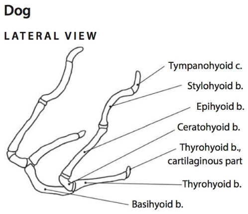
-
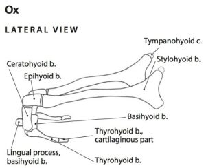
- Dog, Horse & Ox hyoid apparatus. 6
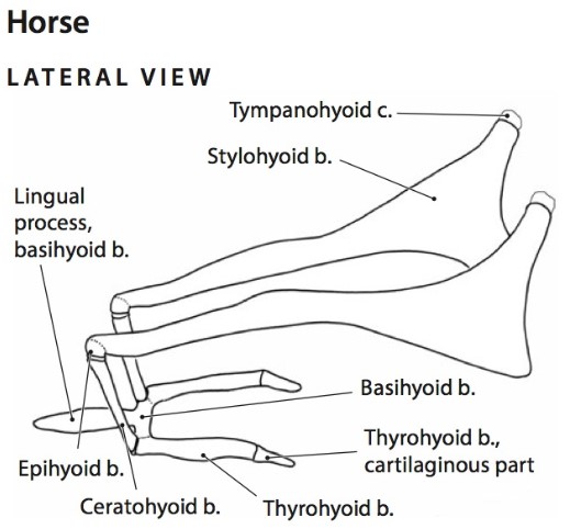
-
- Horse hyoid apparatus. 14

-

- Dog hyoid bones 40
-
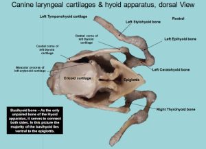
- Dog hyoid bones – dorsal 40

-
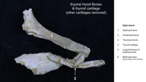
- Horse hyoid bones
-
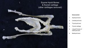
- Horse hyoid bones
-
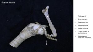
- Horse hyoid bones
Interactive review content:
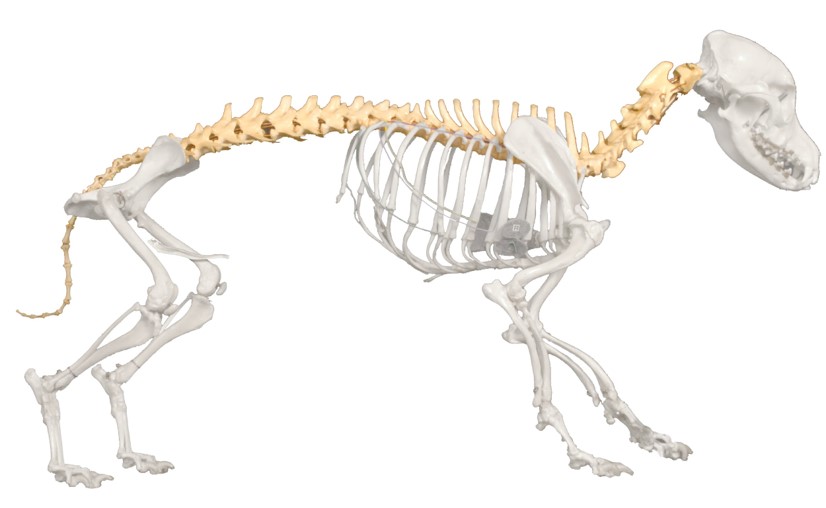
Vertebral column introduction
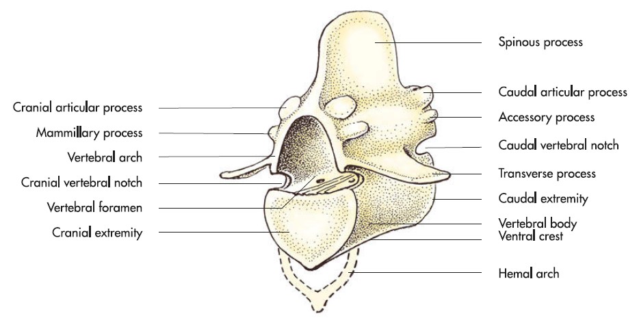
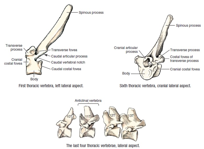
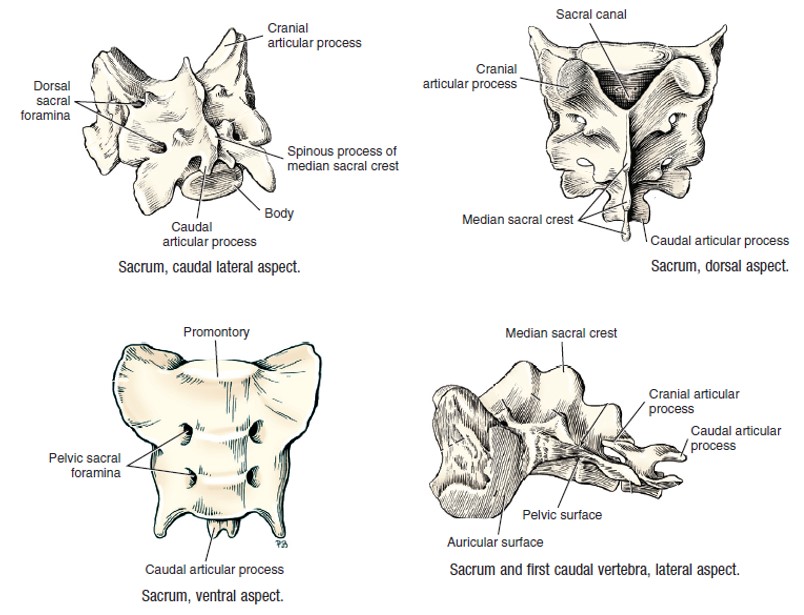
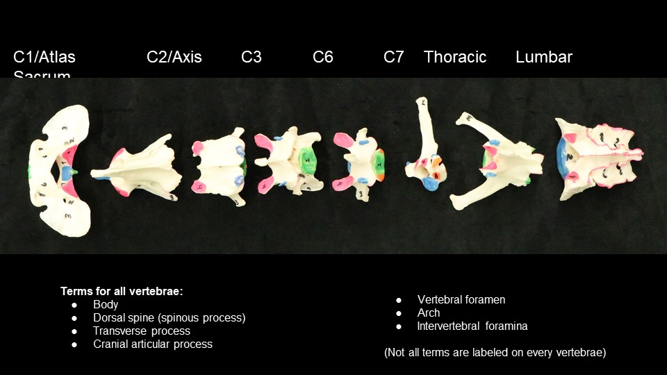
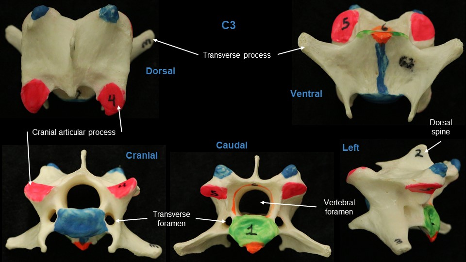
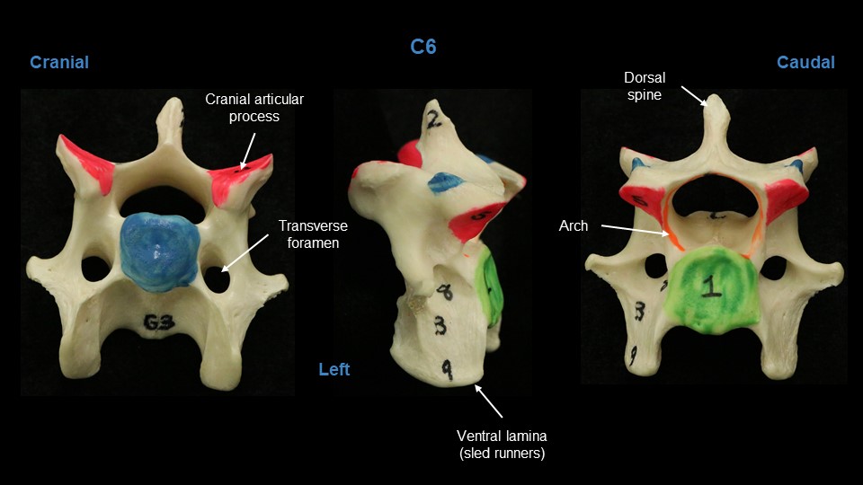
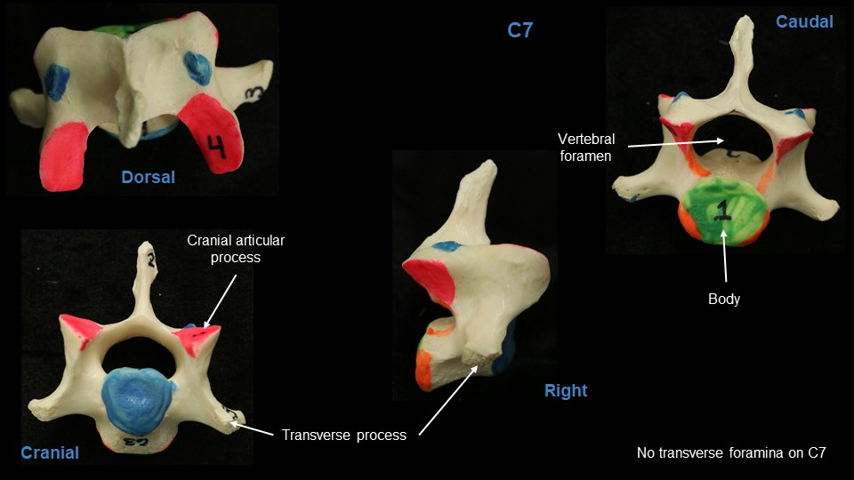
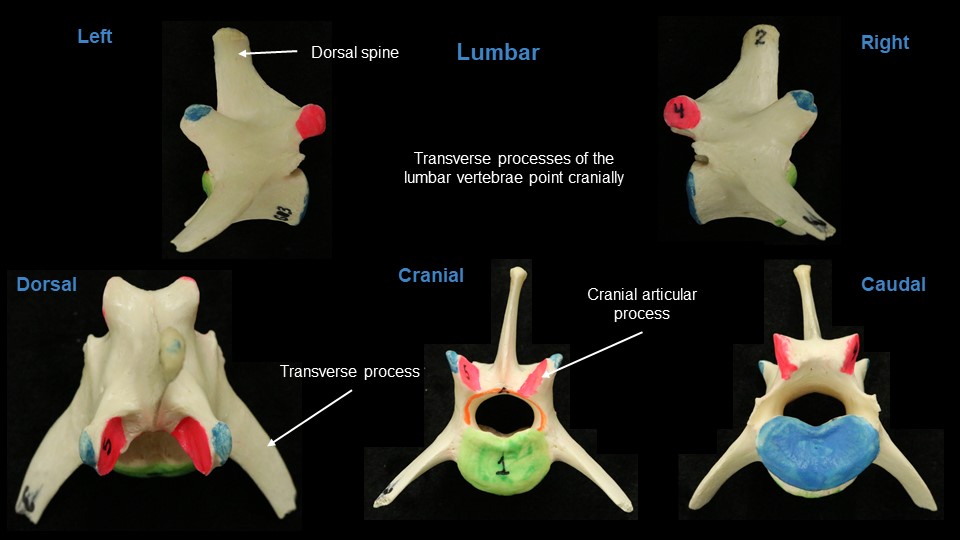
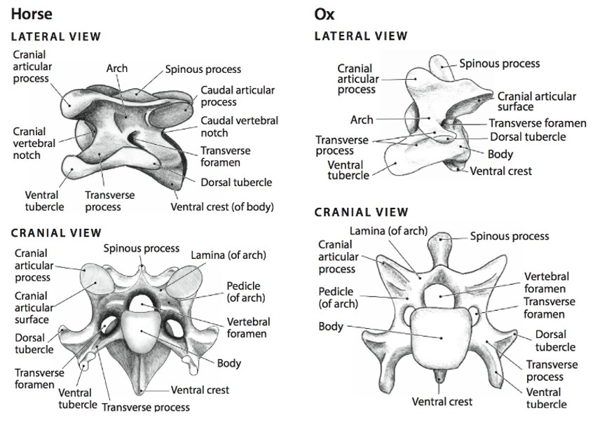
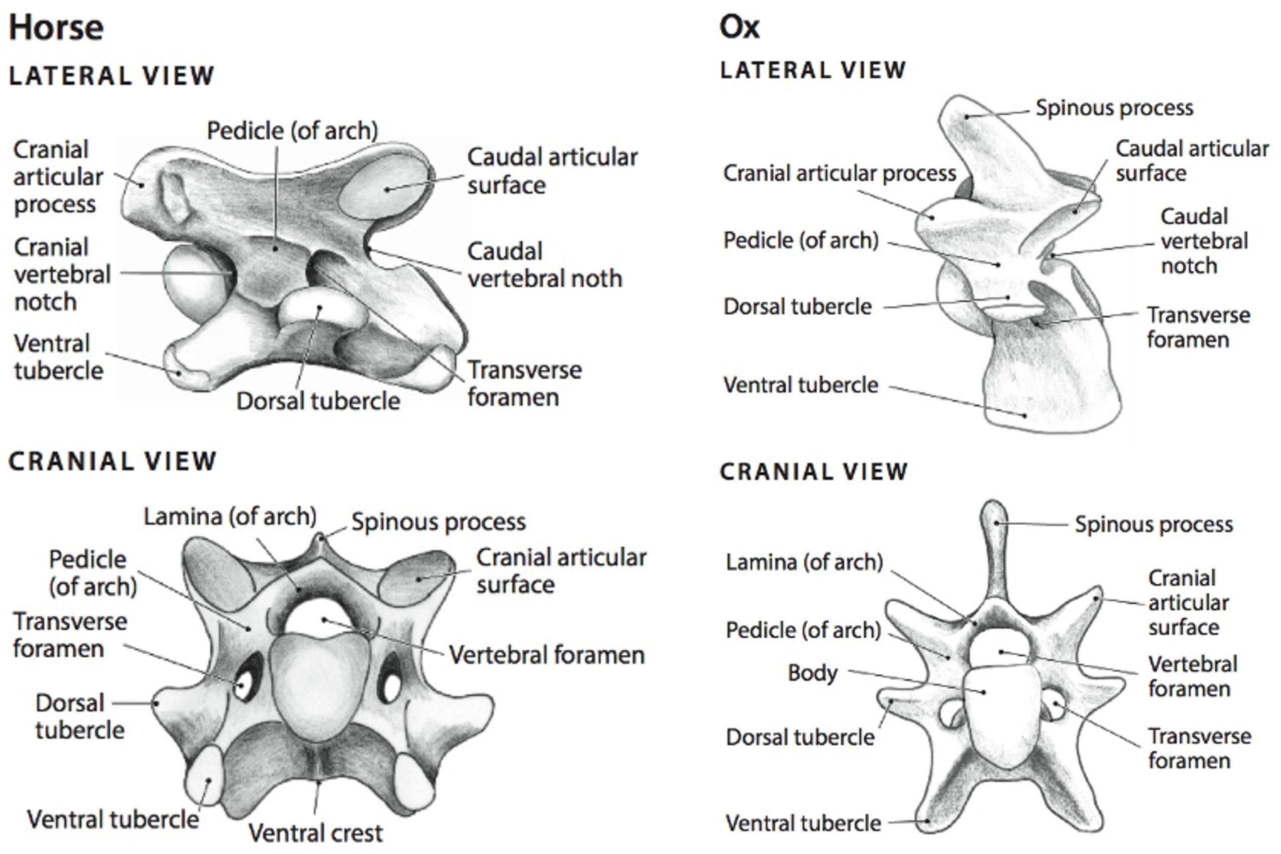
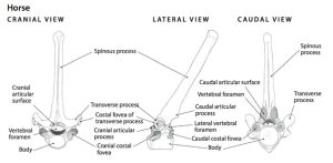
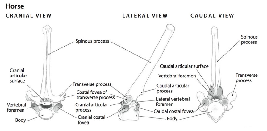
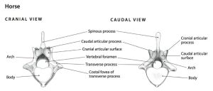
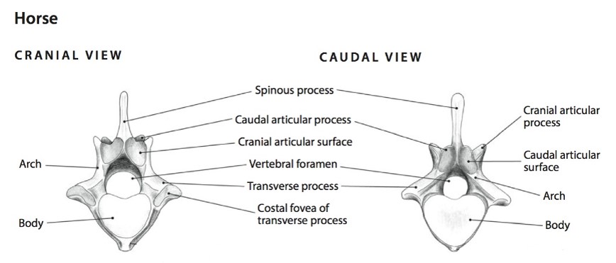
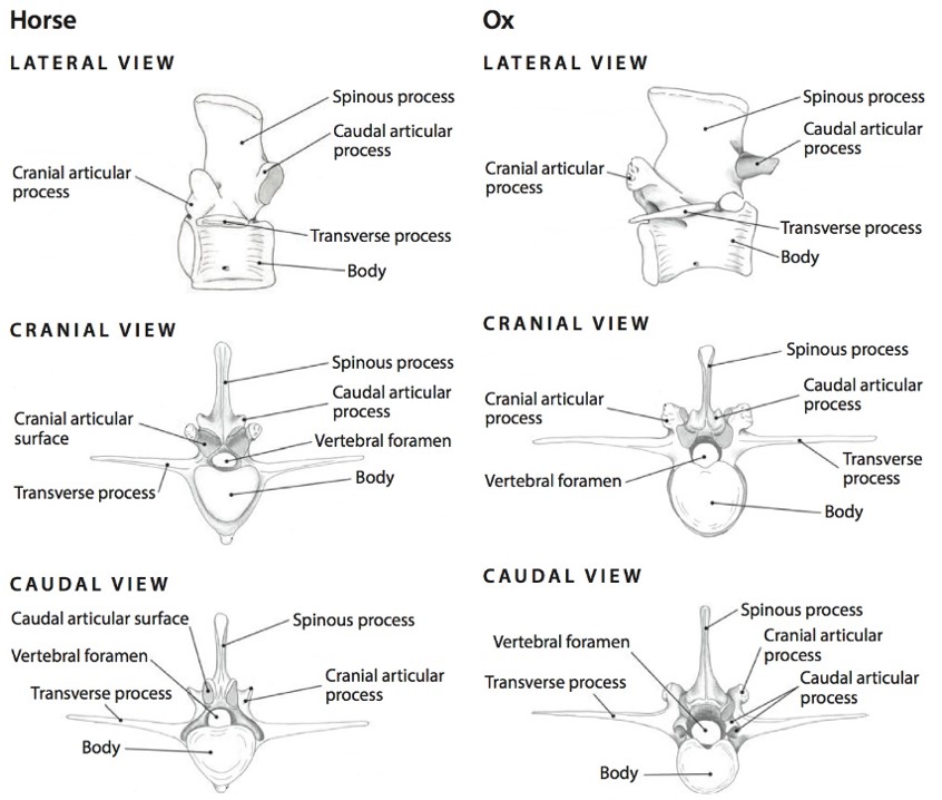
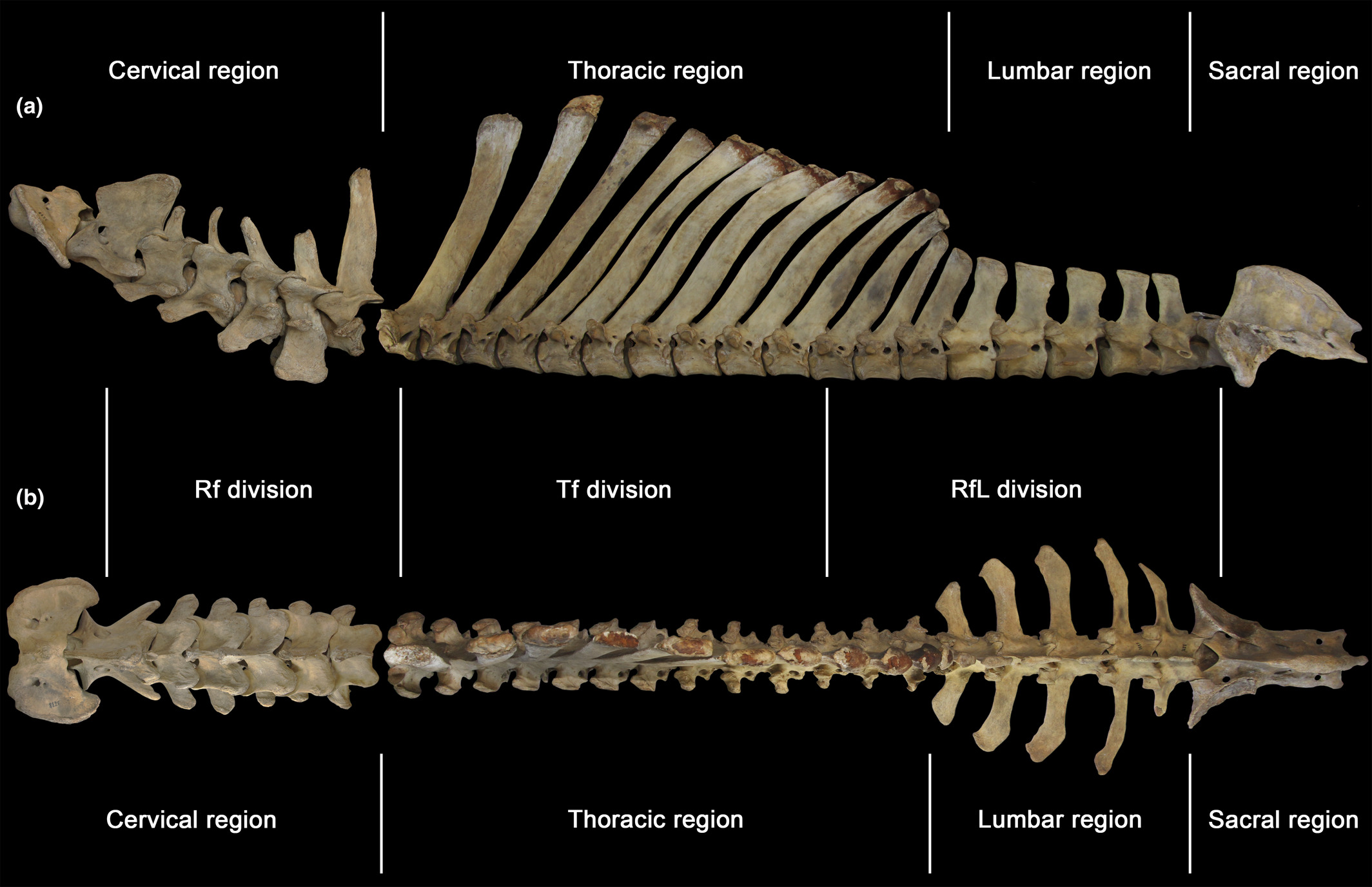
Individual vertebra, joined end to end, form the vertebral column. A vertebra consists of a body, an arch, transverse processes, a (dorsal) spinous process, and articular processes. The vertebral arch is formed by pedicles (the lateral walls) and laminae (the dorsal roof of the arch from which the spinous process projects). The large central opening through each vertebra, bordered by the body and arch, is the vertebral foramen. The continuous bony tunnel of the vertebral column, created by adjacent vertebral foramina, is called the vertebral canal. The spinal cord is located within this canal. Laterally, between consecutive vertebrae, there is a space, the intervertebral foramen, for the passageway of spinal nerves exiting the vertebral canal to pass into the peripheral tissues. The vertebral column has cervical, thoracic, lumbar, sacral and caudal subdivisions. Vertebrae within these subdivisons generally share common features. The number of vertebrae in a subdivision is consistent for the cervical region (7), and then there is variation between species for the remaining divisions. Formulas for the vertebral column are provided in the terms list and use the terms list to guide the learning of the features of vertebrae.
Carnivore vertebral column
-
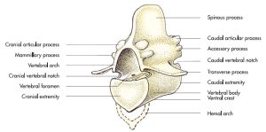
- Basic structure of a vertebra. 8
-
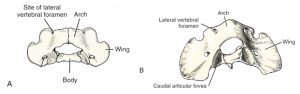
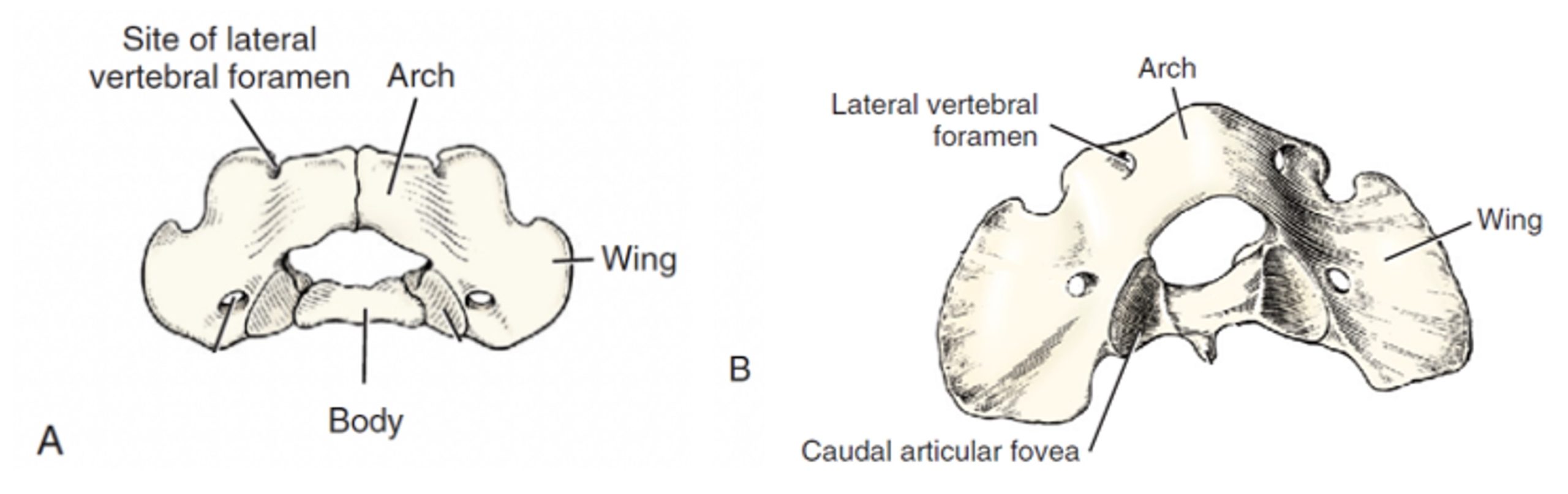
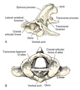
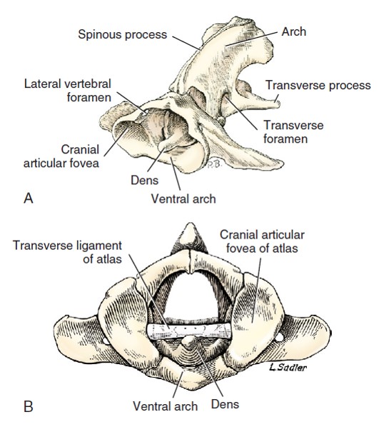
- A. Atlas of a 3.5 month old Beagle, caudodorsal view. B. Atlas of an adult dog, caudodorsal view. 3
-
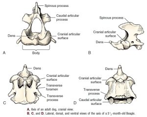
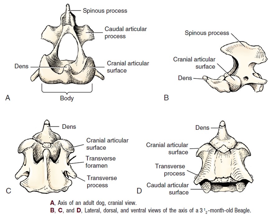
- Dog axis. 3
-
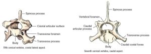
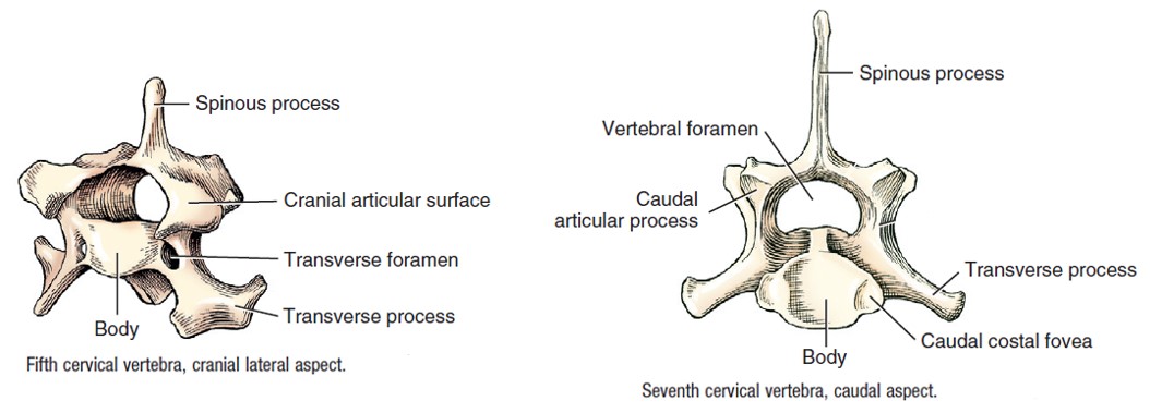
- Dog cervical vertebrae. 3
-
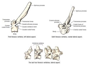
- Dog thoracic vertebrae. 3
-
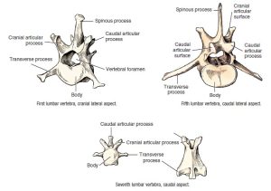
- Dog lumbar and caudal vertebrae. 3

-

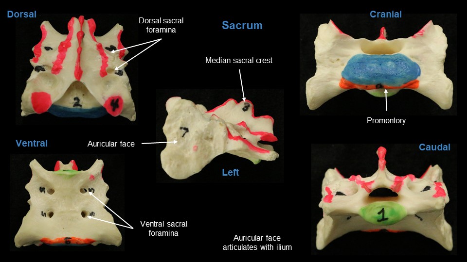
- Dog sacrum. 3
-
- Dog caudal vertebrae. 3

Canine vertebral column:
Ungulate comparative vertebral column
-
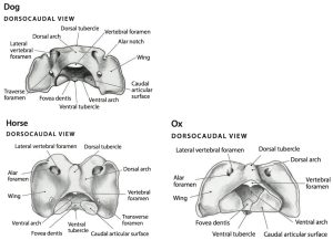
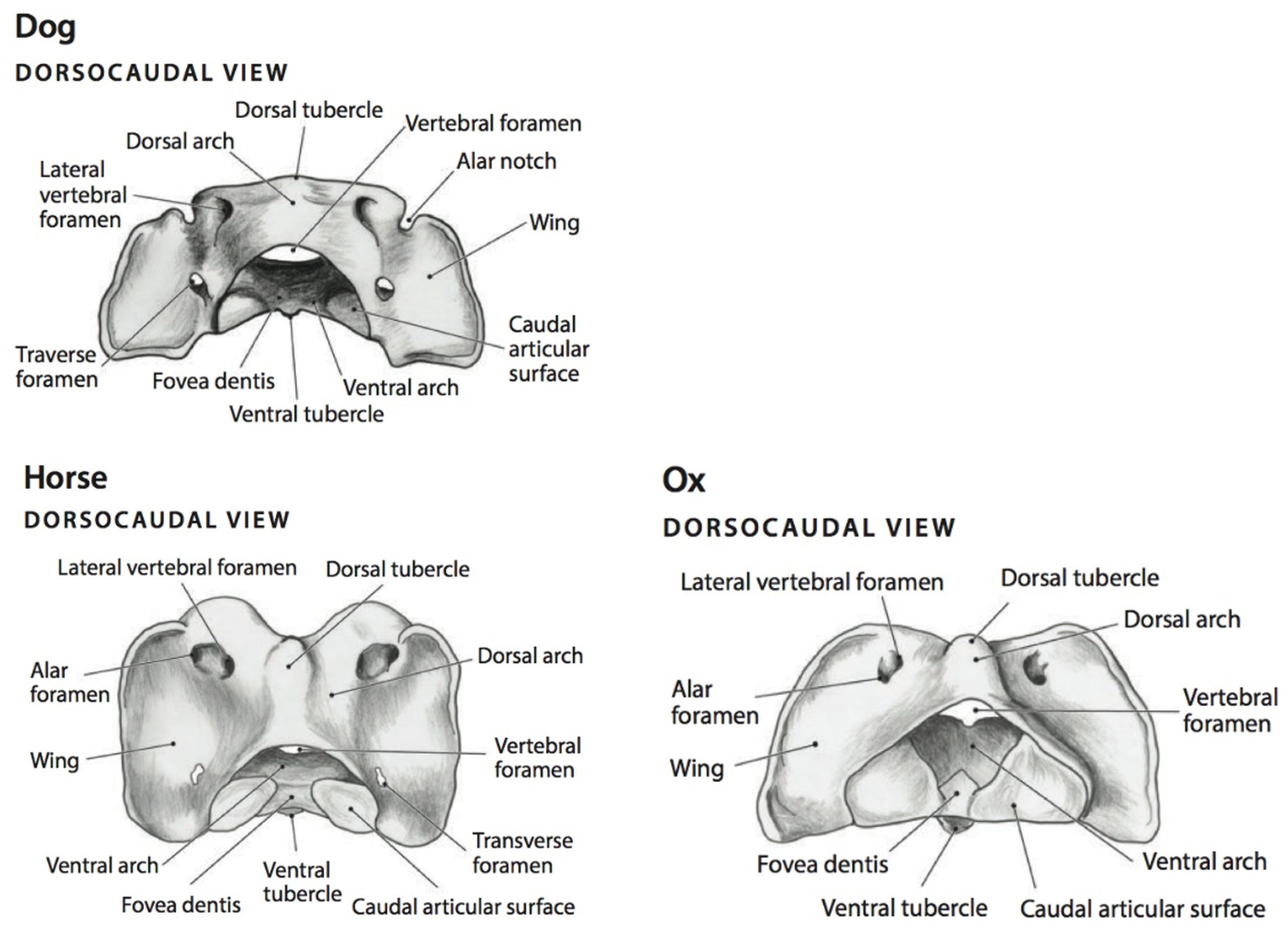
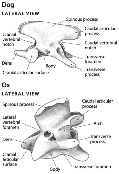
- Dorsocaudal views of the first cervical vertebra (atlas) of the dog, horse, and ox. 6
-

- Lateral views of the second cervical vertebra (axis) of the dog and ox. 6
-
-
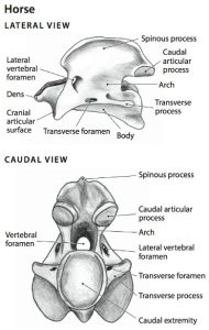
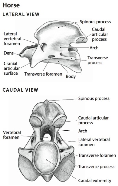
Second cervical vertebra (axis) of the horse, lateral and caudal views.
6
-
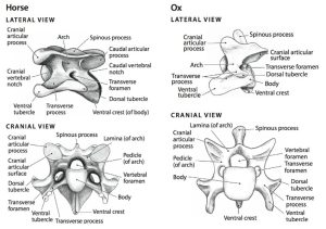
- Fifth cervical vertebra (C5) in the horse and ox.
-
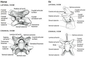
- Sixth cervical vertebra (C6) in the horse and ox. 6
-
-
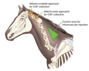
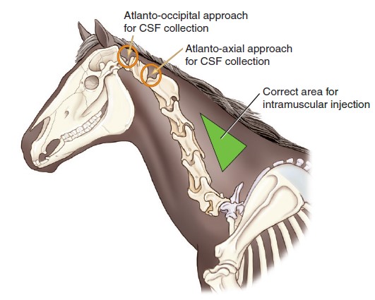
Diagram of the cervical region of the horse depicting the location of the cervical vertebrae, the “safe” area for intramuscular
injection, and the location of 2 possible approaches for cerebrospinal fluid (CSF) collection. 9
-
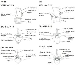
- A cranial thoracic vertebra of the horse; cranial, lateral, and caudal views. 5
-
-
A caudal thoracic vertebra of the horse, cranial and caudal views.
5
-
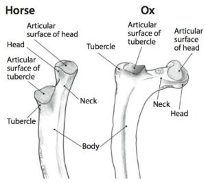
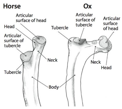
- Proximal portion of a rib of the horse and ox. 5
-
- Lumbar vertebra of the horse and ox; lateral, cranial, and caudal views. 5
-
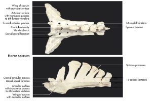
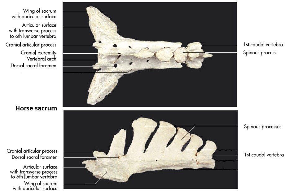
- Horse sacrum. 7
-
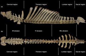
- Regions (cervical, thoracic, lumbar, and sacral) of the ox vertebral column. 20
-

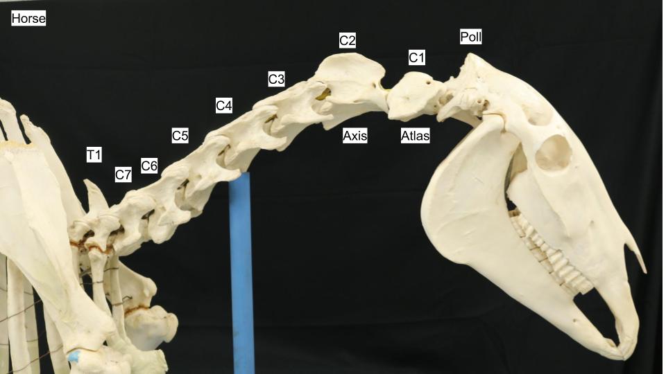
- Horse cervical vertebrae
-

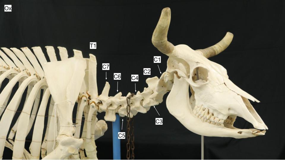
- Ox cervical vertebrae
-

- Ox cervical vertebrae

Interactive review content:
Vertebral column features
Besides muscles, the axial skeleton is supported and connected by many ligaments and specialized and standard joint structures. We will focus on identifying a few major features. The atlanto-occipital and atlantoaxial joints are synovial joints. These joints are continuous with each other through the articulation of the dens of the axis with the ‘floor’ of the atlas vertebral foramen. The dens of the axis is held in place by the transverse ligament of the atlas (present in the carnivore and pig) and a few other ligaments. The atlanto-occipital joint is a hinge joint, moving dorsally and ventrally, i.e. the “yes” joint. The atlantoaxial joint is a rotating joint, moving head side to side, i.e. the “no” joint. A dorsal atlanto-occipital ligament (a membrane anatomically, but will keep it straightforward and call it a ligament) and dorsal atlantoaxial ligament provide fibrous connections between the associated bones. When accessing the vertebral canal at these joint spaces, the dorsal ligament is penetrated.
At the C2-C3 articulation, and moving caudally, adjacent vertebrae articulate via synovial joints at the articular processes and via a fibrous joint between vertebral bodies. The fibrous joint consists of the intervertebral disk. The intervertebral disk is constructed of a central gelatinous core, the nucleus pulposus, and an outer ring of collagenous fibers, the anulus fibrosus. Recall that the developed sacrum is typically a fused unit of multiple sacral vertebra and therefore synovial joints and intervertebral disks are not present between the sacral segments.
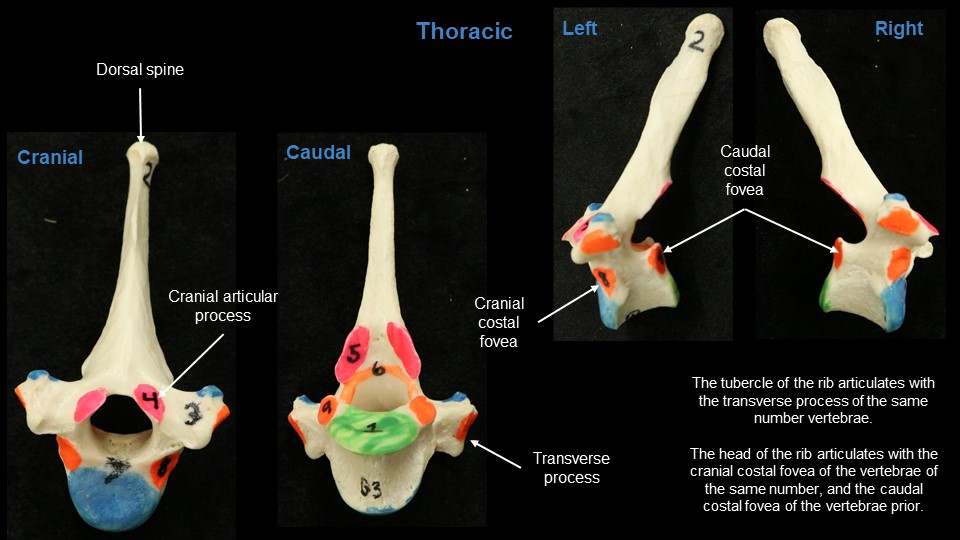
Continuing caudally from the nuchal ligament of the neck region, the supraspinous ligament connects the dorsal aspects of all vertebral spines from T1 vertebra through to the sacral vertebrae. The ligament passes from one spinous process tip to another. A reminder, the vertebral arch consists of the vertebral laminae and pedicles, forming the roof and lateral walls of the vertebral foramen. A ligament connects one vertebral arch to the next along the dorsal margin of the adjacent arches. This is the yellow ligament (ligamentum flava) or sometimes, the interarcuate ligament (‘between arches’). Lastly, in the thoracic region, the intercapital ligament extends between and left and right paired rib heads, holding the rib head to its joint. The intercapital ligament crosses dorsally over the intervertebral disk and therefore possibly helps to keep the disk in its location (i.e. disk protrusion in the thoracic vertebral column is less common where the intercapital ligament is present; for the dog this is ribs 2 to 10).
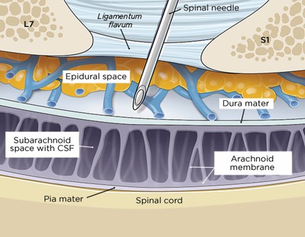
Clinical relevance: accessing the epidural space of the vertebral canal.
The supraspinous ligament is one of a few intervertebral ligaments that are punctured during insertion of a needle for an epidural or a lumbar region cerebrospinal fluid tap. The yellow ligament must also be penetrated during this procedure and this is the critical ‘resistance’ layer felt as the device is being passed into vertebral canal space (the epidural space).
Observe: Be able to describe what the supraspinous and yellow ligaments are and where they exist. Take a look at an articulated vertebral column to ensure your understanding. A few models in the lab show evidence of the ligaments. We are not dissecting this structure on the cadaver.
-
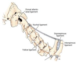
- Ligaments of the cervical region of the dog. 3

-
- A, Atlas and axis of an adult dog, cranial lateral aspect. B, Atlas and axis of a 6-month-old Beagle, cranial view. 3
-

- Lumbar intervertebral disc of a 10-week-old puppy. 3
-
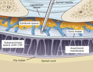
- Epidural block. Needle pierces yellow ligament.Kip Carter
-
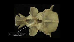
- Transverse ligament of the atlas

Review Videos
Carnivore skull osteology – watch until 22 min, ignore details on foramina for now
Carnivore hyoid bones – 2 min
Ungulate skull osteology – 17 min, ignore details on foramina for now
Cow cervical vertebrae osteology – Watch until 2:40
Terms
| Regions and spaces of the Skull | ||
| Osteology |
Comments | |
| Cranium | Part of skull that encloses the brain. | |
| cranial cavity | Created by the bones of the cranium, brain resides in this cavity | |
| calvaria | Roof of the cranium. | |
| Face (facies) | Part of skull that encloses respiratory and alimentary tracts | |
| Bones of the Skull | |||
| Osteology |
Features |
Species Differences/identify in |
|
| Bones of the cranium | |||
| Frontal bone | Cornual process | Ruminant | |
| Parietal bone | External sagittal crest | Carnivore, horse | |
| Temporal lines | Horse, ruminant | ||
| Interparietal bone – FYI only | FYI only | FYI only | |
| Occipital bone | External occipital protuberance | Carnivore, horse most prominent | |
| Occipital condyles | |||
| Paracondylar processes | |||
| Nuchal crest | |||
| Foramen magnum – where brainstem exits cranial cavity to continue as spinal cord | |||
| Basisphenoid bone | |||
| Pterygoid bone – FYI only | FYI only | FYI only | |
| Temporal bone | Squamous part | Zygomatic process | |
| Mandibular fossa | |||
| Retroarticular process | |||
| Tympanic part | Tympanic bulla | Contains tympanic cavity that is subdivided in cat by septum bulla | |
| External acoustic meatus | |||
| Petrous part | Mastoid process | ||
| Ethmoid bone | Cribriform plate | Seen in an open skull only | |
| Vomer | Singular bone of skull on ventral midline, supports nasal septum | ||
| Bones of the face | |||
| Maxilla | Facial crest | Horse | |
| Facial tuberosity | Ruminant, pig | ||
| Nasal bone | Nasoincisive notch between nasal and incisive bones | ||
| Incisive bone | |||
| Lacrimal bone | |||
| Palatine bone | |||
| Zygomatic bone | Forms zygomatic arch along with portions from temporal and frontal bones | In carnivore, pig: connects to orbital ligament. | |
| Mandible | Body | ||
| Ramus | |||
| Coronoid process | |||
| Condylar process | |||
| Angular process | Carnivore | ||
| Os rostrale (rostral bone) | supports the snout | Pig | |
| Additional features and joints of the Skull | |
| Feature | Description/Species Difference |
| Nasal aperture | bony opening to nasal cavity |
| Nasoincisive notch | created by nasal and incisive bones, prominent in ungulates. |
| Orbit | Orbital margin – consists of multiple bones combining to form rim. |
| Orbital ligament – carnivore, pig; bridges frontal to zygomatic bone, usually fibrous, may ossify in mature cats to form a complete bony rim. | |
| Zygomatic arch | Formed by zygomatic, temporal and sometimes frontal bones; species variation |
| Pterygopalatine fossa | caudoventral to orbit, skull region between maxilla and pterygoid process of basisphenoid bone |
| Temporomandibular joint | synovial joint |
| Intermandibular articulation | (= mandibular symphysis) |
|
Hyoid apparatus |
|
| Bone | Species Difference/comments |
| Tympanohyoid (not seen) | joins stylohyoid to temporal bone |
| Stylohyoid | Divides guttural pouch compartments in horse |
| Epihyoid | Carnivore, ox (vestigial in horse) |
| Ceratohyoid | Surgical removal to treat temporohyoid osteoarthropathy in horse |
| Basihyoid | |
| Lingual process | Horse and ox |
| Thyrohyoid | |
| Vertebral formula | |||||
| Cervical (C) | Thoracic (T) | Lumbar (L) | Sacral (S) | Caudal (Ca) | |
| Carnivore | 7 | 13 | 7 | 3 | 20 |
| Ruminant | 7 | 13 | 6 | 5 | variable |
| Pig | 7 | 14/15 | 6 | 4 | variable |
| Horse | 7 | 18 | 6 | 5 | variable |
| Osteology of the vertebral column | |
| Osteology | Features and comments |
| All vertebrae |
Body (except C1, which has a ventral arch instead) |
| (Dorsal) Spinous process | |
| Transverse process | |
| Articular process | |
| Vertebral foramen | |
| Arch (consists of two pedicles and a lamina) | |
| Vertebral canal | |
| Intervertebral foramen – for spinal nerves to exit vertebral canal. | |
| Cervical vertebrae | |
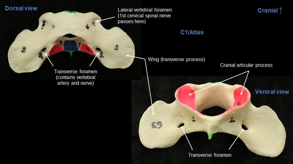
| C1 = Atlas | Wings of atlas (= transverse processes) |
| Transverse foramen – for pathway of vertebral vessels and nerve. | |
| Lateral vertebral foramen – for exit of C1 spinal nerve | |
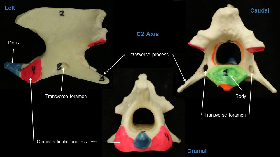
| C2 = Axis | Dens |
| Transverse foramen | |
| C3-C7 | Transverse foramen (except C7) |
| Ventral lamina of C6 (sled runners) | |
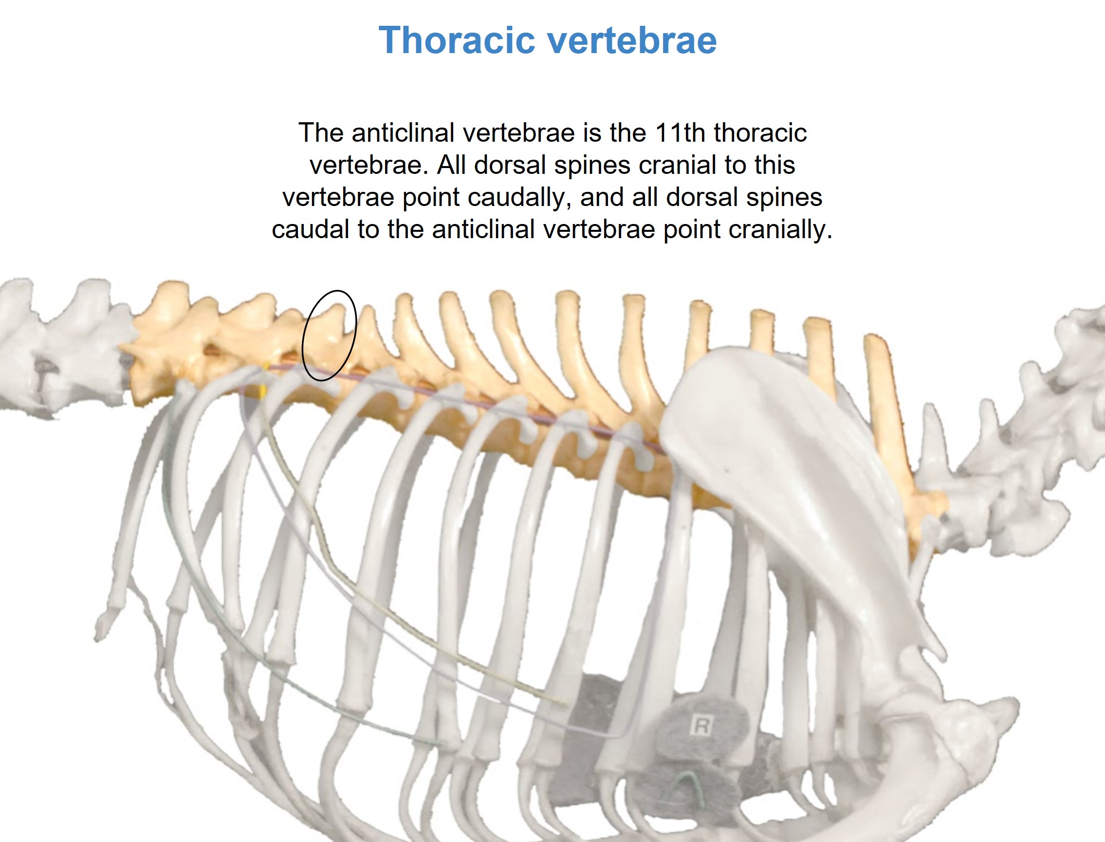
| Thoracic vertebrae |
Costal fovea |
| Anticlinal vertebra | |
| Lumbar vertebrae |
As above for all vertebra |
| Sacral vertebrae (sacrum) |
Sacral foramina – for exiting of branches of sacral spinal nerves. |
| Auricular face – articulates with ilium (to form sacroiliac joint) | |
| Promontory – ventral border of cranial base of sacrum | |
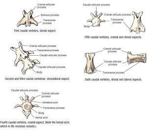
| Caudal (coccygeal) vertebrae |
Hemal arches (V shaped fusion of processes on ventral aspect of caudal vertebra, bovine and carnivore. |
|
Features of the vertebral column |
||
| Feature | Landmark | Species Difference |
|
Atlanto-occipital joint |
the ‘yes’ joint | |
| Dorsal atlanto-occipital ligament (membrane) | ||
| Atlantoaxial joint | the ‘no’ joint | |
| Dorsal atlantoaxial ligament | ||
| Transverse ligament of atlas | Examine in carnivore models. | Present in carnivore and pig. |
| Intervertebral disc | Anulus fibrosus | |
| Nucleus pulposus | ||
| Supraspinous ligament | Between consecutive tips of spinous processes of thoracic and lumbar vertebrae. | |
| Yellow ligament | between adjacent vertebral arches, dorsal margin. | |
| Intercapital ligament | between paired rib heads | for ribs 2-10 in the dog. |
Example Practical Exam Questions
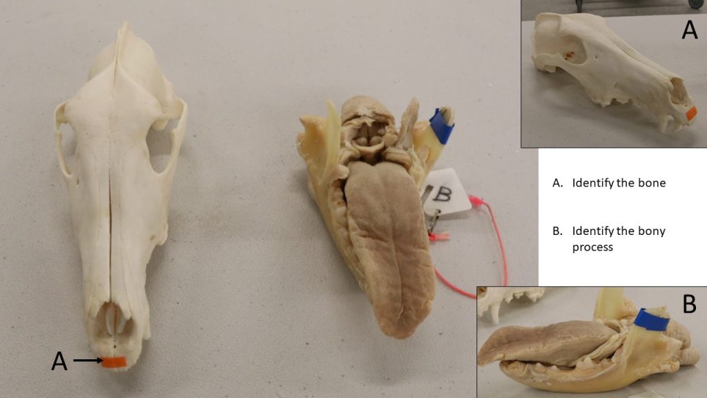
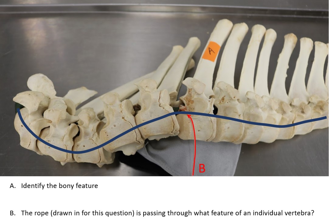
A. Identify the bony feature
B. The looped rope (drawn in) is passing through what feature of an individual vertebra?