
Clinical Application Answers
Clinical Application: Q1
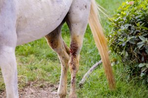
Q1: A horse has a deep laceration across its gaskin. Between what two joints is the gaskin?
A. coxofemoral and stifle
B. stifle and hock
C. tarsus and fetlock
D. pastern and coffin
Answer:
B. stifle and hock
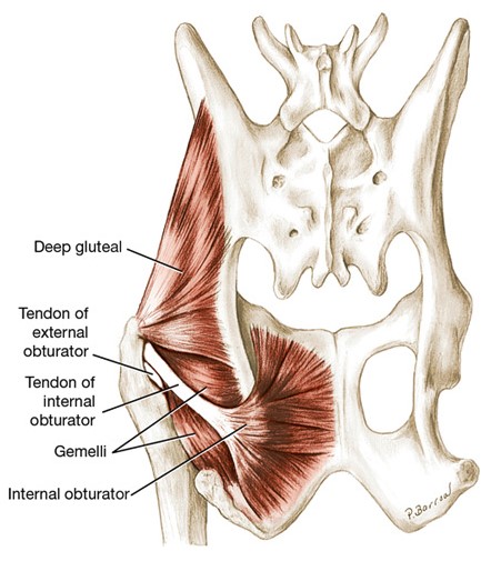
Clinical Application: Q2
One muscle that attaches to the greater trochanter of the femur is the:
A. internal obturator m.
B. superficial gluteal m.
C. deep gluteal m.
D. biceps femoris m.
Answer:
C. deep gluteal m.
Clinical Application: Q3
Q3: what does femoral n. paralysis look like?
Answer:
Femoral n. supplies motor innervation to the quadriceps femoris m. Femoral n. neuropathy (dysfunction) results in an animal’s inability to effectively keep its stifle extended (action of quadriceps femoris m.) and therefore it cannot normally bear weight on the affected limb.
Critical note: A femoral neuropathy or neuromyopathy can occur in horses that are in dorsal recumbency under general anesthesia, and have had their pelvic limb(s) left or held in extension for a prolonged period.
Clinical Application: Q4
Q7: Which muscle and its tendon is partially removed as a surgical aid to managing stringhalt? [see clinical relevance above, refer to lecture notes, see Appendix for answer].
A – long digital extensor m.
B – fibularis tertius m.
C – lateral digital extensor m.
D – gastrocnemius m.
Answer: C – lateral digital extensor m.
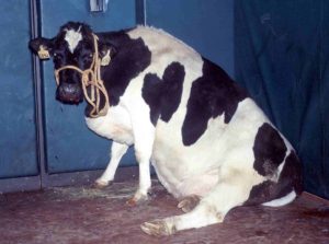
Clinical Application: Q5
Q6: This dairy cow can not rise beyond this point and shows a ‘rabbit leg’ position of both hind limbs. The cow is unable to extend her hocks and weight bear. Rupture of what muscle in the caudal crus results in this clinical presentation?
A: deep digital flexor m.
B: gastrocnemius m.
C: superficial digital flexor m.
D: popliteus m.
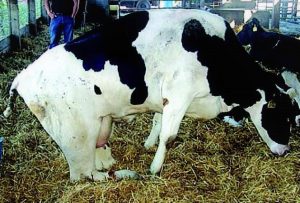
Answer: B. gastrocnemius m. rupture results in ‘rabbit leg’ appearance in this dairy cow.
Clinical Application: Q6
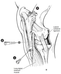
Q6: What stifle joint sac (compartment) has been entered by the needle C, as shown in the image?
A: lateral femorotibial joint
B: medial femorotibial joint
C: femoropatellar joint
Answer: A. lateral femorotibial joint.
How to Perform Arthrocentesis of the Compartments of the Stifle of the Horse – AAEP, Schumacher J., Wilhite, R.
Clinical Application: Q7
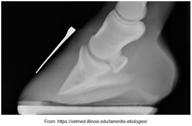
Q7: Review the radiograph above.
Normally the dorsal surface of the distal phalanx is parallel to the dorsal hoof wall.
What is evident in this radiograph?
Why has it occurred?
What is the metallic structure lying on the dorsal hoof wall?
Why was it positioned there before taking the radiograph?
Answer: Evident: The dorsal surface of the distal phalanx diverges from the dorsal hoof wall surface – they are no longer parallel aligned; Why occurred: because of loss of the epidermal/dermal bond between the hoof wall and the underlying dermis, with consequent rotation of the distal phalanx (due to the pull of the deep digital flexor tendon inserted on the distal phalanx sole surface); Metallic structure: a horseshoe nail; Why positioned there: to aid visualization of the dorsal hoof wall surface to help determine if any foot rotation has occurred.