MSK LAB 17A – Pelvic Limb – caudal and medial thigh muscles
Learning Objectives
- Identify muscles of the caudal and medial thigh
- Name the attachments of the muscles of the caudal and medial thigh
- Name the action(s) of the muscles of the thigh, as noted in muscle chart
- Categorize muscles into “muscle action groups”
- Identify the femoral triangle
- Connect structures to the clinically applied anatomy as emphasized.
Lab instructions:
For this lab, teams will focus primarily on dissecting the muscles of the proximal pelvic limb on the caudal and medial aspects, in canine and feline cadavers. This carnivore understanding can then be applied to the prosected large animal cadavers to identify the same muscles (and any extras) as directed.
Before we dissect, a reminder from the Thoracic Limb section:
What do we mean by muscle attachments?
All muscles have attachments. In most instances, the more proximal attachment is considered the origin. The insertion is the more distal attachment, and this end is the limb part that typically moves the most when the muscle contracts. The origin is usually a direct attachment of the muscle cells to the bone. For our purposes, you are not responsible for learning which attachments are the origin versus the insertion. At times, however, you may be asked for the distal or proximal attachment of a muscle or muscles. The insertion often is by a tendon or aponeurosis extending from the muscle cells to the bone. A tendon consists of dense, regularly arranged fibrous connective tissue organized into a small, well-defined bundle. An aponeurosis has the same consistency as a tendon, but the fibrous tissue is arranged as a thin sheet of tissue (i.e. take a round-bodied tendon and flatten it out with a rolling pin like dough rolled out for a pie crust, and you have made an aponeurosis!). A ligament typically refers to a defined dense fibrous connective tissue band between bones, and the term is also used for a variety of thin fibrous connections between organs or between an organ and the body wall (will observe these visceral ligaments in the thoracic and abdominal cavities).
MUSCLE CHARTS: You are responsible for knowing the attachments and action(s) of muscles as described in the muscle action charts. Use additional information in the following descriptions as a reference.
As always, read before you continue dissection! In many instances, during the study of muscles, a description of a specific muscle will be given before the instructions for dissection. Follow instructions as carefully as possible. In each instance clean the exposed surface of the muscle being described, isolate its borders, and verify its attachments. If the muscle is to be “transected,” free it from underlying structures first. As each muscle is defined, try to visualize its attachments and position on the skeleton, and understand its function.
Ready? Then lets go! All muscle dissection is performed on the left side of the body.
The following muscles will be dissected in this lab:
Caudal thigh
- Biceps femoris m.
- Semitendinosus m.
- Semimembranosus m.
Medial thigh
- Sartorius m.
- Gracilis m.
- Pectineus m.
- Adductor m.
Comments on fascia
There are superficial and deep fasciae of the pelvic limb. They cannot always be separated, and, in general, the deep fascia is more dense than the superficial fascia.
The superficial fascia of the trunk continues dorsally on the pelvis as the superficial gluteal fascia. In obese specimens, much fat is present between this fascia and the deep fascia.
A thick deep fascia covers the dorsal muscles of the lumbar area, pelvis, and tail. Called the thoracolumbar fascia (we have already met this fascia in previous sections), it is well developed in the lumbar region and is continued caudally at the iliac crest by the deep gluteal fascia. This distinct glistening fascia covers the muscles of the pelvis and serves in part as origin for the middle and superficial gluteal mucles. The deep gluteal fascia is continued caudally on the tail by the deep caudal fascia. The latter follows the irregularities of the caudal muscles and closely binds these to the vertebrae of the tail.
Distally, the deep gluteal fascia blends with the fascia of the thigh, where it becomes the medial and lateral femoral fasciae. The medial fascia is thin, whereas the lateral femoral fascia, also known as the fascia lata, is thick and serves as an aponeurotic insertion for thigh muscles. The femoral fascia is continued on the leg as the crural fascia.
Dissect: If not done so during the dissection of the epaxial muscles, transect and reflect the superficial and deep gluteal fasciae. For now, keep the medial thigh fascia and the lateral thigh fascia aka the fascia lata intact.
Caudal muscles of the thigh – carnivore dissection
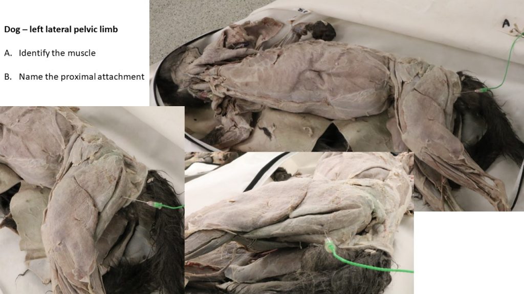
The caudal thigh muscle group consists of three primary muscles: the biceps femoris, laterally; semitendinosus, caudally; and semimembranosus, medially. (A fourth muscle, the slender caudal crural abductor muscle is closely associated with the caudomedial surface of the biceps femoris. It is of passing interest only.)
Biceps femoris m.
The biceps femoris m. is the longest and widest of the muscles of the thigh. The biceps femoris m. attaches proximally on the lateral aspect of the tuber ischii and the sacrotuberous ligament that extends from the tuber ischii to the sacrum. NOTE: The cat does not possess a sacrotuberous ligament. The sacrotuberous ligament will be dissected later in canine cadavers, with the lateral muscles of the pelvis. Cranially and distally, the biceps femoris m. inserts by means of the fascia lata and crural fascia. Caudally, a band of heavy fascia extends from the biceps femoris m. to the tuber calcanei of the tarsus and helps to form the common calcaneal tendon.
Observe: Identify the biceps femoris m.
The lymph node, typically surrounded by fat tissue, at the caudal border of the biceps femoris m. directly caudal to the stifle, is the popliteal lymph node.
The proximal attachments of the biceps femoris will be more apparent after lateral pelvic muscles are dissected.
PROXIMAL ATTACHMENTS: Sacrotuberous ligament in canine (sacrosciatic in ungulates) and ischiatic tuberosity
DISTAL ATTACHMENTS: Lateral stifle, tibia and tuber calcanei
ACTION: Flex the stifle; extend the hip (Secondary: extend stifle and tarsus, abduct hip)
Dissect: Carefully clean the caudal border and the adjacent surface of the biceps femoris m. For now, do not incise the fascia lata insertion on the cranial border of the biceps femoris. It may be incised later in the dissection if needed. Clean the fat around the popliteal lymph node and identify the structure. Undermine and then transect the biceps femoris m. in the middle of the muscle and reflect the transected parts proximally and distally. There is a long thin muscle – the caudal crural abductor m. – directly deep to biceps femoris m. (and often adhered to it) that is not studied. This muscle may be removed as necessary. The large sciatic nerve, now exposed, is interposed between the biceps femoris and the adductor muscles. It will be revisited in the nervous system unit.
-

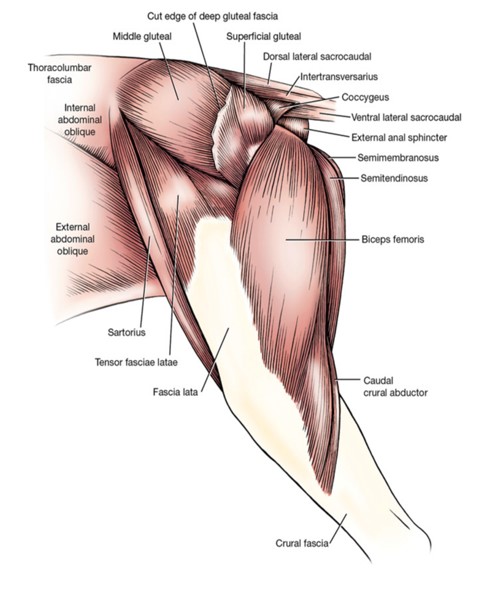
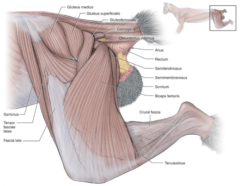
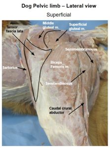
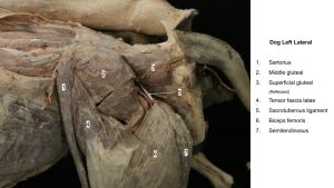
- Dog superficial muscles of left thigh, lateral view. 1
-
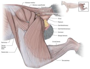
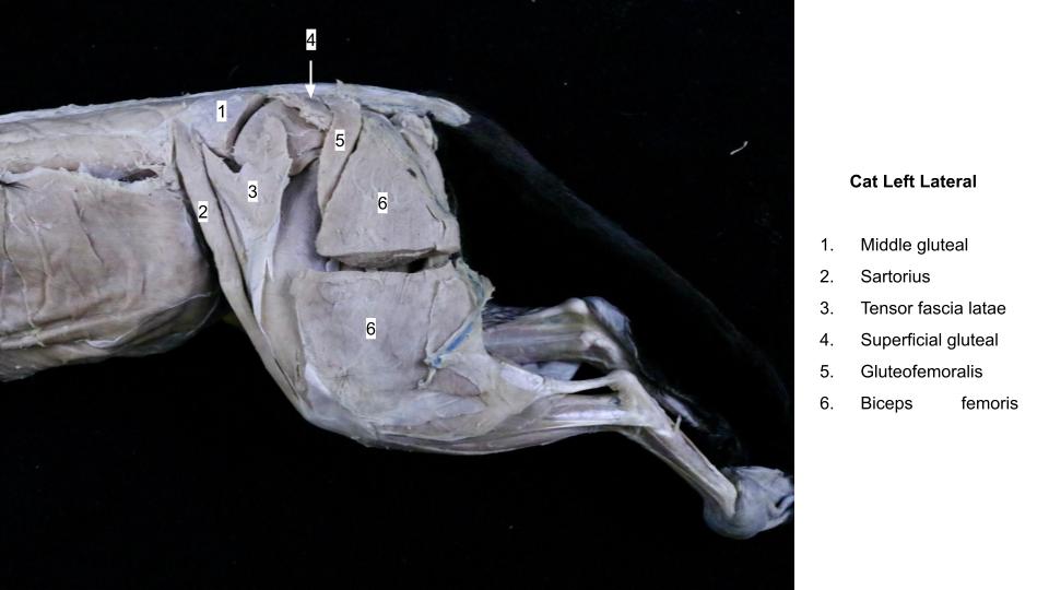
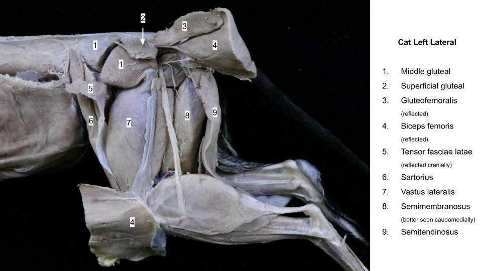
- Superficial muscles of the left hind limb of the cat. 5
-
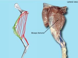
- Dog biceps femoris. 40

-

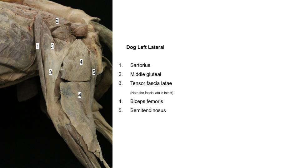
- Dog thigh
-
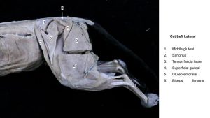
- Cat thigh
-
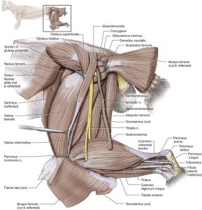
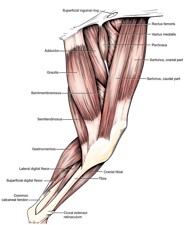
- Dog lateral thigh J. Barnes

Semitendinosus m.
The semitendinosus m., near its proximal attachment, lies between the biceps femoris and semimembranosus mm. Near its distal attachment it lies on the medial head of the gastrocnemius m. and is covered by the gracilis m.. The muscle extends principally from the ischiatic tuberosity to the tibial body. By means of the crural fascia, it also attaches to the tuber calcanei, contributing to the common calcaneal tendon.
PROXIMAL ATTACHMENT: Ischiatic tuberosity (all species) plus vertebrae and sacrosciatic lig. (horse and pig)
DISTAL ATTACHMENT: Tibia and tuber calcanei
ACTION: Flex the stifle; extend the hip. (Secondary: extend the tarsus)
Dissect: Clean the margins and use blunt dissection to free the semitendinosus muscle from surrounding structures.
-
- Dog superficial muscles of left thigh, lateral view. 1
-
- Superficial muscles of the left hind limb of the cat. 5
-
- Deep muscles of the hind limb of the cat in lateral view. 5

-
- Dog thigh
-
- Dog thigh
-
- Cat thigh
Semimembranosus m.
The semimembranosus m. is greater in cross sectional area than the semitendinosus but is not as long. It is wedged between the semitendinosus and biceps femoris mm. laterally and the gracilis and adductor mm. medially. It has two bellies of nearly equal size (do not separate these). This short, thick muscle extends from the ischiatic tuberosity to the medial side of the distal end of the femur and the proximal end of the tibia. The insertions may be seen more adequately after the sartorius and gracilis muscles have been dissected.
PROXIMAL ATTACHMENT: Ischiatic tuberosity
DISTAL ATTACHMENT: Distal femur and medial condyle of the tibia
ACTION: Flex the stifle; extend the hip. (Secondary: extend the stifle)
Dissect: Clean the margins and use blunt dissection to free the semimembranosus muscle from surrounding structures.
-
- Dog superficial muscles of left thigh, lateral view. 1
-
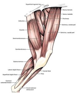
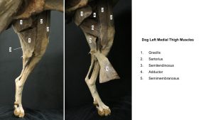
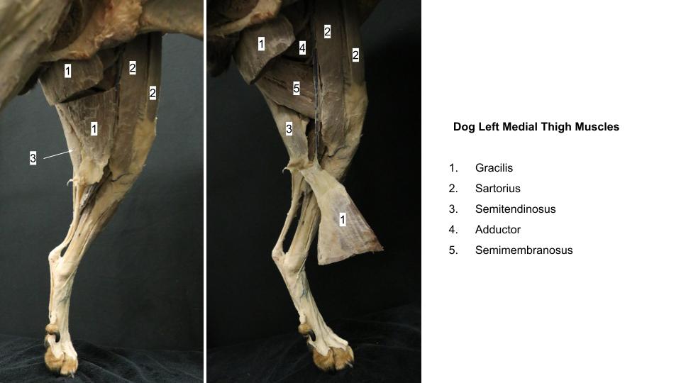
- Medial thigh and crus of the dog. 1
-
- Superficial muscles of the left hind limb of the cat. 5
-
- Deep muscles of the hind limb of the cat in lateral view. 5
-
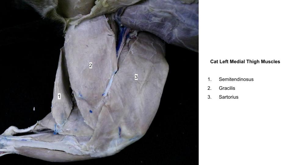
- Superficial muscles of the left hind limb of the cat, medial. 5
-
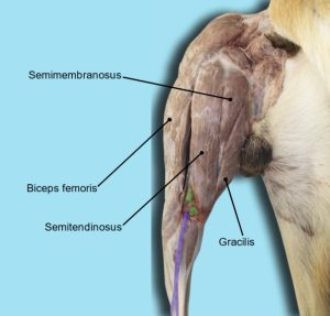
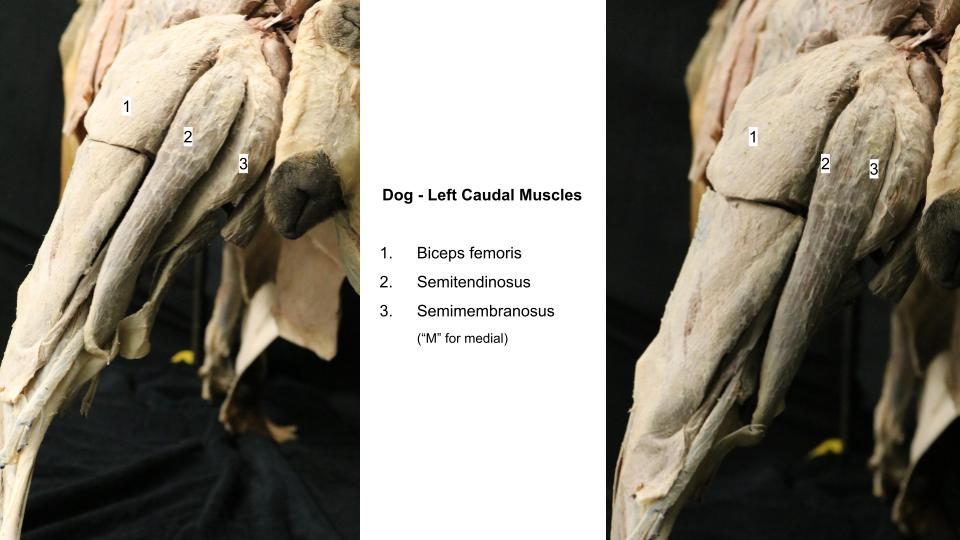
- Dog caudal thigh. 40

-
- Dog thigh
-
- Cat thigh
-

- Cat thigh
Medial muscles of the thigh – carnivore dissection
Sartorius m.
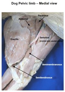
The sartorius m. consists of two strap-like parts in the canine that lie on the cranial and craniomedial surfaces of the thigh. These parts extend from the ilium to the tibia. The cranial part forms the cranial contour of the thigh and is relatively thick. The caudal part is on the medial side of the thigh and is thinner, wider, and longer than the cranial part. Both muscle parts lie predominantly on the medial side of the large quadriceps femoris. In the feline, the sartorius m. is not subdivided into two bellies.
PROXIMAL ATTACHMENT: Ilium
DISTAL ATTACHMENT: Cranial part: patella; Caudal part: tibia
ACTION: Flex the hip (Secondary: cranial part extends the stifle; caudal part flexes the stifle)
Dissect: Clean the margins and use blunt dissection to free the sartorius muscle from surrounding structures.
-
- Dog superficial muscles of left thigh, lateral view. 1
-
- Medial thigh and crus of the dog. 1
-
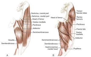
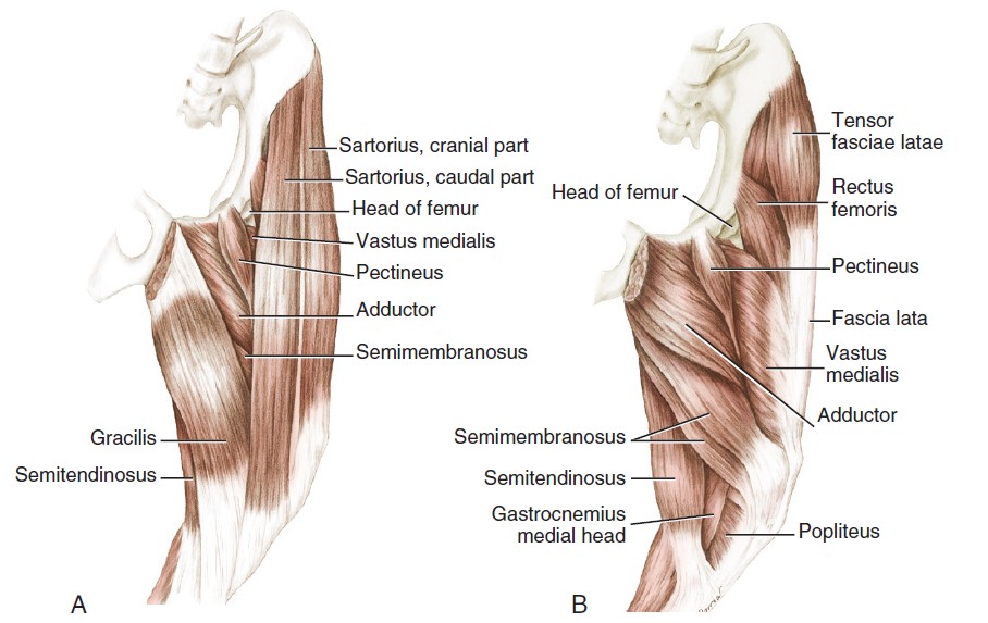
- Muscles of the thigh of the dog. A, Superficial muscles, medial aspect. B, Deep muscles, medial aspect. 3
-
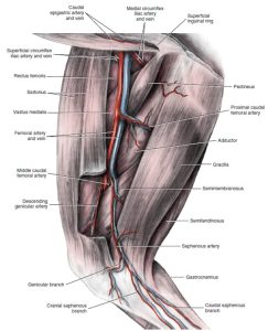
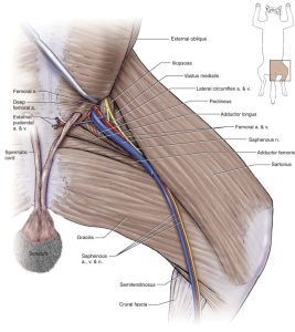
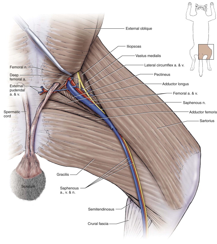
- Arteries and veins of the thigh of the dog, superficial dissection, medial aspect. 1

-
- Superficial muscles of the left hind limb of the cat. 5
-
- Superficial muscles of the left hind limb of the cat, medial. 5
-
- Dog thigh
-
- Dog thigh

-
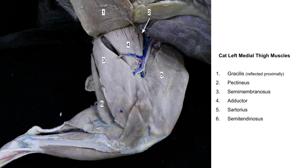
- Cat thigh
-

- Cat thigh
-

- Dog gracilis. 40
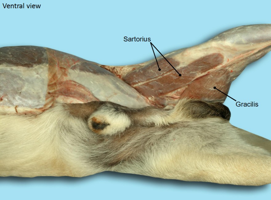
Gracilis m.
The gracilis m. arises from the symphysial tendon, a thick, flat tendon attached ventrally to the pelvic symphysis (i.e. to the ventral midline of pelvis). The proximal aponeurosis of the gracilis m. covers the adductor m.
PROXIMAL ATTACHMENT: Pelvic symphysis
DISTAL ATTACHMENT: Tibia and tuber calcanei
ACTION: Adduct the pelvic limb (Secondary: flex the stifle, extend hip and tarsus)
Dissect: Isolate the muscle borders and then transect the gracilis at its mid-point and reflect the proximal and distal portions.
-
- Medial thigh and crus of the dog. 1
-
- Muscles of the thigh of the dog. A, Superficial muscles, medial aspect. B, Deep muscles, medial aspect. 3
-
- Arteries and veins of the thigh of the dog, superficial dissection, medial aspect. 1
-
- Superficial muscles of the left hind limb of the cat, medial. 5
-
- Dog gracilis. 40
-
- Dog thigh
-
- Cat thigh
-
- Cat thigh
-
- Dog medial thigh J. Barnes

Pectineus m.
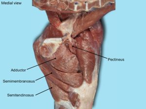
The pectineus m. is a small, spindle-shaped muscle that belongs to the deep medial muscles of the thigh. It lies between the adductor m. caudally and the vastus medialis belly of the quadriceps femoris m. cranially. It has a proximal attachment on the prepubic tendon and iliopubic eminence. There is a small cartilage embedded in the iliopubic origin.
PROXIMAL ATTACHMENT: Ilium and pubis
DISTAL ATTACHMENT: Distomedial femur
ACTION: Adduct the pelvic limb
Dissect: Isolate the thin tendon of insertion of the pectineus muscle on the caudomedial surface of the distal end of the femur and separate the muscle from the adjacent adductor muscle. Note that in the cat, a separate slip of muscle is often inadvertently dissected, lying ‘between’ the adductor and pectineus muscles. This slip is part of the adductor muscle.
-
- Medial thigh and crus of the dog. 1
-
- Muscles of the thigh of the dog. A, Superficial muscles, medial aspect. B, Deep muscles, medial aspect. 3
-
- Arteries and veins of the thigh of the dog, superficial dissection, medial aspect. 1
-
- Superficial muscles of the left hind limb of the cat, medial. 5
-
- Dog medial thigh. 40

-
- Cat thigh
Adductor m.
The adductor m. consists of two muscle bellies (adductor magnus et brevis and adductor longus) that are often not clearly divisible, and we do not attempt to separate them. It is a large pyramidal muscle compressed between the semimembranosus and pectineus, and it extends from the pelvic symphysis to the caudal aspect of the femur. It is partly covered by the biceps femoris m. laterally and gracilis m. medially.
PROXIMAL ATTACHMENTS: Pelvic symphysis, ischium and pubis
DISTAL ATTACHMENT: Distocaudal femur
ACTION: Adduct the pelvic limb (Secondary: extend the hip)
-
- Medial thigh and crus of the dog. 1
-
- Muscles of the thigh of the dog. A, Superficial muscles, medial aspect. B, Deep muscles, medial aspect. 3
-
- Arteries and veins of the thigh of the dog, superficial dissection, medial aspect. 1
-
- Superficial muscles of the left hind limb of the cat, medial. 5
-
- Dog thigh
-
- Cat thigh
Now that we have dissected the medial muscles of the thigh an anatomical feature, the femoral triangle, should be viewed and understood (and it will be revisited in future units).
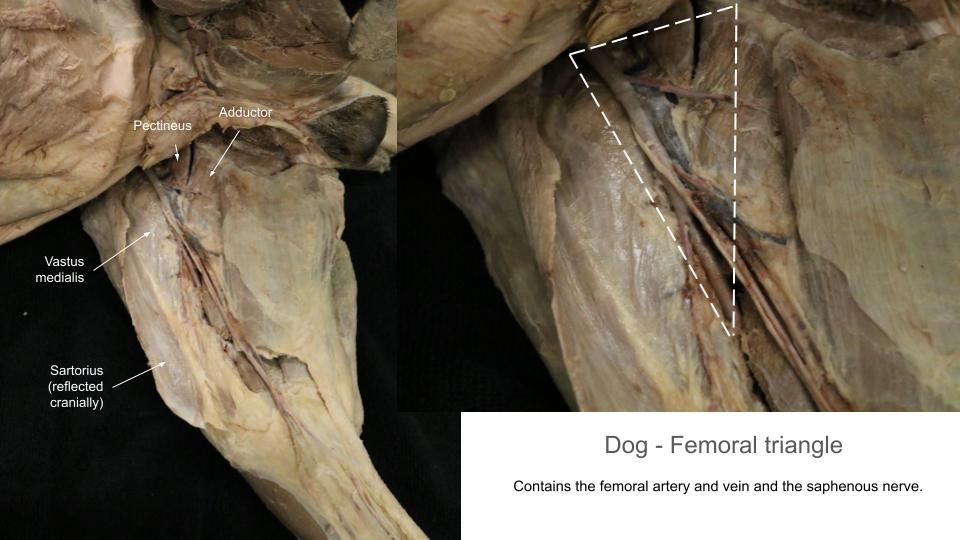
Femoral Triangle
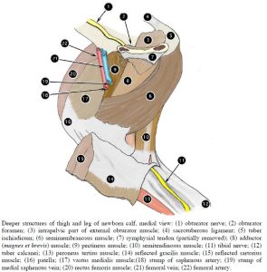
The femoral triangle is a ‘space’, bound mostly by muscles, through which the blood vessels run to and from the pelvic limb. It is located on the proximal medial surface of the thigh with its base at the abdominal wall (at the level of the vascular lacuna) and the apex of the triangle pointed distally. The triangle is bound by the sartorius m. cranially and the pectineus and adductor mm. caudally. The iliopsoas and rectus femoris mm. (to be dissected) form the proximal lateral surface of the triangle. The vastus medialis m. forms the distal lateral part. This triangle contains, among other structures, the prominent femoral artery and vein and the saphenous nerve. In some specimens, the femoral nerve also may be observed in the femoral triangle as it emerges from the iliopsoas muscle.
Observe: Confirm the boundaries of the femoral triangle and, after removing fascia and adipose tissue in the triangle, view the femoral artery and vein and the saphenous nerve. Dissection of these vessels and nerve will be revisited in future units.
Clinical Application: femoral triangle and femoral a. pulse detection.
The pulse of the carnivore may be determined by palpating the femoral artery as it passes through the femoral triangle. Important clinically, when the heart beats and no corresponding femoral pulse is felt, this may be an indication that there is obstruction to the blood flow into the artery (a blood clot at the aortic trifurcation, known as a “saddle thrombus”, may be a cause).
Ungulate comparative anatomy
Observe: Refer to the prosected ungulate cadavers and additional specimens and models to identify and study the following content. Certain muscles have been transected midbody or at their attachments to facilitate viewing deeper structures.
Croup versus Rump
Observe: Identify the gluteal region of the cadavers and relate it to the bony pelvis.
The same area is referred to as the croup in the horse, and the rump in ruminants and the pig.
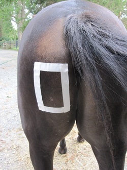
Clinical relevance: croup/rump and intramuscular injection
Intramuscular injections may be performed in the croup or rump region.
Caudal muscles of thigh
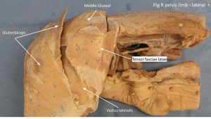
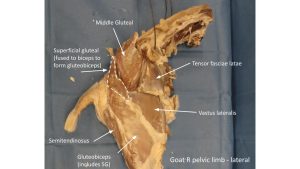
In the ruminant and pig (artiodactyls), the superficial gluteal muscle combines with the biceps femoris muscle to form a large muscle mass, the gluteobiceps muscle. In the horse the superficial gluteal m. remains separate from the biceps femoris m.
Observe: Identify the gluteobiceps m. in the ruminant and pig. Identify the superficial gluteal m. in the horse (attaches to the very prominent third trochanter). These muscles will be revisited in the next lab.
-
-
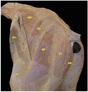
Superficial muscles of equine lateral thigh and gluteal
region . MG middle gluteal; SG superficial gluteal; BF biceps
femoris; ST semitendinosus; SM semimembranosus; TFL tensor fasciae latae; FL fascia lata. 16

-
- Caudolateral horse thigh. 16
-
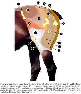
- Superficial structures of croup, thigh, and leg of horse, left lateral view. 2

-
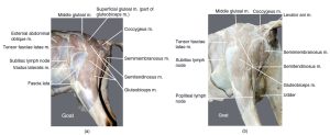
- Hip and thigh muscles of a goat, left lateral view (a). Hip and thigh muscles of a goat, left caudolateral view (b). 12
-
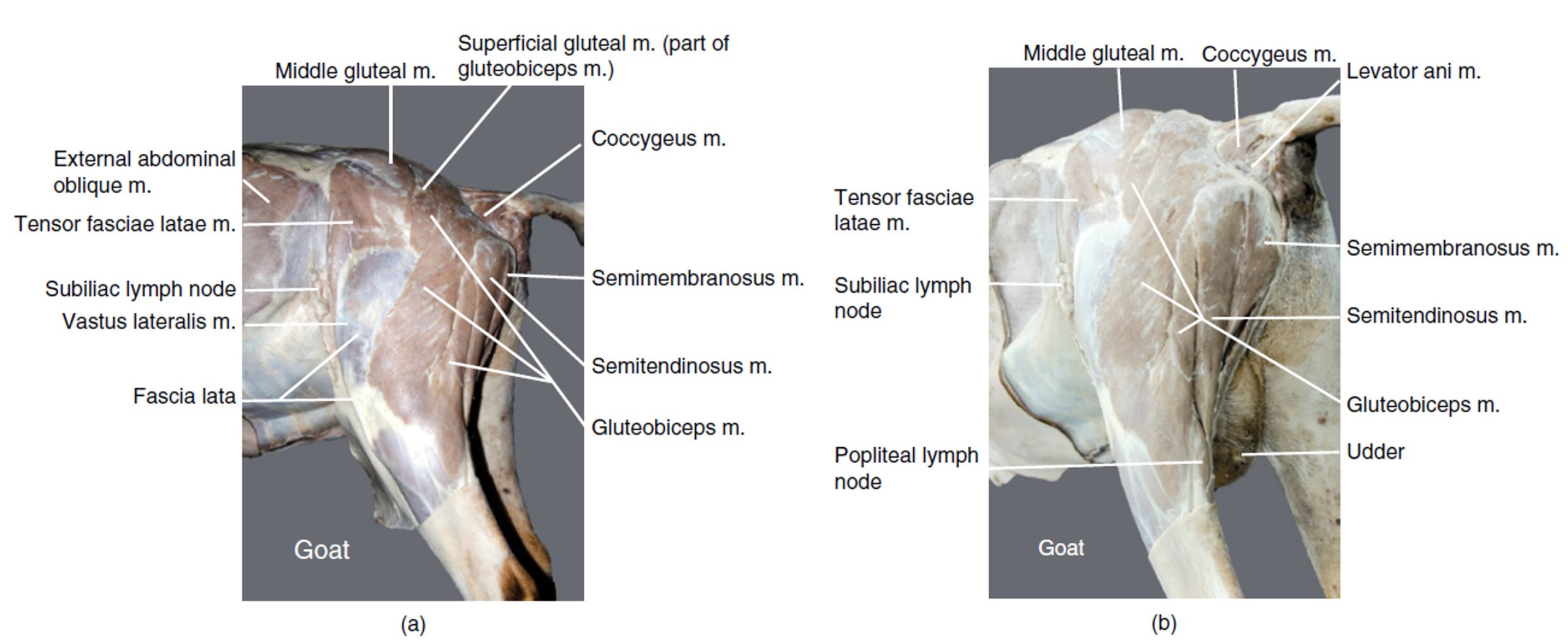
- Superficial structures of rump, thigh, and leg of newborn calf, right lateral view. 2

-
- Pig thigh

-
- Goat thigh

Clinical relevance: third trochanter fracture in horse
Fracture of third trochanter in horse.
FYI – From AVMA: Fractures of the third trochanter in horses: 8 cases (2000–2012)
Observe: We will return to the middle and deep gluteal mm. in the next lab. For now we will continue with the caudal thigh region.
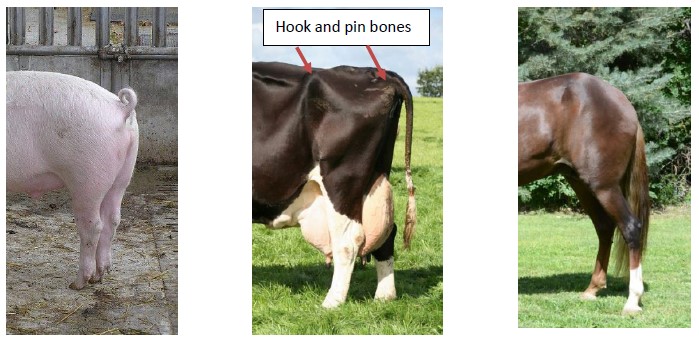
The gluteobiceps m. in artiodactyls has a cranial and caudal division in the biceps femoris part of the muscle. In the horse, the biceps femoris m. has three divisions: cranial, middle, and caudal. The divisions in the biceps are notable distally. Caudal to the biceps muscle mass, the semitendinosus m. and semimembranosus m. are present. These latter two muscles have different proximal attachments in the horse and pig (ischium and vertebrae) versus the ruminant (ischium only), which accounts for the rounded croup of the horse and rump of the pig, versus the angular rump of the ruminant.
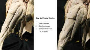
Observe: Identify the borders of the biceps femoris m. (gluteobiceps m. in artiodactyls) and its distal subdivisions. Identify the semitendinosus m. (ST) and semimembranosus m. (which lies “M” for medial to the ST), noting the different proximal attachments between horse/pig and ruminants.
-
-
Superficial muscles of equine lateral thigh and gluteal
region . MG middle gluteal; SG superficial gluteal; BF biceps
femoris; ST semitendinosus; SM semimembranosus; TFL tensor fasciae latae; FL fascia lata. 16
-
- Caudolateral horse thigh. 16
-
- Superficial structures of croup, thigh, and leg of horse, left lateral view. 2
-
- Hip and thigh muscles of a goat, left lateral view (a). Hip and thigh muscles of a goat, left caudolateral view (b). 12
-
- Superficial structures of rump, thigh, and leg of newborn calf, right lateral view. 2
Clinical relevance: intramuscular injections in the caudal thigh; fibrotic myopathy in the horse
The semitendinosus and semimembranosus mm. are an intramuscular injection site; fibrotic myopathy in the horse is a scarring of the hamstring muscles, most often affecting the semitendinosus m. A characteristic gait anomaly arises with fibrotic myopathy.
-

- “How to Administer Intramuscular (IM) Injections” from Dover Equine Veterinary
-
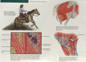
- Hip and thigh disorders 18

Medial muscles of the thigh
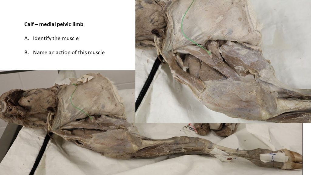
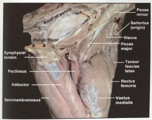
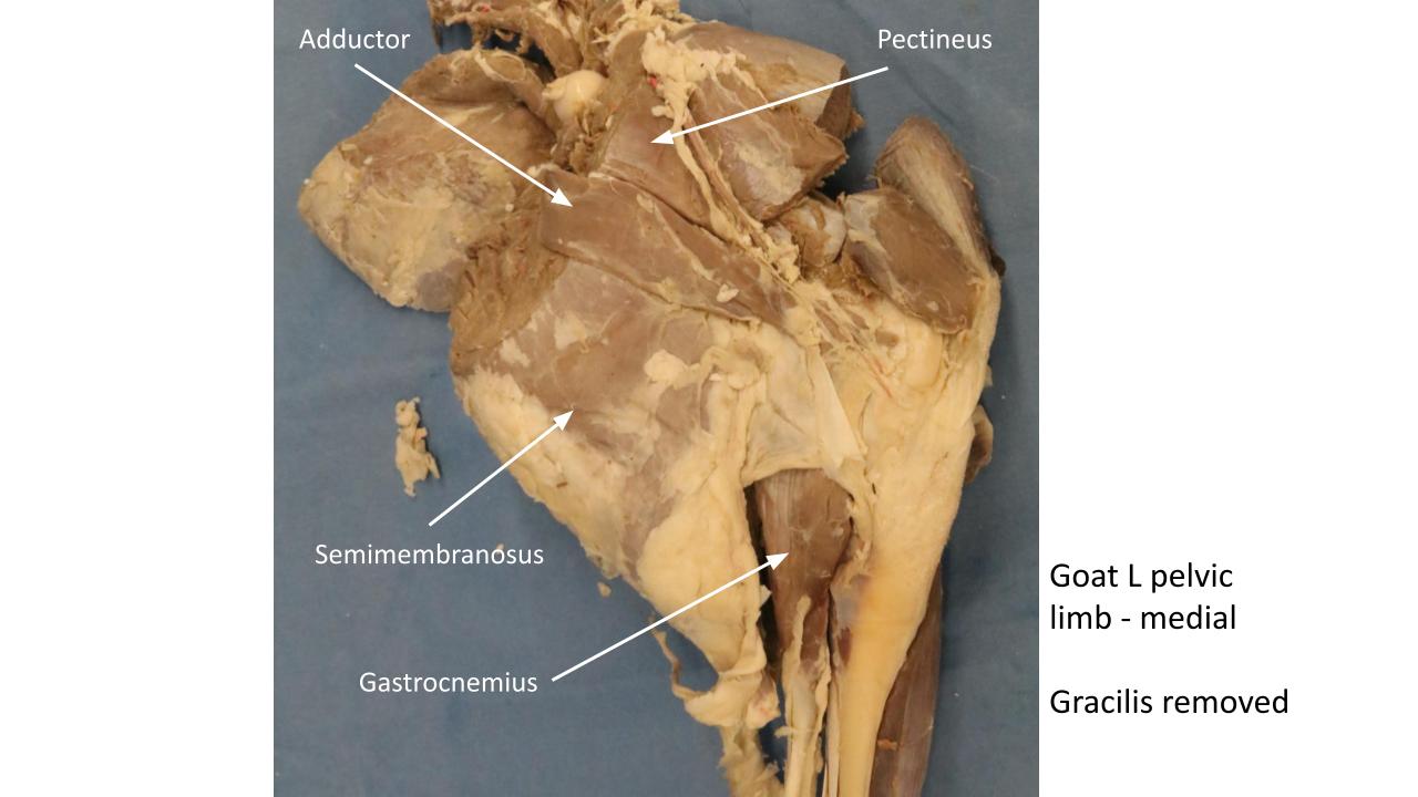
Observe: These muscles are primarily viewed on detached limb models in the horse (if provided) and otherwise can be viewed on the goat, calf and pig cadavers. The gracilis m. can be viewed on the intact equine cadaver. The same muscles identified in the carnivore dissections are present in ungulates. Identify the sartorius m.(which has two tendons of origin in the artiodactyls), gracilis m., adductor m. and pectineus m. Identify the femoral triangle, bound by the sartorius m. cranially and the pectineus m. caudally. If lymph nodes are noted in the horse and goat cadavers, in the femoral triangle region, these are the proximal femoral lymph nodes (to be considered in a later unit).
Clinical relevance: splayed legs when muscles of adduction are not functioning.
Medial muscles of the thigh are adductors of the coxal (hip) joint; if the adductors of the hip don’t function (maybe due to nerve damage) the animal may exhibit splayed out hindlimbs and an inability to stand on slippery floors because the animal can’t keep its legs adducted (i.e. can’t prevent legs from sliding away from body).
Take a moment to consider/recall the muscle that attaches to the lesser trochanter of the femur, what is its action? Injury to this muscle may cause lameness – it can be palpated per rectum in horses. HINT: this muscle is formed (and named accordingly) by two separate muscles combining before their attachment on the femur.
-
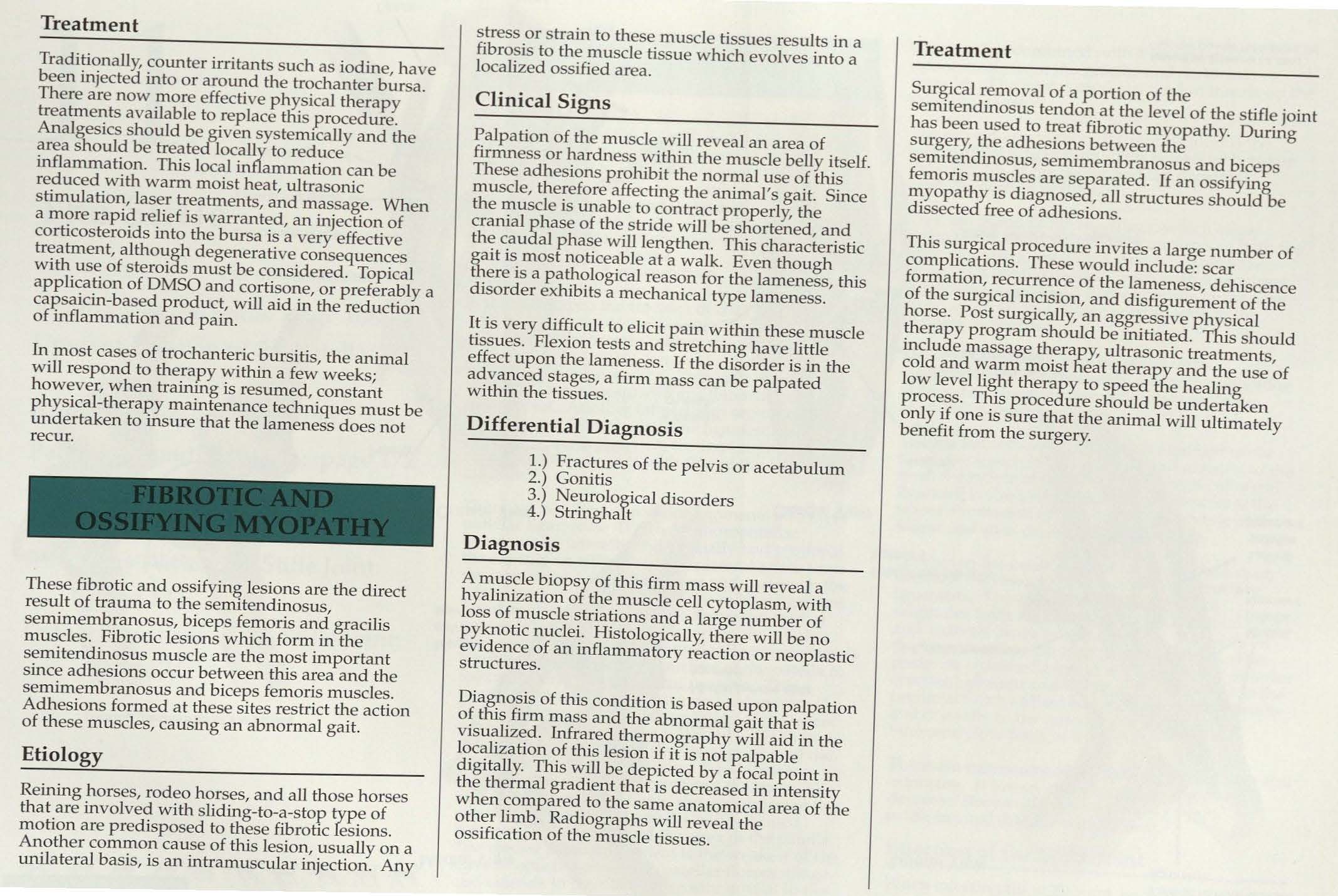
- Superficial structures of thigh and leg of newborn calf, medial view. 2

-
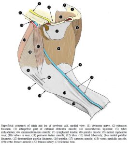
- Deeper structures of thigh and leg of newborn calf, medial view. 2

-
- Left hind limb of the horse, superficial, medial view. 16

-
- Left hind limb of the horse, deep, medial view. 16

-

- Goat thigh
Review Videos
Dog Pelvic Limb Muscles – 21 minutes (whole limb)
Horse gluteal muscles – 3 minutes
Horse proximal muscles – 35 minutes (whole limb)
Horse passive stay apparatus – 22 minutes
Calf pelvic limb muscles – 24 minutes (whole limb)
Goat pelvic limb muscles – watch until 18 minutes (whole limb)
Pig pelvic limb muscles – watch until 16 minutes
Muscle Chart
| Muscle Action Groups | |
| Flexors of the stifle and extensors of the hip |
|
| Flexors of the hip |
|
| Adductors of the pelvic limb |
|
| Muscle/structure | Attachments/comments | Action/comments |
| Biceps femoris m.
Gluteobiceps m. (in artiodactyls) |
In artiodactyls, biceps femoris and superficial gluteal are fused to form gluteobiceps m. (see next lab) |
Flex the stifle; extend the hip
(Secondary: extend stifle and tarsus, abduct hip) |
| Semitendinosus m. |
|
Flex the stifle; extend the hip.
(Secondary: extend the tarsus) The vertebral and ligament attachments in the horse and pig provide for the rounded contour of their croup/rump. |
| Semimembranosus m. |
|
Flex the stifle; extend the hip.
(Secondary: extend the stifle) The vertebral and ligament attachments in the horse and pig provide for the rounded contour of their croup/rump. |
| Sartorius m. |
|
Flex the hip
(Secondary: cranial part extends the stifle; caudal part flexes the stifle) |
| Gracilis m. |
|
Adduct the pelvic limb
(Secondary: flex the stifle, extend hip and tarsus) |
| Pectineus m. |
|
Adduct the pelvic limb |
| Adductor m. |
|
Adduct the pelvic limb
(Secondary: extend the hip) |
| Popliteal lymph node | Identify in carnivore | Site for lymph node biopsy (eg fine needle aspiration). |
| Fascia lata | Identify very thick fascia on cranial and lateral thigh | Related to biceps femoris and tensor fasciae latae mm. |
| Femoral Triangle | Identify and recall muscular boundaries of this region | Recall clinical application for femoral pulse palpation in carnivores. |
| Croup/Rump | Horse = croup
Artiodactyls = rump |
Croup of horse and rump of pig have a rounded contour due to additional vertebral origins of hamstring muscles.
In the carnivore and ruminants the ST and SM muscles arise from the ischiatic tuberosity but not the sacral or caudal vertebrae. |