
MSK LAB 19A – Pelvic Limb – lateral pelvis, caudal hip, and cranial thigh muscles
Learning Objectives
- Identify muscles of the lateral pelvis, caudal hip, and cranial thigh
- Name the attachments of the muscles of the lateral pelvis, caudal hip, and cranial thigh
- Name the action(s) of the muscles of the lateral pelvis, caudal hip and cranial thigh
- Categorize muscles into “muscle action groups”
- Connect structures to the clinically applied anatomy as emphasized.
For this lab we will dissect and learn the muscles (and other highlighted structures) of the lateral pelvis region, the caudal hip, and the cranial hip and thigh region. The muscles to be considered are listed below. Follow the lab guide to dissect these muscles on the carnivore specimen for your team. Then move to identifying the comparative anatomy of these muscles, and other structures highlighted, on the ungulate specimens.
Lateral pelvis (croup/rump)
- Tensor fasciae latae m.
- Gluteofemoralis m. (cat)
- Superficial gluteal m.
- Middle gluteal m.
- Deep gluteal m.
Caudal hip
- Internal obturator m.
- Gemelli m.
- Quadratus femoris m.
- External obturator m.
Cranial thigh
- Quadriceps femoris m. (4 bellies)
- Rectus femoris m.
- Vastus medialis m.
- Vastus lateralis m.
- Vastus intermedius m.
- Iliopsoas m.
- Sartorius m. (also reviewed on medial thigh in Lab 17A)
Lateral muscles of the pelvis
Tensor Fasciae Latae m.
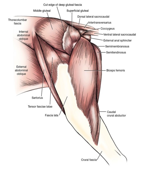
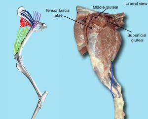
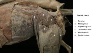
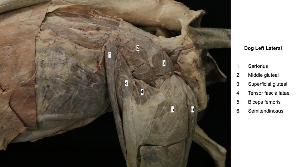
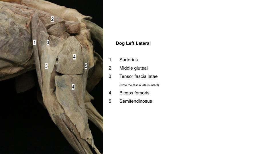
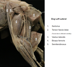
The tensor fasciae latae m. is a triangular muscle (looks a bit like an upside down V shape) that attaches proximally to the tuber coxae. It lies between the sartorius m. cranially, the middle gluteal m. caudodorsally, and the quadriceps femoris m. distomedially. Part of its caudodorsal surface is attached to the middle gluteal m. near its origin. The muscle can be divided into two portions (two arms of the V). The cranial, more superficial portion is inserted on the fascia lata (lateral femoral fascia), which radiates over the quadriceps and blends with the fascial insertion of the biceps femoris m. The deeper caudal portion is inserted on a layer of fascia lata that runs deep to the biceps femoris m. toward the stifle on the lateral surface of the vastus lateralis m. (part of the quadriceps femoris m.)
PROXIMAL ATTACHMENT: Ilium, gluteal fascia
DISTAL ATTACHMENT: Fascia lata
ACTION: Flex the hip, extend the stifle
Observe: Identify the muscle and note its borders. At this time, do not cut the cranially lying fascia lata, which serves as part of its insertion. You may need to do so when isolating the quadriceps femoris m.
-

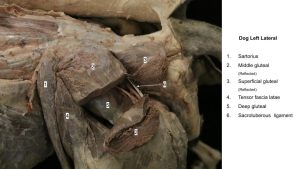
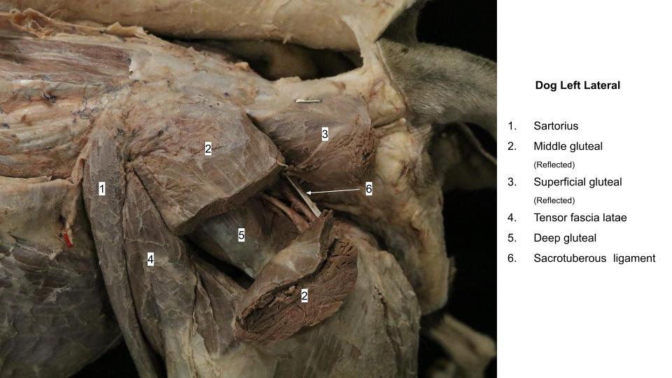
- Dog superficial muscles of left thigh, lateral view. 1
-
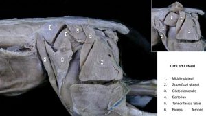
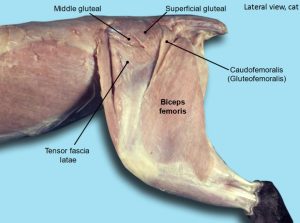
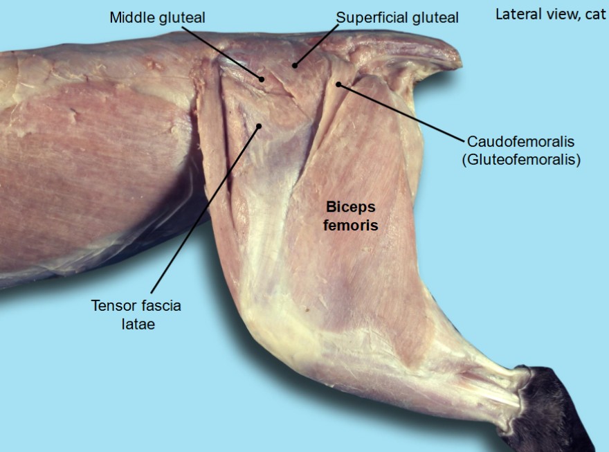
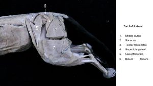
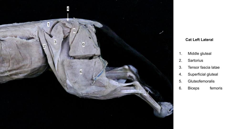
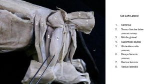
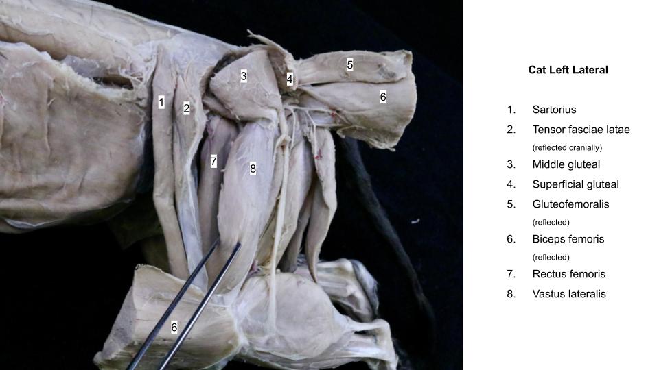
- Superficial muscles of the left hind limb of the cat. 5
-
- Dog lateral hip. 40

-
- Dog thigh
-

- Dog thigh
-
- Cat thigh

Gluteofemoralis m.
In the cat, the gluteofemoralis m., which is a long thin muscle caudal to the superficial gluteal m. and lying along the cranial margin of the biceps femoris m., is an additional muscle not present in the dog or ungulates.
PROXIMAL ATTACHMENT: Caudal vertebrae
DISTAL ATTACHMENT: Fascia lata
ACTION: Abduct hip or move tail laterally
Dissect: In the cat, clean and isolate the gluteofemoralis m. and transect it about a centimeter proximal to its distal attachment. Reflect the proximal portion of the muscle.
-
- Superficial muscles of the left hind limb of the cat. 5
-
- Cat lateral hip. 40
-
- Cat thigh
Superficial Gluteal m.
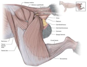
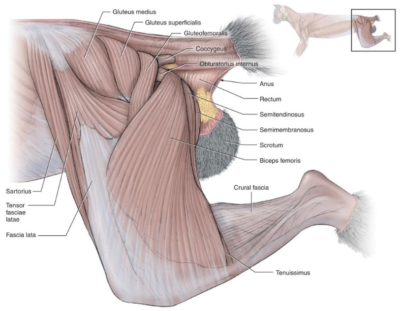
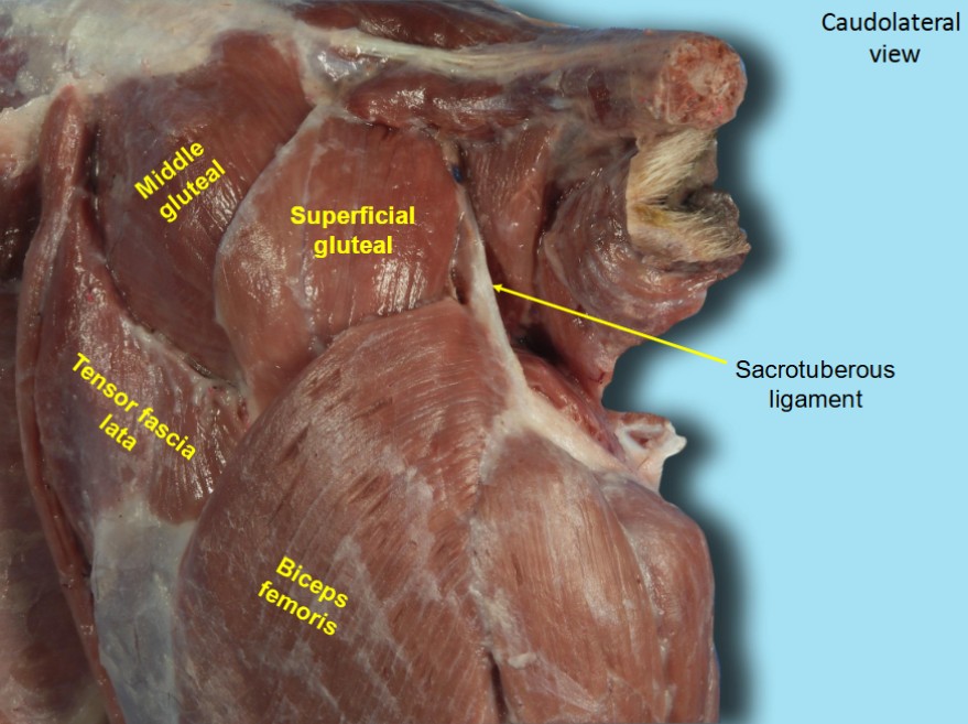
The superficial gluteal m. is small and lies caudal to the middle gluteal. Its fibers run distally, from the deep gluteal fascia that covers the middle gluteal and from the sacrum and the first caudal vertebra to the level of the greater trochanter of the femur, where the fibers converge before forming an aponeurosis that runs under the biceps to attach to the third trochanter.
PROXIMAL ATTACHMENT: Pelvis
DISTAL ATTACHMENT: Third trochanter (of femur)
ACTION: Extend the hip and abduct the pelvic limb
Dissect: Clean and isolate the superficial gluteal m. and transect it 1 cm proximal to its aponeurosis of distal attachment to the third trochanter. In the dog, be aware of the closely related sacrotuberous ligament at the proximal attachments of the superficial gluteal and avoid transecting it.
-
- Dog superficial muscles of left thigh, lateral view. 1
-
- Superficial muscles of the left hind limb of the cat. 5
-

- Dog gluteal muscles. 40
-
- Dog thigh
-
- Dog thigh
-
- Cat thigh
Sacrotuberous Ligament
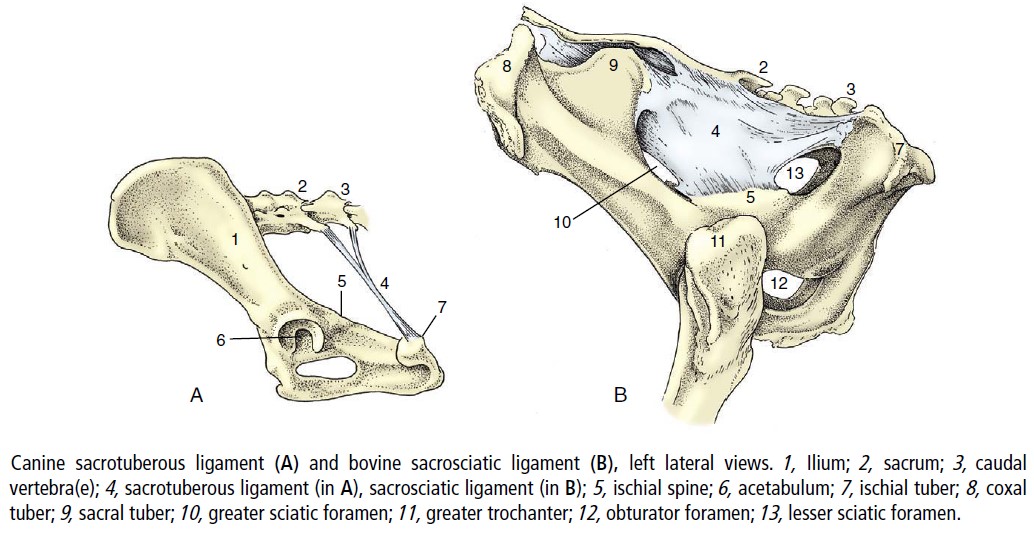
The sacrotuberous ligament is a fibrous band that runs from the sacrum to the lateral angle of the ischiatic tuberosity (tuber ischii) in the dog. Recall, it is absent in the cat. It is a site of attachment for muscles.
Observe: View and palpate this ligament in the dog cadavers, and view it on prosections (wet and dry).
-
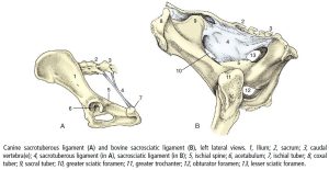
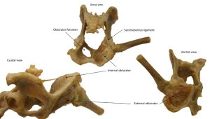
- Canine sacrotuberous ligament and bovine sacrosciatic ligament. 8
-
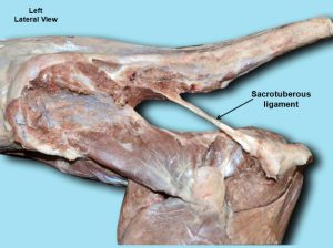
- Dog sacrotuberous ligament 40

-
- Dog hip
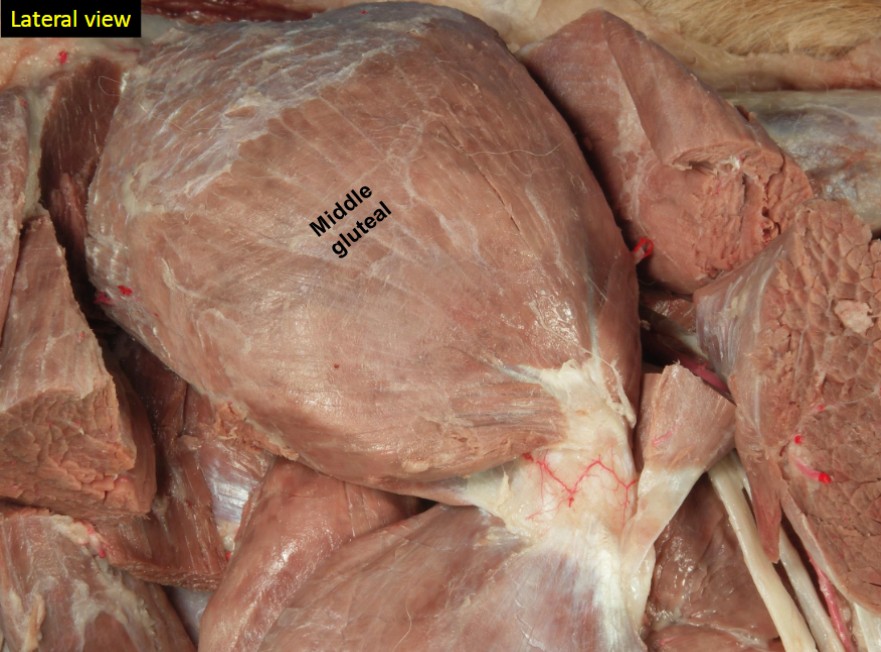
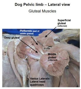
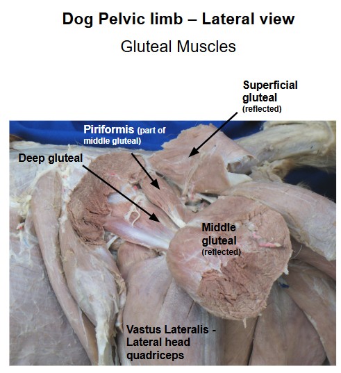
Middle Gluteal m.
The middle gluteal m. is a large, ovoid muscle that lies between the tensor fasciae latae and the superficial gluteal. The caudodorsal border of the middle gluteal m. is covered by the superficial gluteal m. A deep caudal portion of the middle gluteal m. can be isolated from the main muscle mass and this part is called the piriformis muscle (fyi). Its separate tendon inserts at the same location as the rest of the muscle mass.
PROXIMAL ATTACHMENT: Ilium
DISTAL ATTACHMENT: Greater trochanter (of femur)
ACTION: Extend the hip, abduct the pelvic limb, and rotate femur medially at hip
Dissect: Clean the surface of the muscle by reflecting the deep gluteal fascia and its attached transected superficial gluteal muscle craniodorsal to the iliac crest; this reveals the fibers of the middle gluteal. Carefully separate by blunt dissection the cranioventral border of the middle gluteal from the underlying deep gluteal m. Then, starting at the middle of the cranioventral border of the middle gluteal, transect the entire muscle and reflect the distal half toward its insertion on the greater trochanter of the femur to reveal the underlying deep gluteal m. Sometimes the piriformis m. is left behind during this reflection and it lies on the caudal aspect of the deep gluteal m.
-
- Dog superficial muscles of left thigh, lateral view. 1
-
- Superficial muscles of the left hind limb of the cat. 5
-

- Dog deep gluteal. 40
-
- Dog gluteal muscles J. Barnes
-
- Dog thigh
-
- Cat thigh
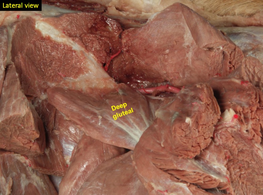
Deep Gluteal m.
The deep gluteal m. is fan-shaped and completely covered by the middle gluteal m. Its fibers converge to insert on the cranial face of the greater trochanter.
PROXIMAL ATTACHMENT: Ilium and ischiatic spine
DISTAL ATTACHMENT: Greater trochanter (of femur)
ACTION: Extend the hip, abduct the pelvic limb, and rotate femur medially at hip
Observe: Identify the deep gluteal m.
-
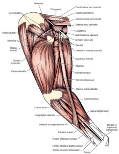
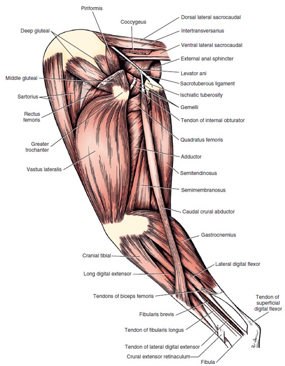
- Deep Muscles of the Left Pelvic limb of the dog, lateral view. 1
-
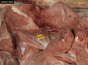
- Dog deep gluteal. 40
-
- Dog hip
Caudal muscles of the hip
The four muscles of this group are important because of their proximity to the hip. They lie caudal to the hip and extend from the ventral and dorsal surfaces of the ischium to the femur. All rotate the limb laterally. This action opposes the medial rotation by the gluteal muscles so that the thigh moves in a sagittal plane at the hip.
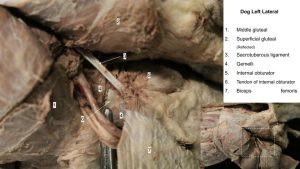
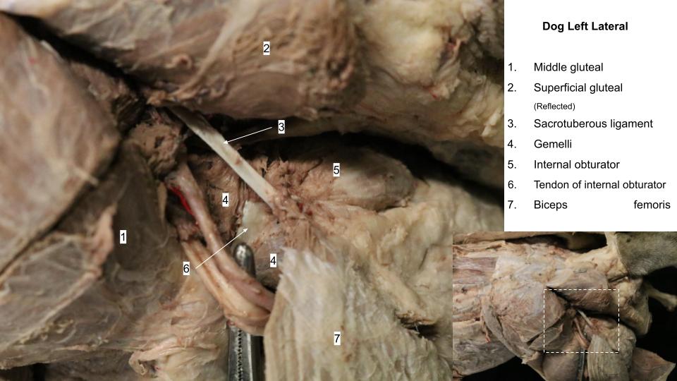
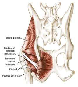
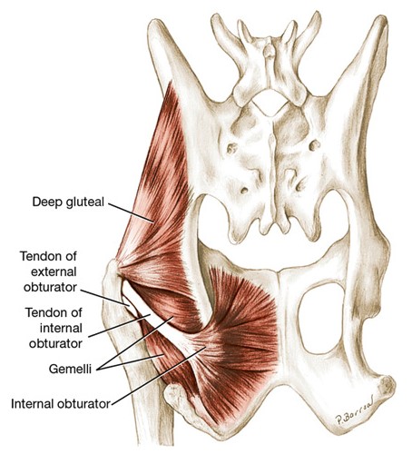
Internal Obturator m.
The internal obturator m. is fan-shaped on the dorsal surface of the ischium and pubis. It is so named as it lies within the confines of the pelvic cavity. It covers the obturator foramen and its muscle fibers converge toward the lesser ischiatic notch where its tendon of attachment begins. The tendon of the internal obturator m. passes over the lesser ischiatic notch (and ventral to the sacrotuberous ligament in the dog).
PROXIMAL ATTACHMENT: Dorsal ischium and pubis
DISTAL ATTACHMENT: Trochanteric fossa
ACTION: Lateral rotation of femur at the hip.
Dissect: Isolate the tendon of insertion of the internal obturator m. by removing fat and fascia covering its surface [and working caudomedial to the sacrotuberous ligament in the dog]. The tendon will be located caudal to the caudal border of the deep gluteal muscle. Abduction of the limb releases tension on the tendon making it a little easier to elevate. In the cat, the internal obturator m. tendon is typically buried in the gemelli mm. (see next muscle) and requires more dissection to locate.
Now, with your finger, you may be able to follow the tendon to palpate the dorsal surface of the muscle belly on the dorsal surface of the ischium and pubis. Make sure to visualize this muscle on plastinated models.
-
- Muscles of left hip joint of the dog, dorsal aspect. 1
-
- Deep muscles of the hind limb of the cat in lateral view. 5

-
- Dog hip
Clinical Application: Q2
Q2: One muscle that attaches to the greater trochanter of the femur is the:
A. internal obturator m.
B. superficial gluteal m.
C. middle gluteal m.
D. biceps femoris m.
Gemelli mm.
The gemelli mm., two muscles fused together, lie under and adjacent the tendon of the internal obturator. They are interposed between the quadratus femoris m. (not quadriceps femoris! this is a different muscle) and external obturator m. distally and the deep gluteal m. proximally and cranially. The gemelli are deeply grooved by the tendon of the internal obturator m. so that their edges overlap this tendon and they appear as a small muscle belly on either side of the tendon.
PROXIMAL ATTACHMENT: Ischium
DISTAL ATTACHMENT: Trochanteric fossa
ACTION: Lateral rotation of femur at the hip.
Observe: Observe the gemelli mm. on either side of the internal obturator’s tendon of insertion. Note that these muscles share an attachment with the internal obturator muscle on the trochanteric fossa of the femur.
-
- Muscles of left hip joint of the dog, dorsal aspect. 1
-
- Deep Muscles of the Left Pelvic limb of the dog, lateral view. 1
-
- Deep muscles of the hind limb of the cat in lateral view. 5
-
- Dog hip
Quadratus femoris m.
Later, the quadriceps femoris m. is studied. Note the subtle spelling difference that distinguishes these very different muscles!
The quadratus femoris m. is short and thick. It lies deep to the biceps femoris m., where it is interposed between the adductor m. medially, the biceps femoris m. laterally, and the external obturator m. and gemelli mm. dorsally. Its fibers are at right angles to the long axis of the thigh. The dorsal border of the quadratus femoris m. lies closely applied to the ventral border of the gemelli mm.
PROXIMAL ATTACHMENT: Ischium
DISTAL ATTACHMENT: Intertrochanteric crest
ACTION: Lateral rotation of femur at the hip. (Secondary: extend the hip)
Dissect: Reflect the proximal part of the transected biceps femoris m. in order to dissect and isolate the margins of the exposed portion the quadratus femoris m. from the lateral side. Dissect it directly ventral to the caudal border of the gemelli mm. What is viewed is only a small section of this relatively elongated rectangular muscle. View this muscle on platinated models, to appreciate its full extent and attachments. It should be examined from both the medial and lateral sides.
-
- Deep Muscles of the Left Pelvic limb of the dog, lateral view. 1
-
- Deep muscles of the hind limb of the cat in lateral view. 5
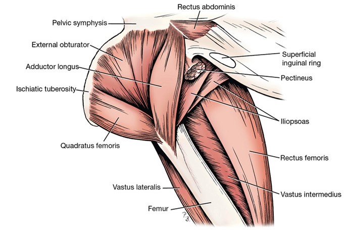
External obturator m.
The external obturator m. is fan-shaped (like its internal partner) and arises on the ventral surface of the pubis and ischium. It is so names because it lies on the outside of the pelvic cavity. It also covers the obturator foramen. Its caudal border is covered by the quadratus femoris m., whereas its cranial border is hidden by the adductor m.
PROXIMAL ATTACHMENT: Ventral ischium and pubis
DISTAL ATTACHMENT: Trochanteric fossa
ACTION: Lateral rotation of femur at the hip.
Observe: Observe the external obturator muscle on plastinated models. It is not dissected on the cadaver, however, its caudal lateral margin may be noted between the quadratus femoris m. and gemelli mm.
-
- Deep muscles medial to left hip joint of the dog. 1
-
- Dog obturator mm.

Cranial muscles of the thigh
Quadriceps femoris m.
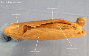
The quadriceps femoris m. has four heads of origin (hence its name), which are fused distally. They are a) the rectus femoris m., b) the vastus lateralis m., c), the vastus intermedius m., and d) the vastus medialis m., all of which will be dissected independently. The vastus muscles attach proximally to the femur and the rectus femoris muscle attaches to the ilium. All heads functionally attach distally to the tibial tuberosity. The patella lies within the ‘tendon’ of insertion. The quadriceps femoris m. is the most powerful extensor of the stifle joint and its action is necessary for the animal to support its weight normally.
The patella, a large sesamoid bone (the human ‘kneecap’), is intercalated in the insertion of the quadriceps. The muscle heads attach to the patella. The patella articulates with the trochlea of the femur (forming the femoropatella joint). From the patella, the patellar ligament extends distally to attach to the tibial tuberosity. It represents the functional tendon of insertion of the quadriceps femoris.
Dissect: At this time, the fascia lata will likely need to be transected and reflected to expose the quadriceps femoris heads laterally. Keep the fascia lata attached to the tensor fasciae latae muscle proximally. The sartorius muscle may need to be transected to access the rectus femoris and medial head of the quadriceps. If it is transected, cut both parts midway between the proximal and distal attachments and reflect the distal parts to their insertions on the patella and cranial border of the tibia.
Read the muscle descriptions below and dissect the margins of the quadriceps femoris m. sharply and bluntly until the four heads are identified.
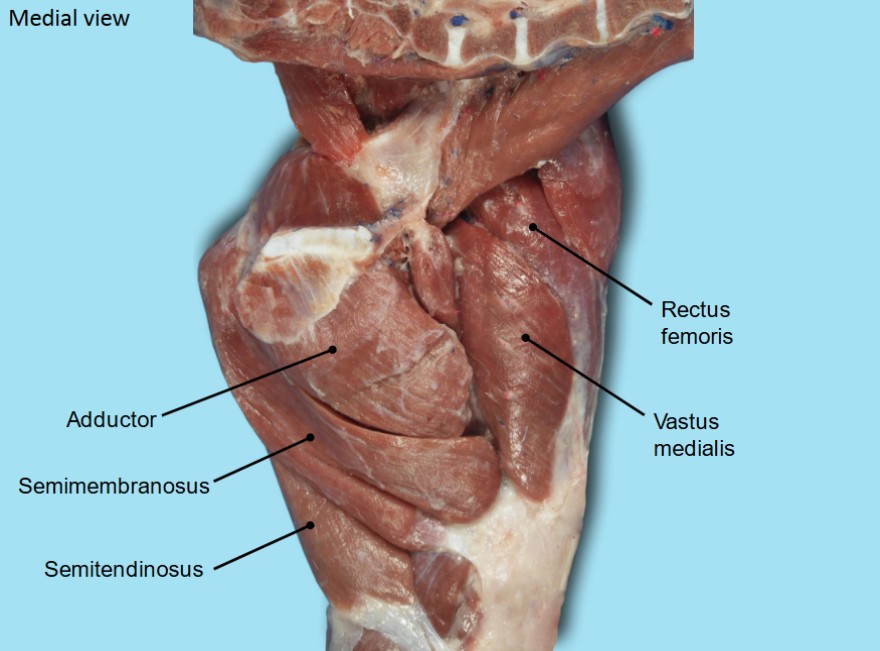
The rectus femoris m. is the most cranial component of the quadriceps femoris. Proximally, it is circular in transverse section and it is sandwiched between the vastus muscles. Because the rectus femoris arises from the ilium it also functions as a flexor of the hip, along with its stifle extension action shared with the other 3 heads.
The vastus lateralis m. lies lateral and caudal to the rectus femoris, to which it is fused distally. The vastus lateralis is partly separated from the vastus intermedius by an intermuscular septum. Notice that the vastus lateralis arises from the proximal part of the lateral lip of the caudal rough surface of the femur.
The vastus intermedius m. lies directly on the cranial surface of the femur and is quite intimately fused with the other two vasti. It arises with the vastus lateralis, from the lateral side of the proximal end of the femur.
Dissect: Push the rectus femoris m. laterally or medially so that the deeper lying vastus intermedius m. can be visualized, and palpated. If it cannot be seen, the rectus femoris muscle may be transected. Due to the fusion of the 3 vasti muscles at their deep margins, a clear distinction may not be apparent from lateral to intermediate to medial. When directly palpating the muscle covering the cranial surface of the shaft of the femur, the vastus intermedius m. is being palpated.
The vastus medialis m. arises from the medial side of the proximal end of the cranial surface of the femur and the proximal end of the medial lip of the caudal rough surface. It is inserted with the other heads of the quadriceps on the tibial tuberosity.
PROXIMAL ATTACHMENTS: Rectus femoris = ilium; Vastus mm. = proximal femur
DISTAL ATTACHMENT: Tibial tuberosity
ACTION: Extend the stifle (Secondary: rectus femoris m. also flexes the hip)
-
- Deep Muscles of the Left Pelvic limb of the dog, lateral view. 1
-
- Deep muscles of the hind limb of the cat in lateral view. 5
-
- Dog medial thigh

-
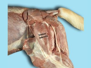
- Dog vastus lateralis. 40

-

- Dog medial thigh 40
-
- Dog thigh

-
- Dog quad mm.

-
- Cat thigh
Iliopsoas m.
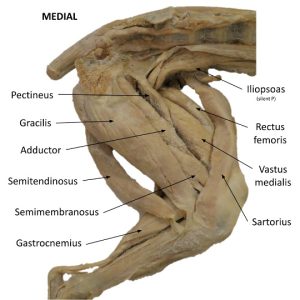
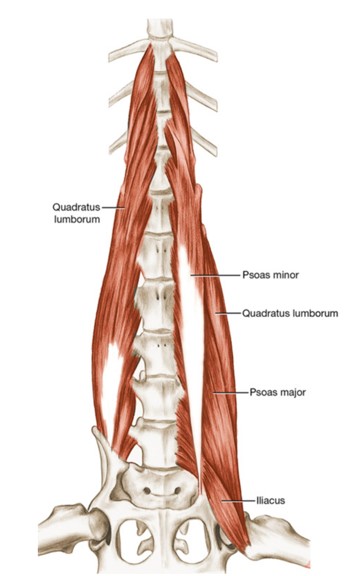
The iliopsoas m., pronounced with a silent “p”, is a sublumbar muscle now visible at its insertion on the lesser trochanter of the femur. This end of the muscle lies between the pectineus m. medially and the rectus femoris m. laterally. The iliopsoas m. represents a fusion of the psoas major and iliacus muscles. The psoas major m. is long and arises from the ventral aspect of the transverse processes and bodies of lumbar vertebrae. It passes caudally and ventrally under the cranioventral aspect of the ilium, where it joins the iliacus m. The iliacus m. is short and arises from the smooth ventral surface of the ilium between the arcuate line and the lateral border of the ilium. The two muscle masses continue caudoventrally as the iliopsoas m. to their conjoined insertion on the lesser trochanter. The action of the iliopsoas m. is to flex the hip joint and the lumbar vertebral column. It is the major flexor of the hip joint. The femoral nerve emerges from the muscle as it passes through the fibers of the iliopsoas m. This is one of the few places to readily observe the femoral nerve on the lab cadavers (revisited in the nervous system).
PROXIMAL ATTACHMENT: Ilium and lumbar vertebrae
DISTAL ATTACHMENT: Lesser trochanter (of femur)
ACTION: Flex the hip
Dissect: Dissect as needed to observe the iliopsoas m. between the pectineus and rectus femoris mm. Observe the femoral nerve as it passes through the fibers of this muscle. Examine the muscle on plastinated specimens.
-

- Sublumbar muscles, deep dissection, ventral aspect. The psoas major and psoas minor have been removed on the right side; they are present on the left. The combined iliacus and psoas major form the iliopsoas m. (circled), which inserts on the lesser trochanter of the femur. 1
-
- Dog medial thigh
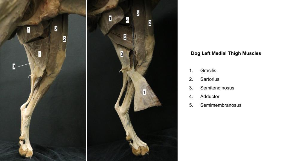
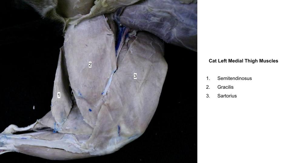
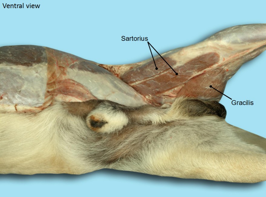
Sartorius m.
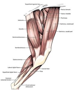
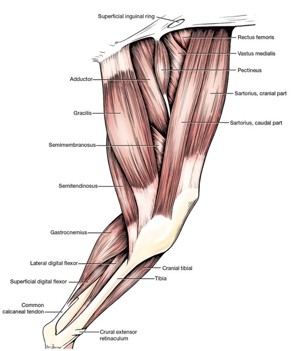
The sartorius m. gets a brief mention here again because its cranial belly in the dog passes along the cranial margin of the thigh region. It has been considered in detail in Lab 17A, the medial thigh section.
-
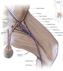
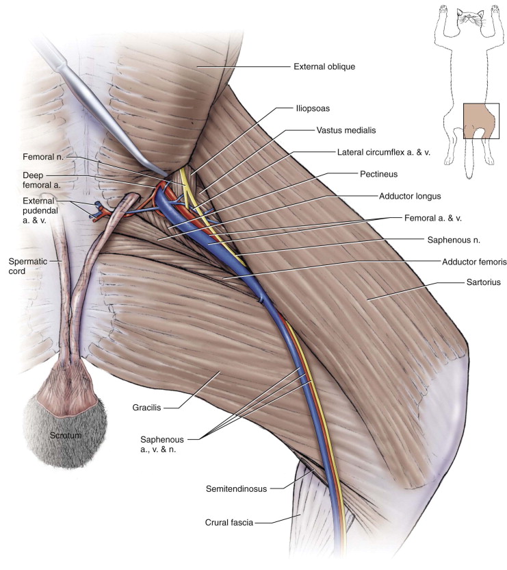
- Medial thigh and crus of the dog. 1
-
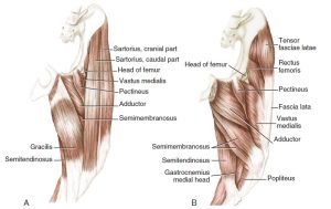
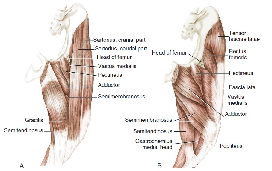
- Muscles of the thigh of the dog. A, Superficial muscles, medial aspect. B, Deep muscles, medial aspect. 3
-
- Superficial muscles of the left hind limb of the cat, medial. 5
-
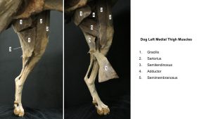
- Dog thigh
-

- Cat thigh
-

- Dog gracilis. 40
Ungulate comparative anatomy
Observe: Refer to the prosected ungulate cadavers and additional specimens and models to identify and study the following content. Certain muscles have been transected midbody or at their attachments to facilitate viewing deeper structures.
Lateral muscles of the pelvis
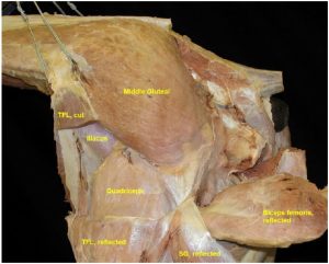
The middle gluteal m. is a substantial bulk of muscle. It is a powerful extensor of the hip joint, attaching to the greater trochanter (and specifically, the caudal part of the greater trochanter in the horse).
Observe: Identify the middle gluteal m. and the previously noted superficial gluteal (horse) or gluteobiceps (artiodactyls). The sacral attachments of the superficial gluteal (or gluteobiceps) are cut so the muscle can be reflected ventrally to expose the termination of the middle gluteal m. on the greater trochanter of the femur.
-
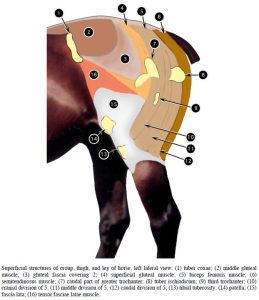
- Superficial structures of croup, thigh, and leg of horse, left lateral view. 2

-
-
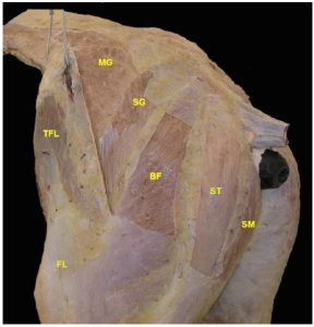
Superficial muscles of equine lateral thigh and gluteal
region . MG middle gluteal; SG superficial gluteal; BF biceps
femoris; ST semitendinosus; SM semimembranosus; TFL tensor fasciae latae; FL fascia lata. 16

-
- Muscles of equine lateral thigh and gluteal region . SG superficial gluteal; TFL tensor fasciae latae. 16

-
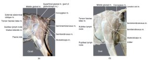
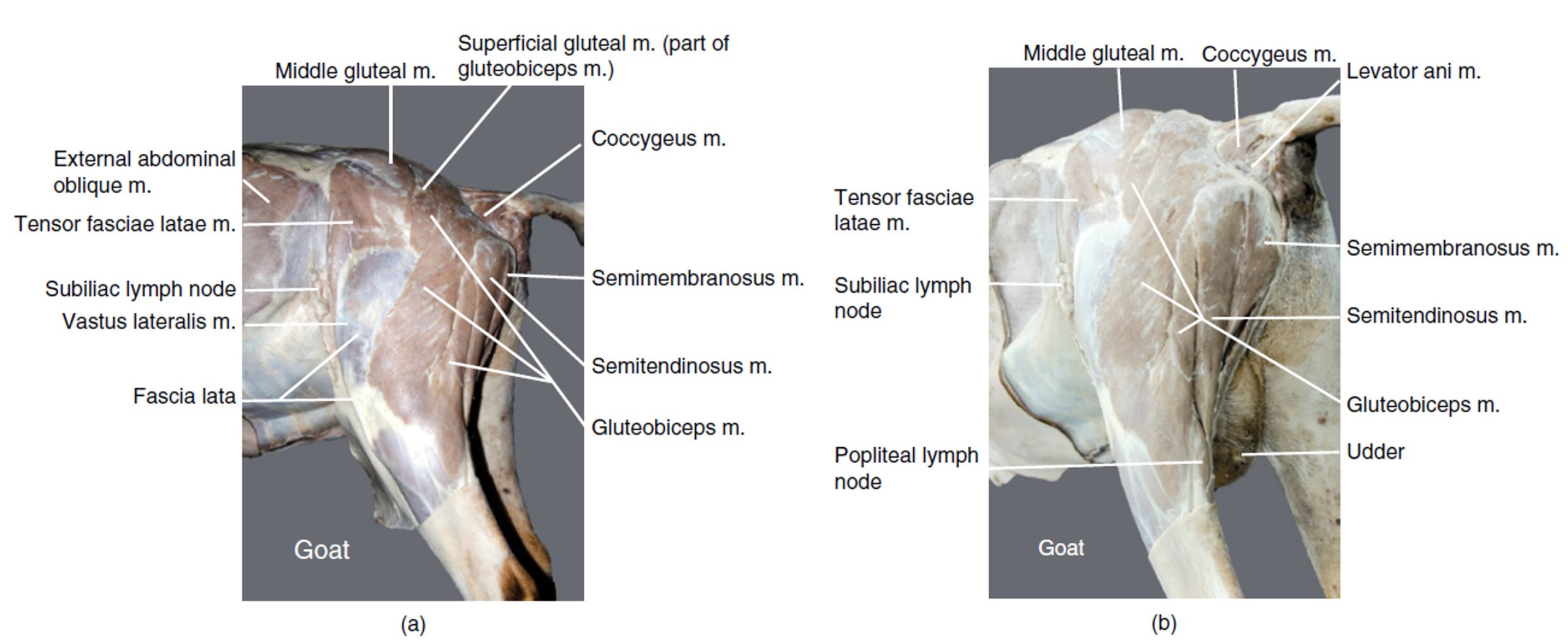
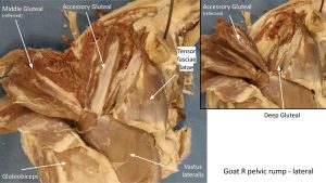
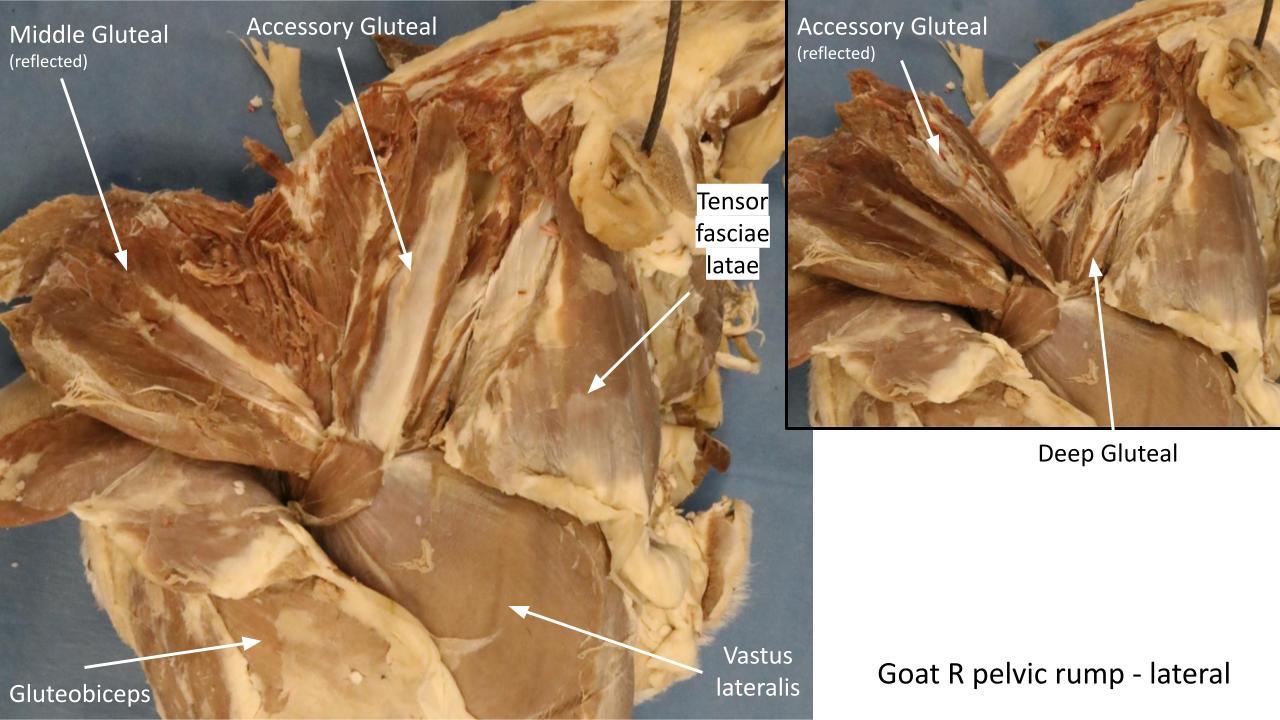
- Hip and thigh muscles of a goat, left lateral view (a). Hip and thigh muscles of a goat, left caudolateral view (b). 12
-
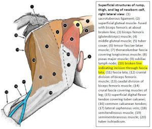
- Superficial structures of rump, thigh, and leg of newborn calf, right lateral view. 2

-
- Horse hip
-
- Pig hip

-
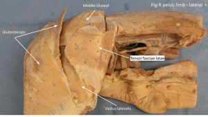
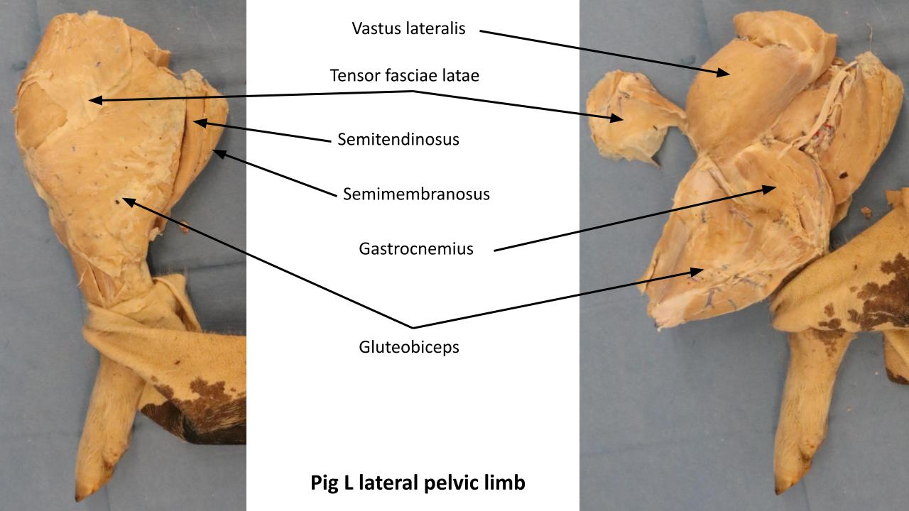
- Pig thigh

-
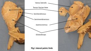
- Pig limb
-
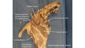
- Goat thigh

-
- Goat thigh
The tensor fasciae latae m. and fascia lata lie craniolaterally on the thigh with the muscle partially covering the middle gluteal.
Observe: Identify the tensor fasciae latae and fascia lata. It has been transected to allow the deeper dissection.
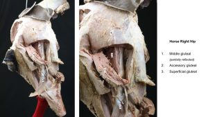
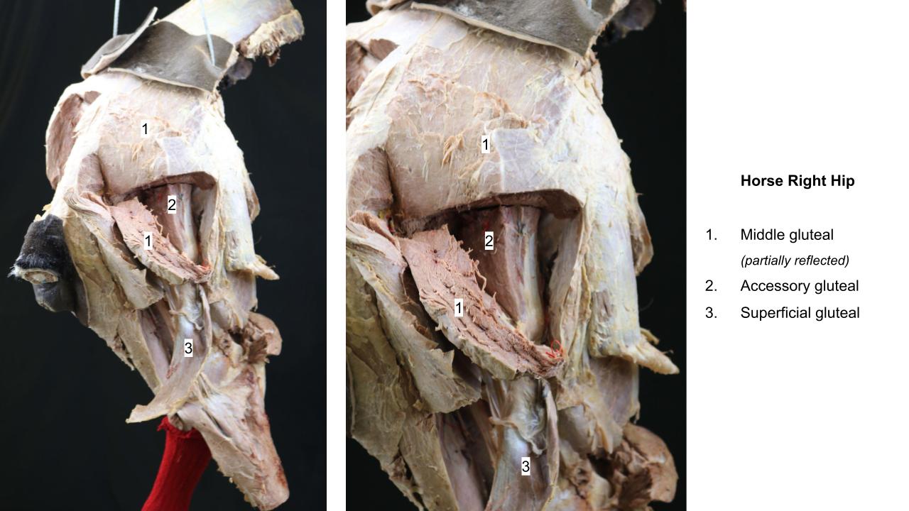
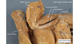
Deep to the middle gluteal m. is an extra gluteal muscle in ungulates called the accessory gluteal m.. The accessory gluteal m. has a distinct tendon of attachment to the greater trochanter (and specifically, the cranial part of the greater trochanter in the horse). Deep to the tendon of attachment of the accessory gluteal m. to the greater trochanter is the trochanteric bursa. A deep gluteal m. is also present, to finish out the gluteal muscle story in ungulates.
Observe: Identify the gluteal muscles in the ungulate prosections, and the trochanteric bursa in the horse The middle gluteal m. has been transected and elevated to provide a view of the accessory and deep gluteal mm. As a point of reference, the sciatic nerve crosses over the superficial surface of the deep gluteal m. at its caudodorsal margin. The accessory gluteal m. has been transected – elevate its distal tendinous attachment to view the trochanteric bursa. Appendix on synovial structures.
Clinical relevance: trochanteric bursitis.
Trochanteric bursitis (‘whorlbone’ lameness): inflammation of the bursa is a cause of lameness in performance horses. There is a whorl in the hair coat in the region of the bursa, hence the name ‘whorlbone lameness’ may be used in racehorse parlance.
-

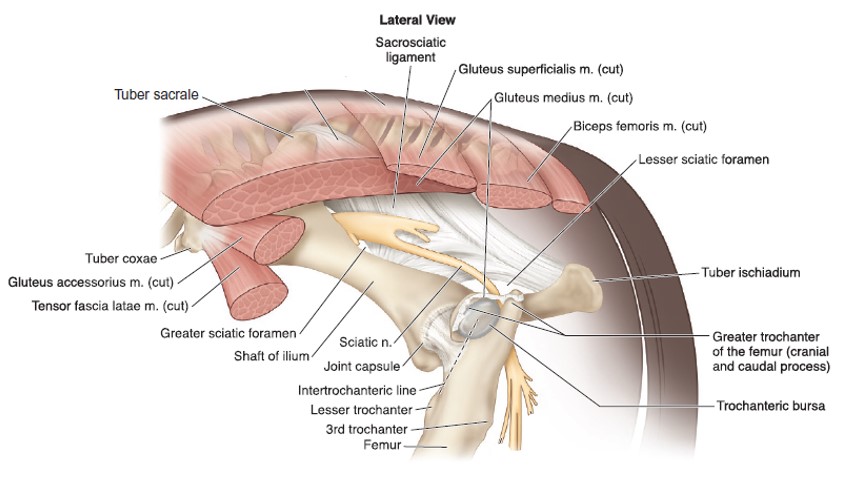
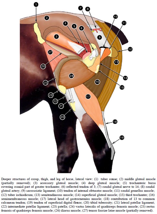
- Deeper structures of croup, thigh, and leg of horse, lateral view. 2
-
- Muscles of equine lateral thigh and gluteal region . SG superficial gluteal; TFL tensor fasciae latae. 16
-
- Hip and thigh muscles of a goat, left lateral view (a). Hip and thigh muscles of a goat, left caudolateral view (b). 12
-
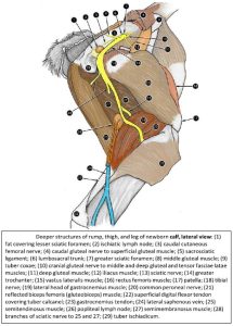
- Deeper structure of rump, thigh, and leg of newborn calf, lateral view. 2

-
- Horse hip
-
- Pig hip
-
- Pig thigh
-
- Pig limb
-
- Goat thigh
-
- Goat thigh
Cranial muscles of the thigh
Observe: Once again, refer to the prosected ungulate cadavers and additional specimens and models to identify and study the following content. Detached prosected limbs are useful to review. Certain muscles have been transected midbody or at their attachments to facilitate viewing deeper structures.
The bulk of the cranial thigh muscles is represented by the four(4)-bellied quadriceps femoris m. muscle unit. This muscle is critical for normal weight bearing in the pelvic limb, with its primary action being stifle extension.
Observe: Identify the dissected quadriceps femoris m. by proximally separating the rectus femoris m. from the vastus lateralis m. and vastus medialis m. Reflect the tensor fasciae latae m. out of the way to view the quadriceps femoris bellies. Elevate and retract the rectus femoris head laterally or medially to expose, or at least to palpate, the vastus intermedius m. lying on the cranial surface of the femur.
-
- Deeper structures of croup, thigh, and leg of horse, lateral view. 2
-
- Muscles of equine lateral thigh and gluteal region . SG superficial gluteal; TFL tensor fasciae latae. 16
-
- Hip and thigh muscles of a goat, left lateral view (a). Hip and thigh muscles of a goat, left caudolateral view (b). 12
-
- Deeper structure of rump, thigh, and leg of newborn calf, lateral view. 2
-
- Pig thigh
-
- Pig limb
-
- Goat thigh
-
- Goat thigh
Clinical relevance: quadriceps femoris dysfunction
Quadriceps femoris m. is the major extensor of the stifle, innervated by the femoral n.
Q3: What does femoral n. paralysis look like?
Review Videos
These are the same video links as the prior lab.
Dog Pelvic Limb Muscles – 21 minutes (whole limb)
Horse gluteal muscles – 3 minutes
Horse proximal muscles – 35 minutes (whole limb)
Horse passive stay apparatus – 22 minutes
Calf pelvic limb muscles – 24 minutes (whole limb)
Goat pelvic limb muscles – watch until 18 minutes (whole limb)
Pig pelvic limb muscles – watch until 16 minutes
Muscle Chart and Additional Structures
| Muscle Action Groups | |
| Flexors of the hip |
|
| Extensors of the hip |
|
| Lateral rotators of the femur |
|
| Extensors of the stifle |
|
| Muscle/structure | Attachments/comments | Action/comments |
| Tensor fascia latae m. |
|
Flex the hip, extend the stifle |
| Gluteofemoralis m. (cat only) |
|
Abduct hip or move tail laterally |
| Superficial gluteal m. |
|
Extend the hip and abduct the pelvic limb |
| Sacrotuberous ligament (canine feature) |
Fibrous band connecting sacrum to ischiatic tuberosity | Attachment site for muscles in canine. |
| Middle gluteal m. (and piriformis, FYI) |
|
Extend the hip, abduct the pelvic limb, and rotate femur medially at hip |
| Accessory gluteal m. (ungulates only) |
|
Extend the hip, abduct the pelvic limb – ungulate only. |
| Trochanteric bursa (horse feature) |
lies deep to tendon of accessory gluteal as it inserts on greater trochanter. | Identify in horse specifically. |
| Deep gluteal m. |
|
Extend the hip, abduct the pelvic limb, and rotate femur medially at hip |
| Internal obturator m. (carnivore only) |
|
Lateral rotation of femur at the hip. |
| Gemelli mm. (carnivore only) |
|
Lateral rotation of femur at the hip. |
| Quadratus femoris m. (carnivore only) |
|
Lateral rotation of femur at the hip. (Secondary: extend the hip) |
| External obturator m. (carnivore only) |
|
Lateral rotation of femur at the hip. |
Quadriceps femoris m.
|
|
Extend the stifle. (Secondary: rectus femoris also flexes hip) |
| Iliopsoas m. (carnivore only) |
|
Flex the hip |
| Sartorius m. |
|
Flex the hip
(Secondary: cranial part extends the stifle; caudal part flexes the stifle) |