MSK LAB 20A – Pelvic Limb Crus muscles
Learning Objectives
- Identify muscles of the crus
- Name the attachments and action(s) of the muscles of the crus
- Categorize muscles into “muscle action groups”
- Connect structures to the clinically applied anatomy as emphasized
Lab overview:
The muscles of the crus, or ‘leg’, the region between the stifle and tarsus, are divided into craniolateral and caudal groups. Note that the term leg technically refers only to the crus, (Crus [L] = leg or shin) not to the entire pelvic limb! There are considerable comparative differences between the species we are studying and these differences will be highlighted along the way. Once again, teams will dissect these muscles on carnivore cadavers and the muscles of interest are listed below, for all the species we are studying.
Craniolateral crus muscles:
-
Cranial tibial
-
Long digital extensor
-
[Short digital extensor – not dissected]
-
Lateral digital extensor – dissected in ungulates only (but is present in dog and cat).
-
Fibularis longus – not present in horse
-
Fibularis (Peroneus) tertius – ungulates only, not present in dog and cat.
-
[Fibularis brevis – not dissected in dog and cat, not present in ungulates.]
Caudal crus muscles:
-
Gastrocnemius – medial and lateral heads
-
Superficial digital flexor
-
Soleus – feline and ungulates only, not present in dog.
-
Deep digital flexor – 3 heads
-
Popliteus
Before we isolate and identify muscles we will consider the fascia of the region, a large tendon created by the combination of smaller tendons, and a fibrous band on the cranial distal crus that helps keep tendons bound in their pathway.
Comments on fascia of the crus and distal limb
The superficial crural, tarsal, metatarsal, and digital fasciae are similar to the superficial fasciae of the corresponding regions of the forelimb. Cutaneous vessels and nerves course in the superficial fascia and will be studied in later units.
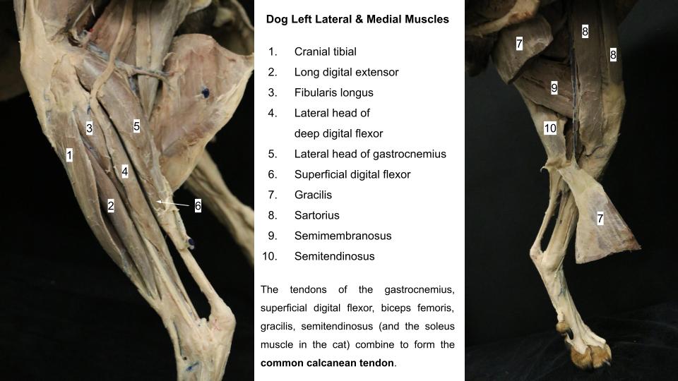
The medial and lateral femoral fasciae blend over the stifle and are continued distally in the leg as the deep crural fascia. The deep crural fascia covers the muscles of the leg and surfaces of the crural skeleton not enveloped by muscle (e.g. the medial tibia). Septa from this fascia extend between the muscles to attach to the bones. Laterally, the fibers of the caudal part of the biceps femoris m. radiate into the fascia. Medially, the semitendinosus and gracilis mm. are continuous with this fascia. These connections are located caudally where the crural fascia contributes to the common calcaneal tendon. The tendons of the gastrocnemius m., superficial digital flexor m., and tarsal tendons of the biceps femoris, gracilis, and semitendinosus mm. (and the soleus m. in the cat) combine to form the common calcaneal tendon (FYI = the Achilles tendon in the human).
Observe: Observe and palpate the common calcaneal tendon. Name the muscle tendons which contribute to it.
In the distal crus, on the cranial surface, the deep crural fascia is thickened to form an oblique transverse band of about 0.5 cm width (in the dog), called the crural extensor retinaculum. As it stretches obliquely from the distal third of the fibula to the medial malleolus of the tibia, it binds down the tendons of the long digital extensor and the cranial tibial muscles. The deep crural fascia decreases in thickness as it passes over the tarsus, where it becomes the deep tarsal fascia. A thickening of the tarsal fascia forms another (tarsal) extensor retinaculum that binds down the long digital extensor tendon. The deep fascia extends into the metatarsal and digital pads and closely joins these pads with the skeletal and ligamentous structures.
Observe and dissect: Identify extensor retinacula (singular = retinaculum) on prosected models. On the cadavers, when ready to, the extensor retinacula may be transected to aid following tendon pathways.
Craniolateral muscles of the crus
Dissect: To identify and clean margins of the crus muscles the deep fascia must be sharply excised and discarded. Have a go now. Cut and peel away the fascia to expose the muscle belly, trimming it free at the margins of muscles where the fascial septa create a division between neighboring muscles.
Cranial tibial m.
The cranial tibial m. is the most cranial muscle of the craniolateral group. Its medial margin is in contact with the tibia. It arises from the cranial border and the adjacent proximal articular margin of the tibia. Its tendon inserts on the plantar surface of the base of the first and second metatarsals. The tendon of the cranial tibial m. runs under the crural extensor retinaculum and is provided with a synovial sheath over most of the flexor surface of the tarsus.
PROXIMAL ATTACHMENT: Proximal tibia
DISTAL ATTACHMENT: Proximal metatarsals (species variation)
ACTION: Flex the tarsus
Dissect: After excising the thick crural fascia (above), identify, clean and separate the cranial tibial muscle from surrounding muscles.
-
- Muscles of the left crus of the dog. 1

-
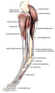
- Muscles of left pelvic limb of the dog, lateral view. 1

-
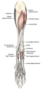
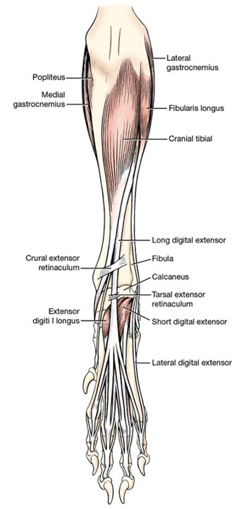
- Muscles of left pelvic limb of the dog, cranial view. 1
-

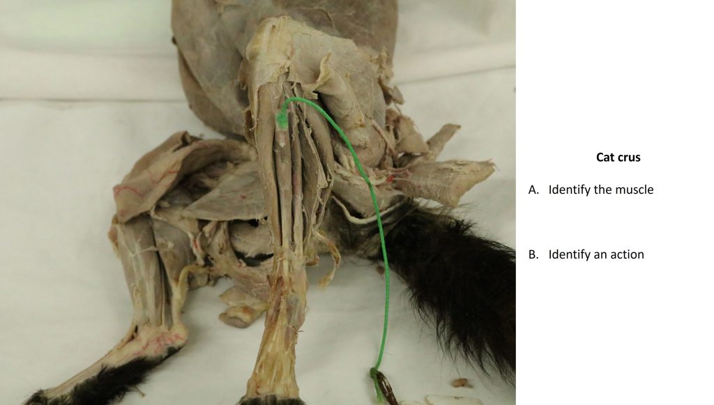
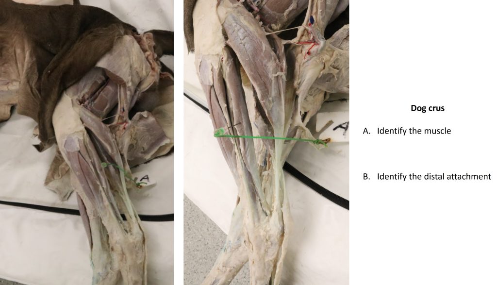
- Dog crus
-
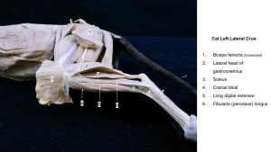
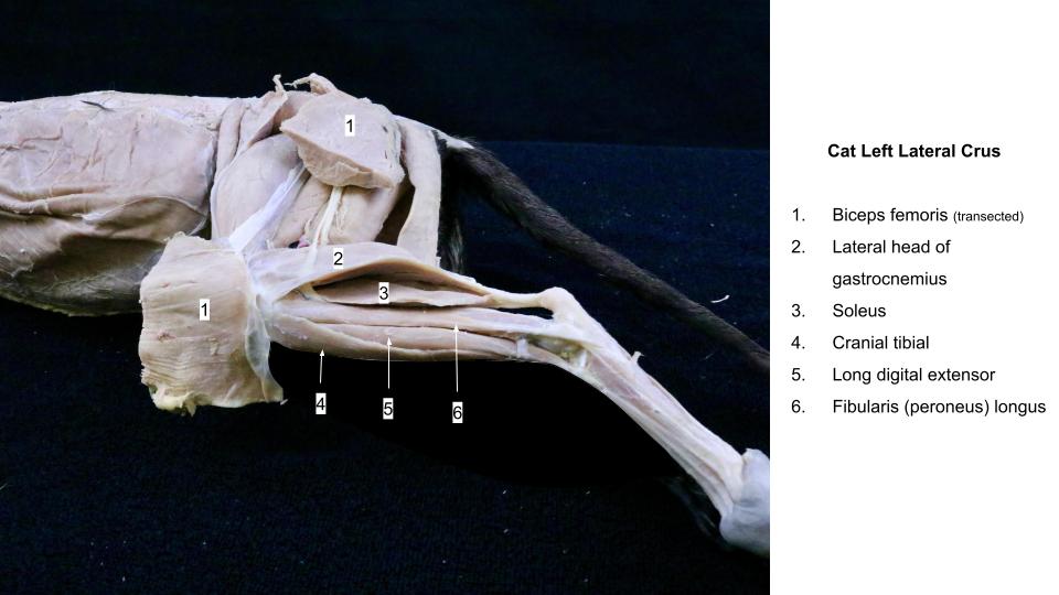
- Cat crus
Long digital extensor m.
The long digital extensor m. is a spindle-shaped muscle that is partly covered by the cranial tibial m. medially and the fibularis longus m. laterally. The proximal attachment is via a tendon to the extensor fossa of the femur. This tendon runs distally over the articular margin of the tibia in the extensor groove of the tibia and is lubricated by an extension of the stifle joint capsule. In the distal crus the tendon runs under the crural extensor retinaculum along with the cranial tibial tendon.
The long digital extensor tendon continues over the dorsal tarsus, surrounded by a synovial sheath and is held in place by the tarsal extensor retinaculum. At the tarsometatarsal junction the four tendons of the long digital extensor m. diverge toward their respective digits, where they insert on the extensor process of the distal phalanx.
PROXIMAL ATTACHMENT: Extensor fossa of the femur
DISTAL ATTACHMENT(S): Extensor process(es) of distal phalanx(ges) – species variation
ACTION: Flex the tarsus, extend the digits
Dissect: Clean and observe the long digital extensor m. and its four tendons of distal insertion to the level of the metatarsophalangeal joints (or at least the distal metatarsus).
-
- Muscles of the left crus of the dog. 1
-
- Muscles of left pelvic limb of the dog, lateral view. 1
-
- Muscles of left pelvic limb of the dog, cranial view. 1
-
- Dog crus
-
- Cat crus
-

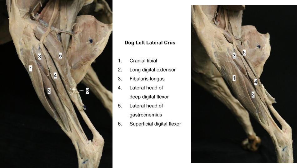
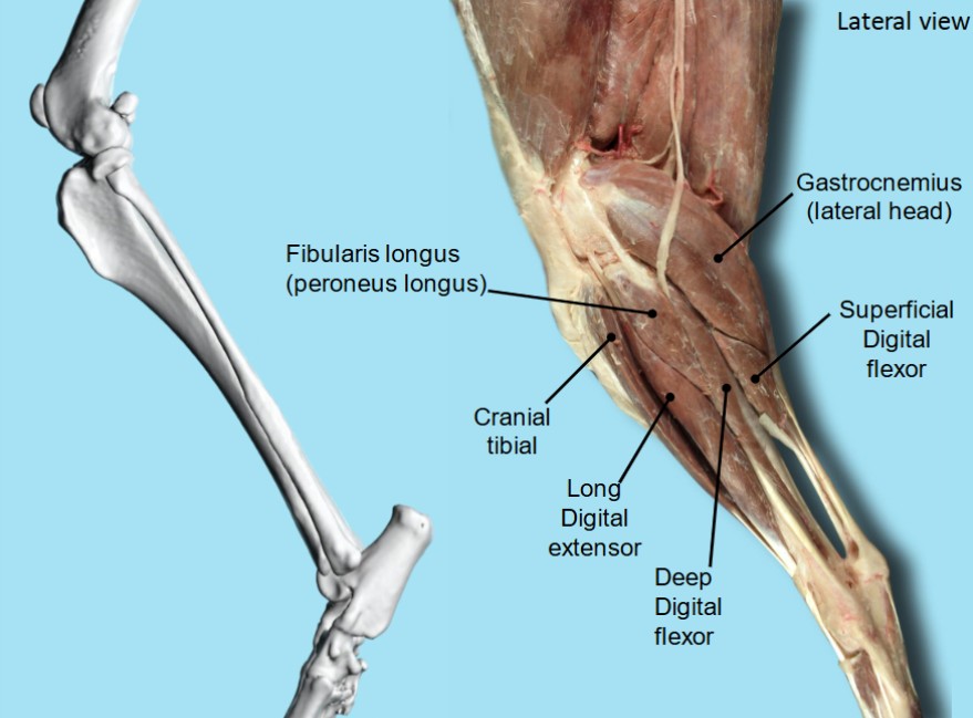
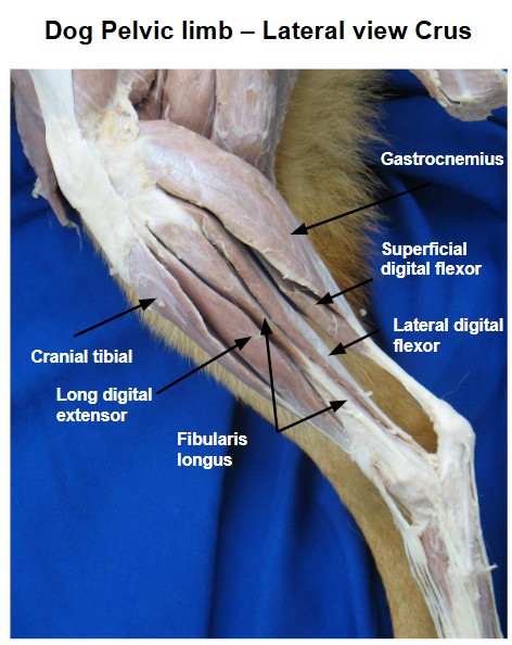
- Dog lateral crus 40
Fibularis longus m.
The fibularis longus m. lies just caudal to the long digital extensor m., where a triangular portion of its short belly lies directly under the crural fascia. It is a short, thick, wedge-shaped muscle that lies in large part cranial to the fibula. It arises from the lateral collateral ligament of the stifle and the adjacent parts of the tibia and fibula. Its stout tendon courses distally on the lateral side of the fibula caudal to the crural extensor retinaculum. It has a long synovial sheath that begins at a plane through the crural extensor retinaculum and extends to its insertion on the fourth tarsal bone and the plantar surfaces of the base of all metatarsals.
Dissect: Preserving all ligaments and tendons that lie superficial to the tendon of the fibularis longus m., trace it to its insertion on the fourth tarsal bone. Do not dissect the tendon beyond this attachment.
The tendon lies in a sulcus of the lateral malleolus of the fibula (check a skeleton) and it then makes a right angle turn medially around a groove in the fourth tarsal bone and courses to the plantar side of the tarsus.
PROXIMAL ATTACHMENT: Proximal tibia and fibula
DISTAL ATTACHMENT: Proximal metatarsals and T4
ACTION: Flex the tarsus
FYI: The lateral digital extensor m. and the fibularis brevis m. are located deep to the fibularis longus m. on the lateral aspect of the leg and need not be dissected in the carnivore. Because of the clinical significance of the lateral digital extensor m. in the horse, we are identifying this muscle in the horse and ruminant.
-
- Muscles of the left crus of the dog. 1
-
- Muscles of left pelvic limb of the dog, lateral view. 1
-
- Muscles of left pelvic limb of the dog, cranial view. 1
-
- Dog crus
-
- Cat crus
-
- Dog lateral crus 40
-
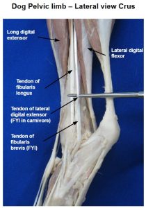
- Dog lateral crus J. Barnes

Caudal muscles of the crus
Gastrocnemius m.
The gastrocnemius m. consists of two heads that sandwich the superficial digital flexor m. between them. These muscles form the caudal bulge of the leg (i.e., the ‘calf’ muscles) and contribute the major component of the common calcaneal tendon.
The two heads of the gastrocnemius arise from the medial and lateral supracondylar tuberosities of the femur. In each tendon of origin there is a sesamoid bone that articulates with the caudodorsal aspect of the femoral condyle. These sesamoids are known clinically as the medial and lateral fabella, and anatomically they are termed the sesamoids of the gastrocnemius m. The sesamoids are readily apparent on radiographs and may be used as anchor points in surgical procedures of the stifle.
PROXIMAL ATTACHMENTS: Supracondylar tuberosities of the femur
DISTAL ATTACHMENT: Tuber calcanei
ACTION: Extend the tarsus (secondary – flex the stifle)
Dissect: Identify and clean the margins of the lateral and medial heads of the gastrocnemius and follow them to their union as a common tendon that inserts on the proximal dorsal surface of the tuber calcanei. See the superficial digital flexor m. description below for more understanding of the relationship between the two muscles and their tendons.
-
- Muscles of the left crus of the dog. 1
-
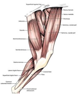
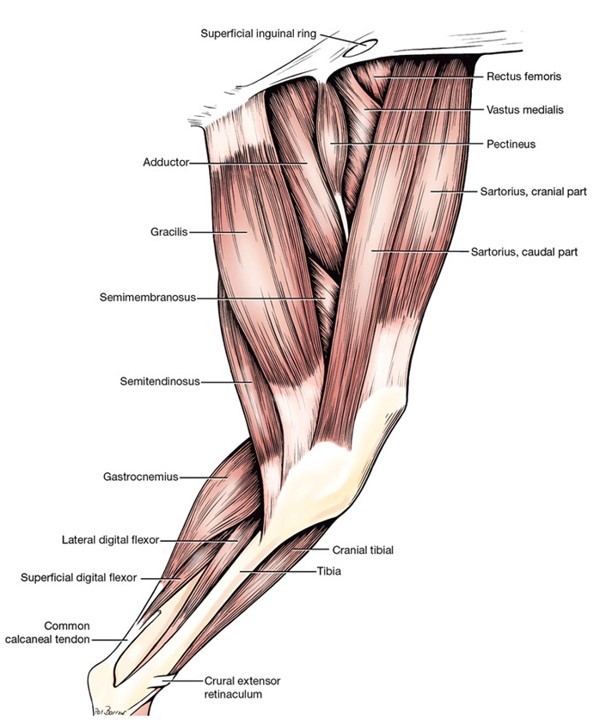
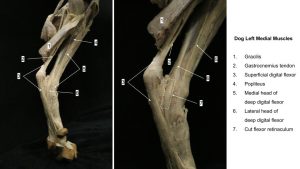
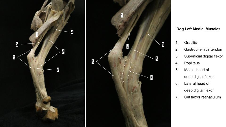
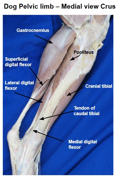
- Medial thigh and crus of the dog. 1
-
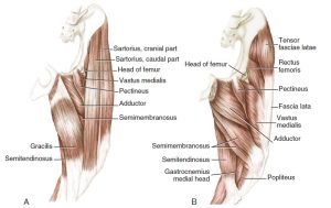
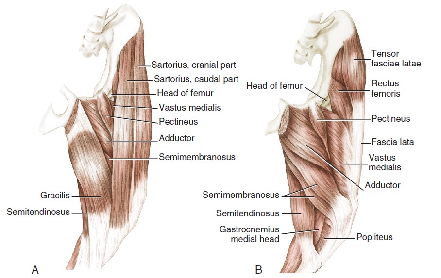
- Muscles of the thigh of the dog. A, Superficial muscles, medial aspect. B, Deep muscles, medial aspect. 3
-
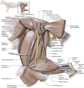
- Muscles of left pelvic limb of the dog, lateral view. 1
-
- Muscles of left pelvic limb of the dog, cranial view. 1
-
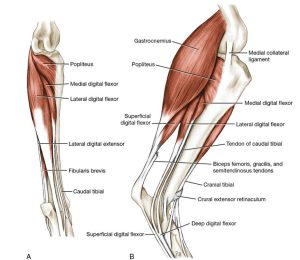
- A, Deep muscles of left crus of the dog, caudal view. B, Muscles of left crus, medial aspect. 1

-

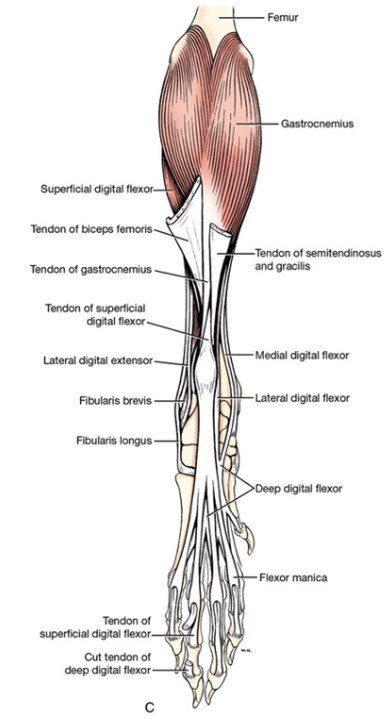
- Muscles of left pelvic limb of the dog, caudal view. 1
-
- Deep muscles of the hind limb of the cat in lateral view. 5

-

- Dog lateral crus J. Barnes
-
- Dog lateral crus 40
-
- Dog crus
-
- Cat crus
Superficial digital flexor m.
The superficial digital flexor (SDF) m. is a spindle-shaped muscle that arises from the lateral supracondylar tuberosity of the femur with the lateral head of the gastrocnemius. Its deep surface apposes the deep digital flexor and the popliteus mm., whereas its other surfaces are largely covered by the gastrocnemius muscle bellies (the SDF m. enclosed by the gastrocnemius muscle bellies is somewhat akin to a hot dog wrapped in its bun!). Proximal to the tuber calcanei, the SDF tendon passes from a deep to superficial location by crossing the medial surface of the gastrocnemius tendon (spiraling around it). The SDF tendon then widens, forms a cap over the tuber calcanei and attaches to each side, and then continues distally. As the SDF tendon passes over the tuber calcanei it is protected by a subtendinous calcaneal bursa – we will revisit this bursa more closely in the horse for its relevance to penetrating trauma in the region. At the distal plantar surface of the tarsus/proximal metatarsal level, the tendon bifurcates; each of these branches in turn bifurcates, thus forming four tendons of nearly equal size that attach to the middle phalanx of their respective digit.
PROXIMAL ATTACHMENT: Lateral supracondylar tuberosity – carnivore; Supracondylar fossa – ungulate
DISTAL ATTACHMENTS: Tuber calcanei and middle phalanx(ges) (plus proximal phalanx in horse)
ACTION: Extend the tarsus, flex the digit(s)
Dissect: With a probe, separate the gastrocnemius tendon from the superficial digital flexor tendon by entering between their tendons just proximal to the calcaneus. Proximally, bluntly separate the superficial digital flexor m. as much as possible from the heads of the gastrocnemius.
-
- Medial thigh and crus of the dog. 1
-
- Muscles of left pelvic limb of the dog, lateral view. 1
-
- A, Deep muscles of left crus of the dog, caudal view. B, Muscles of left crus, medial aspect. 1
-
- Muscles of left pelvic limb of the dog, caudal view. 1
-
- Deep muscles of the hind limb of the cat in lateral view. 5
-
- Dog lateral crus 40
-
- Dog crus
-
- Dog crus
-

- Cat crus
Soleus m. – feline
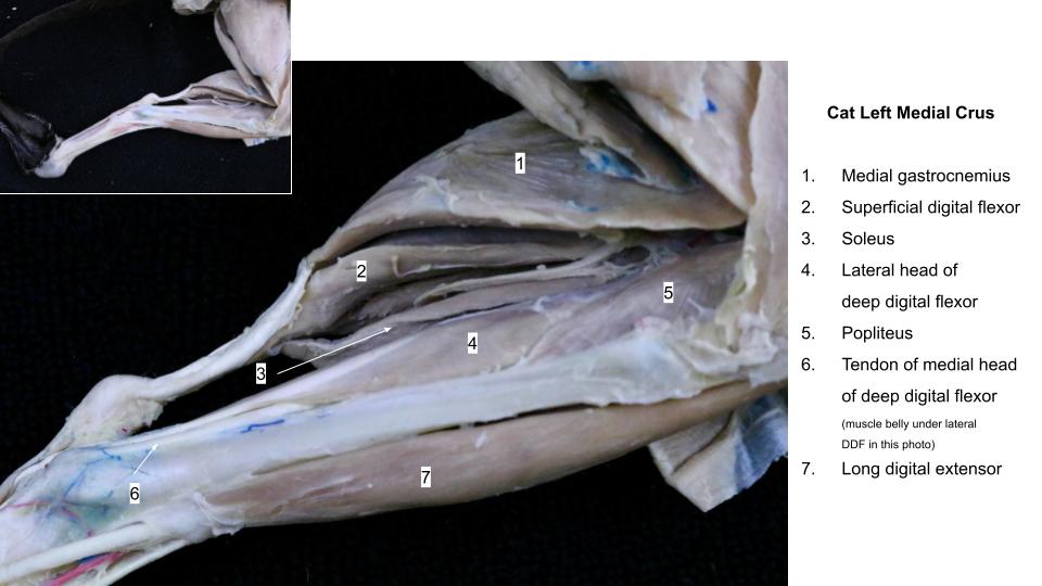
In the cat, the soleus m. is located caudolaterally, adjacent the lateral head of the gastrocnemius m. It originates on the proximal fibula and terminates on the common calcaneal tendon. Its action is extension of the tarsus. It is innervated by the tibial n. (revisited in nervous system unit).
PROXIMAL ATTACHMENT: Proximal fibula
DISTAL ATTACHMENT: Common calcaneal tendon
ACTION: Extend the tarsus
Dissect: Cat cadavers – identify and clean the margins of the soleus m., being sure to distinguish it from the gastrocnemius lateral head, and the superficial digital flexor m., located caudally to it and between the gastrocnemius heads.
-
- Deep muscles of the hind limb of the cat in lateral view. 5
-
- Cat crus
Deep digital flexor m.
The deep digital flexor m. (as for the thoracic limb) typically has 3 heads in the pelvic limb, however in the carnivore, the very small caudal tibial head does not contribute to the deep digital flexor combined tendon and is therefore considered an independent muscle (that we have little cause to dissect in the carnivore, however we will identify and dissect it in ungulates). The two heads of note in the carnivore are the lateral and medial heads of the deep digital flexor. The caudal tibial head is listed for completeness at this time.
- The lateral head of the deep digital flexor m. is also more simply known and labeled as the lateral digital flexor m.. It arises from the caudolateral border of the proximal two thirds of the tibia, most of the proximal half of the fibula, and the adjacent interosseous membrane. Its tendon begins as a wide expanse on the caudal side of the muscle but condenses distally. The tendon is surrounded by the tarsal synovial sheath as it passes over the sustentaculum tali of the calcaneus, through the flexor canal of the tarsus. The flexor retinaculum binds the tendon in the flexor canal of the tarsus.
- The medial head of the deep digital flexor m. is also more simply known and labeled as the medial digital flexor m. It is smaller and lies between the lateral digital flexor m. and the popliteus m. From the proximal end of the tibia, it runs distomedially. Its tendon lies on the caudomedial side of the tibia and then the tendon passes in its own dedicated synovial sheath tunnel over the medial tarsus. The tendon angles plantarly and at the proximal metatarsus it unites with the larger tendon of the lateral digital flexor to form the singular deep digital flexor tendon at this proximal level.
- Caudal tibial m. or caudal tibial head. – very fine independent muscle of caudal crus in the carnivore and not to be identified. It remains a functional head of the DDF in ungulates, where it will be identified.
Subsequently, at the plantar mid-metatarsus level, the deep digital flexor tendon divides into four branches, that each travel to their respective digit and attach to the distal phalanx (similar to the thoracic limb).
PROXIMAL ATTACHMENTS: Proximal caudal tibia and fibula – species variation
DISTAL ATTACHMENT: Plantar distal phalanx(ges)
ACTION: Extend tarsus, flex digit(s)
Dissect: Vertically transect the flexor retinaculum, the thick fibrous tissue which forms the caudomedial border of the flexor canal of the tarsus, to access the tendon of the lateral head of the deep digital flexor, in the canal. Clean the tendons of the medial and lateral heads of the deep digital flexor m., and at the level of the distal tarsus/proximal metatarsus, observe the smaller tendon of the medial head joining the larger tendon of the lateral head, of the deep digital flexor, to form a common tendon.
-
- A, Deep muscles of left crus of the dog, caudal view. B, Muscles of left crus, medial aspect. 1
-
- Muscles of left pelvic limb of the dog, caudal view. 1
-
- Dog lateral crus 40
-
- Dog crus
-
- Dog crus
-
- Cat crus
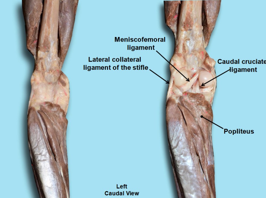
Popliteus m.
The popliteus m. is covered by the gastrocnemius m. and the superficial digital flexor m. and lies on the stifle joint capsule and caudal surface of the proximal tibia. It arises from the lateral epicondyle of the femur by a long tendon. The tendon courses caudally, deep to the lateral collateral ligament of the stifle. Where the tendon transitions into the muscle belly, there is a sesamoid that articulates with the caudal aspect of the lateral condyle of the tibia. The muscle portion starts at the lateral condyle of the tibia and extends around to attach to the proximal third of the tibia on the medial side of the leg, related along its distal margin to the deep digital flexor m. heads.
PROXIMAL ATTACHMENT: Lateral epicondyle of the femur
DISTAL ATTACHMENT: Proximal caudal tibia
ACTION: Medial rotation of leg at the stifle
Dissect: On the proximal, caudomedial aspect of the tibia, identify and clean the margin of the popliteus m. to separate it from the deep digital flexor m. heads. Observe the popliteus muscle on prosected models too. Note that the sesamoid of the popliteus muscle is located deep to its proximal tendon of attachment. The sesamoid is seen radiographically and should not be misinterpreted as a fracture fragment.
-
- A, Deep muscles of left crus of the dog, caudal view. B, Muscles of left crus, medial aspect. 1
-

- Dog medial crus J. Barnes
-

- Dog popliteus 40
-
- Cat crus
Ungulate comparative anatomy
Observe: Refer to the prosected ungulate cadavers and additional specimens and models to identify and study the following content. Below is a horse section and a ruminant section. We are not identifying crus muscles in the pig. Certain muscles have been transected midbody or at their attachments to facilitate viewing deeper structures. In the following lab further exploration of the ungulate limb anatomy will be available through dissection of the horse limb.
Craniolateral crus – horse
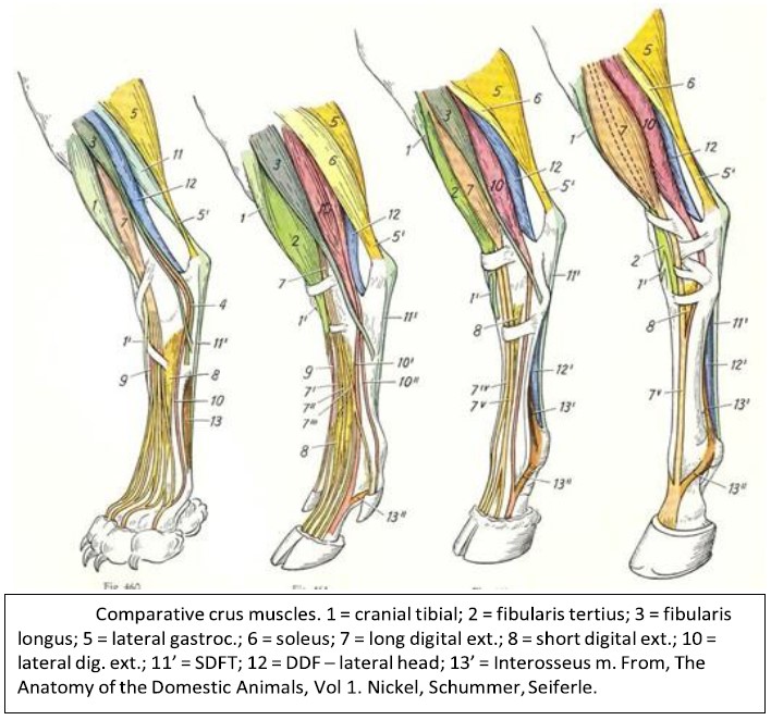
The very dense crural fascia has been excised to isolate and identify the muscles and tendons of the craniolateral crus. Crus muscles show comparative variation between the species and the color-coded figure below is a useful resource for understanding the differences.
Notably the ungulates have a fibularis tertius m. that is not present in carnivores (peroneus tertius is an older term for this muscle and it is still commonly used in clinical language and in the literature). The fibularis tertius m. is a fleshy muscle belly in artiodactyls and is represented as a fibrous band in the horse. This is a very distinct difference between the horse and artiodactyls! The prominent fleshy muscle in the cranial crus of the horse is the long digital extensor m. What looks to be the same muscle in the ruminant is the fibularis tertius m. Be sure to identify the lateral digital extensor m. – it is significant in the surgical treatment of Stringhalt in the horse. The soleus m., identified in the cat, is present in all ungulates, and is most developed in the pig.
Observe: Refer to horse prosections and figures to identify the cranial tibial m., long digital extensor m., fibularis (peroneus) tertius m. (a distinct fibrous band in the horse, and a fleshy muscle in artiodactyls), and lateral digital extensor m. Review the attachments of the muscles (see comparative muscle chart).
-

- Comparative crus muscles. 23
-
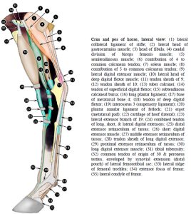
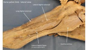
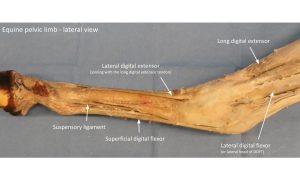
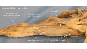
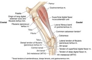
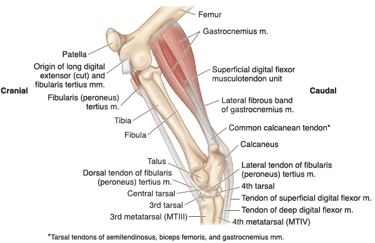
- Crus and pes of the horse, lateral view. 2

-
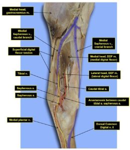
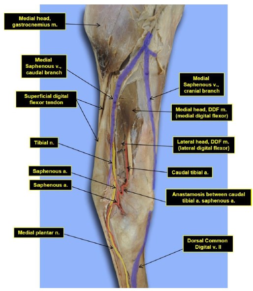
- Medial crus and tarsus of the horse. 16
-
- Horse crus

-
- Horse crus

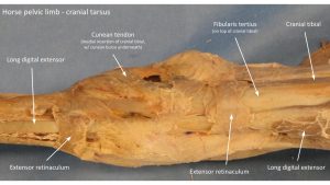
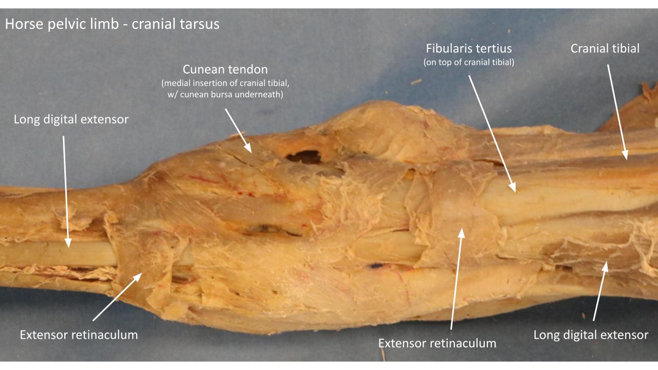
Tendons on the craniolateral crus are bound down to the limb by three (3) horizontal bands of fibrous tissue as they cross over the tarsal region – as in the carnivore, each band is called an extensor retinaculum (crural, tarsal, and metatarsal bands). At the distal attachments of the cranial tibial and fibularis tertius mm. both of these muscles have two tendons of insertion that are closely related. The medial tendon of insertion of the cranial tibial m. is referred to as the cunean tendon and it passes over the cunean bursa.
Observe: Observe the extensor retinaculum (pl: -a) on the prosected limbs and models. Observe the dissected distal attachments of the fibularis (peroneus) tertius and cranial tibial mm. at the tarsus. Note the transected medial insertion tendon of the cranial tibial m. (= cunean tendon) and reflect it to expose the cunean bursa.
-
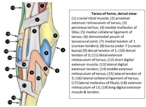
- Tarsus of horse, dorsal view. 2

-
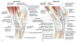
- Equine tarsus. (A) Lateral view of the tarsus depicting the muscles, tendons, ligaments, synovial sheaths, and bursae. (B) Medial viewof the same region. 9

-
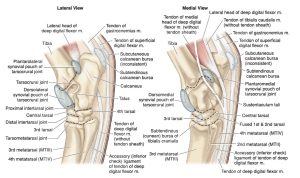
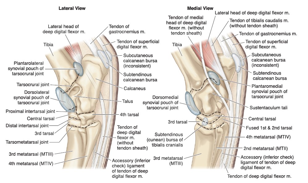
- Joints and bursae of the equine tarsus. 9
-
- Horse crus

-
- Horse crus
Clinical relevance: Q4
“Stringhalt” and the lateral digital extensor m. in the horse; clinical signs of ruptured fibularis (peroneus) tertius (part of reciprocal apparatus, see below); cunean tenectomy to treat bone spavin (not used as commonly for treatment now).
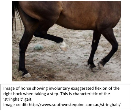
Q4: Which muscle and its tendon is partially removed as a surgical aid to managing stringhalt? [see clinical relevance above, refer to lecture notes too].
A – long digital extensor m.
B – fibularis tertius m.
C – lateral digital extensor m.
D – gastrocnemius m.
Caudal crus – horse
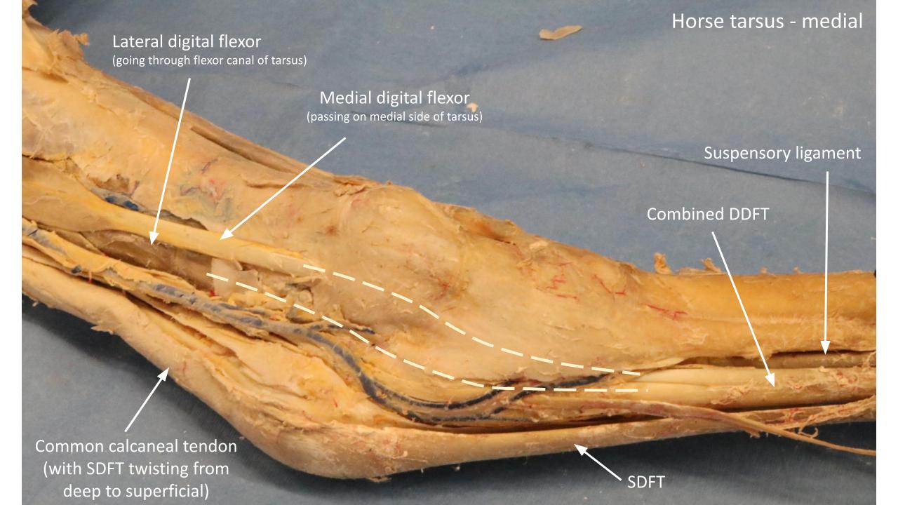
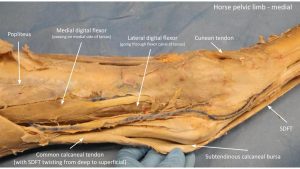
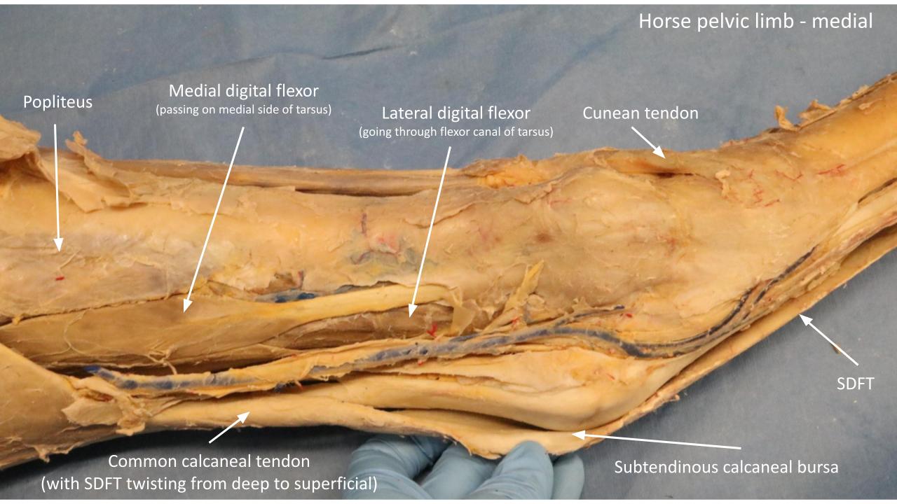
The caudal crus muscles are much the same as in carnivores with a few highlights. As previously mentioned, the soleus m., identified in the cat, is present in all ungulates, and is most developed in the pig. The superficial digital flexor muscle in the horse is primarily a fibrous band along its length, with only a small amount of muscle present proximally. A very strong common calcaneal tendon is present. The deep digital flexor has the typical three (3) heads and all three contribute to the DDF tendon (unlike in carnivores, where the caudal tibial head remained a separate structure). The tendons of the lateral and caudal tibial heads of the deep digital flexor pass through the flexor canal of the tarsus, wrapped in a synovial sheath. The flexor canal is bound plantaromedially by the fibrous flexor retinaculum. This retinaculum is incised to view the tendons passing through the canal. The tendon of the medial head of the deep digital flexor passes over the medial surface of the tarsus within its own tendon sheath, and unites with the other DDF tendons in the proximal metatarsus. These structures will be considered in detail in the next lab.
Observe: On horse prosections and models, and referring to figures, identify the medial and lateral heads of the gastrocnemius m., superficial digital flexor m., deep digital flexor m. (3 heads: lateral head of DDF aka lateral digital flexor, medial head of DDF aka medial digital flexor, and caudal head or caudal tibial m.), common calcaneal tendon (includes contributions from biceps femoris, semitendinosus), and popliteus m. Separate the division in the common calcaneal tendon to note how the SDFT passes from deep to superficial by spiraling around the tendon of the gastrocnemius m. Review the attachments of these muscles (see muscle chart).
-
- Comparative crus muscles. 23
-
- Crus and pes of the horse, lateral view. 2
-
- Horse crus
-
- Horse crus
-

- Horse crus
-
- Horse crus
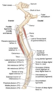
Reciprocal apparatus – horse
The very fibrous fibularis tertius (FT) and superficial digital flexor (SDF) mm. act in tandem as mechanical connectors between the stifle and tarsus, entirely a function of their location and attachments. Whenever the stifle of a horse is extended, tension on the fibrous superficial digital flexor results in an obligatory (i.e. reciprocal) extension of the tarsus. If the stifle is flexed, so then is the tarsus. One joint is flexed or extended and the other follows with the same action i.e. reciprocates. Thus, the stifle and tarsus work in unison because of their SDF and FT mechanical connections and these two fibrous structures together make up the reciprocal apparatus of the horse. It follows then, that when the stifle is locked in extension by the patellar locking mechanism (received a mention back in Lab 16A), the tarsus is fixed in extension, and this is an important contribution to the pelvic limb passive stay apparatus (hang on, the what??). For further understanding of this stay apparatus and locking mechanism, hold on a tic because the next lab takes a deep dive into these concepts and anatomy. After Lab 21A, pop back to this paragraph and it ideally makes more sense.
-
- Lateral view of the stay apparatus of the horse pelvic limb. 9

-
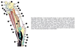
- Crus of the horse, lateral view. 2

-
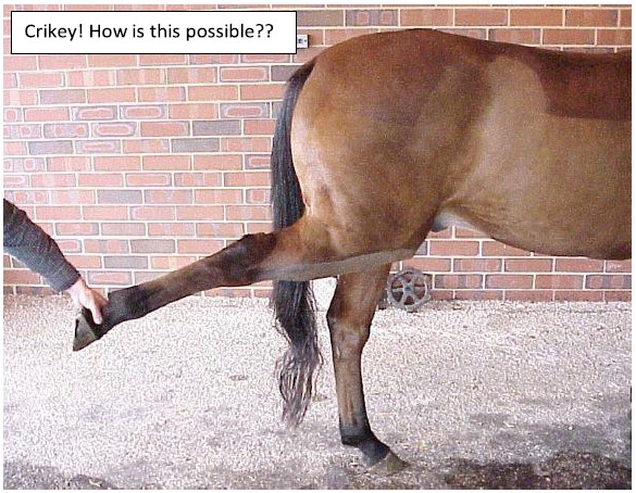
- Lateral view of the reciprocal apparatus of the horse pelvic limb. 9
Clinical relevance: reciprocal apparatus and rupture of fibularis tertius m.
Reciprocal apparatus is part of the pelvic limb passive stay apparatus. When the fibularis tertius is ruptured, the tarsus can be extended while the stifle is flexed, a distinct clinical finding!
Craniolateral crus – ruminant
Dense crural fascia has been excised to isolate and identify the muscles and tendons of the craniolateral crus. Crus muscles show comparative variation between the species and the color-coded figure below is a useful resource for understanding the differences.
Notably the ungulates have a fibularis tertius m., that is not present in carnivores (peroneus tertius is an older term for this muscle and it is still commonly used in clinical language and in the literature). The fibularis tertius m. is a fleshy muscle belly in artiodactyls and is represented as a fibrous band in the horse. This is a very distinct difference between the horse and artiodactyls! The prominent fleshy muscle in the cranial crus of the horse is the long digital extensor m. What looks to be the same muscle in the ruminant is the fibularis tertius m. The long digital extensor m. in the ruminant has two identifiable muscles bellies, supplying tendons to the two digits they bear weight on. The soleus m., identified in the cat, is present in all ungulates, and is most developed in the pig. Like carnivores, ruminants also have the fibularis (peroneus) longus m., a muscle that is not present in the horse.
-
- Comparative crus muscles. 23
-
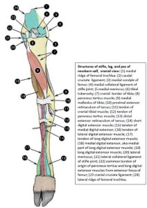
- Structures of stifle, leg, and pes of newborn calf; cranial view. 2

-
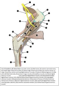
- Crus of the newborn calf; lateral view. 2

Tendons on the craniolateral crus are bound down to the limb by two (2) horizontal bands of fibrous tissue as they cross over the tarsal region – as in the carnivore, each band is called an extensor retinaculum (crural and metatarsal bands).
Observe: Observe the extensor retinacula (sing: -um) on the prosected limbs and models.
Caudal crus – ruminant
The caudal crus muscles are much the same as in carnivores with a few highlights. As previously mentioned, the soleus m., identified in the cat, is present in all ungulates, and is most developed in the pig. The deep digital flexor has the typical three (3) heads and all three contribute to the DDF tendon (unlike in carnivores, where the caudal tibial head remained a separate structure). A very strong common calcaneal tendon is present.
-

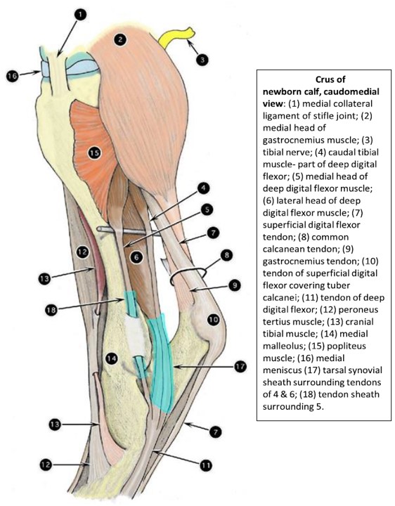
- Crus of the newborn calf; caudomedial view. 2
Clinical relevance: Q5
Ruptured gastrocnemius mm. or tendons; progressive hindlimb paralysis; spastic paresis (Elso Heel).
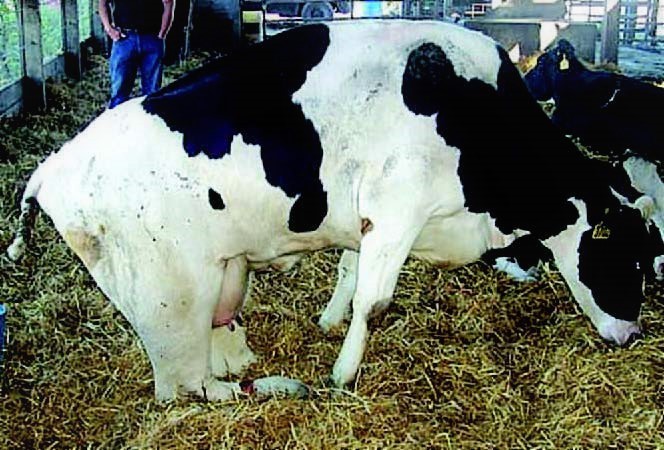
Q5: This dairy cow can not rise beyond this point and shows a ‘rabbit leg’ position of both hind limbs. The cow is unable to extend her hocks and weight bear. Rupture of what muscle in the caudal crus results in this clinical presentation? [review muscle chart for answer].
A: deep digital flexor m.
B: gastrocnemius m.
C: superficial digital flexor m.
D: popliteus m.
Review videos
These are the same videos listed in lab 2, just linked to begin at the crus.
Dog crus muscles – 7 minutes
Horse crus muscles – 18 minutes
Calf crus muscles – 14 minutes
Goat crus muscles – 10 minutes
Horse passive stay apparatus – 19 min
Muscle and Structures Chart
| Crus Muscle Action Groups | |
| Flexors of the tarsus |
|
| Flexors of the tarsus and extensors of the digit(s) |
|
| Extensors of the tarsus |
|
| Extensors of the tarsus and flexors of the digit(s) |
|
| Muscle/structure | Attachments/comments | Action/comments |
| Cranial tibial m. |
|
Flex tarsus |
| Cunean tendon – horse | Medial tendon of insertion of Cranial tibial m. attaches to fused T1/T2 bones | Horse only |
| Cunean bursa – horse | Subtendinous bursa of cunean tendon | Horse only |
| Long digital extensor m. |
|
Flex tarsus, extend digit(s). |
| Extensor retinaculum | Fibrous band binding down tendons – species variation | |
| Lateral digital extensor m. – horse and ruminant |
|
Flex tarsus, extend digit(s) |
| Fibularis longus m.
(not present in horse) |
|
Flex tarsus |
| Fibularis tertius m. – horse and ruminant |
|
Flex tarsus |
| Gastrocnemius m. – medial and lateral heads |
|
Extend tarsus
(Secondary: flex the stifle) |
| Superficial digital flexor m. |
|
Extend tarsus, flex digit(s) |
| Soleus m. – identify in cat |
|
Extend tarsus |
| Common calcaneal tendon | Powerful combined tendon on caudal crus | Recall the contributing muscles. |
| Deep digital flexor m.
3 heads: Lateral digital flexor Medial digital flexor Caudal tibial (ungulates) |
|
Extend tarsus, flex digit(s) |
| Popliteus m. |
|
Medial rotation of leg at the stifle |
| Reciprocal apparatus – horse | Fibularis tertius and SDF mm. working together, part of PL passive stay apparatus in horse. | Stifle and tarsus extend and flex in unison in horse. |