Lab 5A: Nerves of the Ungulate Thoracic Limb
Learning Objectives
- Name and identify nerves and their branches in the extrinsic and intrinsic mm. of the TL.
- Understand innervation of muscle action groups.
- Identify the relevant nerves to perform a distal limb nerve block in the equine.
Lab Instructions:
Ungulate Proximal Limb
While obviously much larger and longer, the nerves of the limbs generally follow the same path and innervate the same muscles as we observed in the carnivores. (These patterns exist despite 60+ million years of evolutionary history separating carnivorans from perissodactyls and artiodactyls. Evolutionary conservation is pretty amazing!).
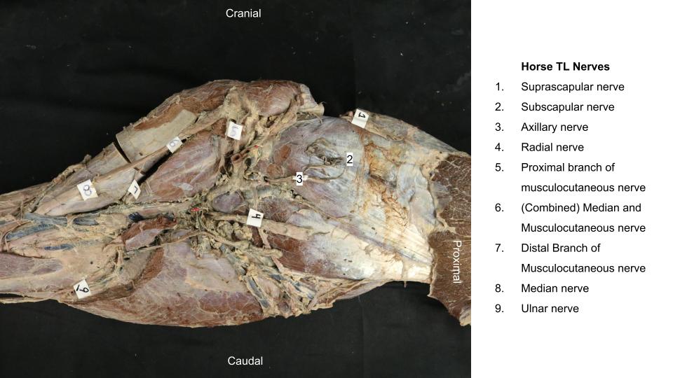
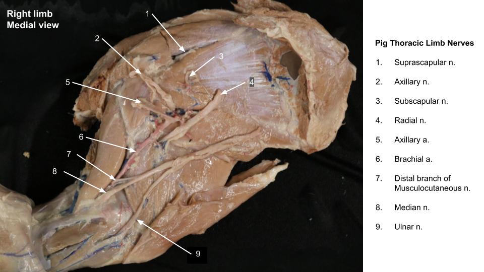
Observe: Identify the following nerves in the instructors’ ungulate prosections:
- Suprascapular n. (passes between supraspinatus and subscapularis mm. supplies supraspinatus and infraspinatus mm.)
- Subscapular n. (stumps of nerve passing into subscapularis m.)
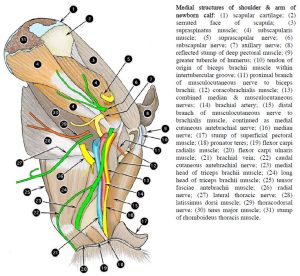
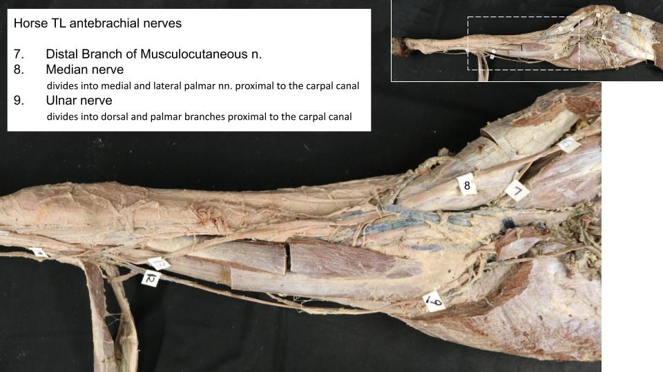
- Musculocutaneous n. (proximal branch to biceps brachii m., distal branch to brachialis m.; frequently combined with median in ungulate brachium)
- Medial cutaneous antebrachial n. (horse)
- Axillary n. (supplies flexors of shoulder, passes proximal to insertion of teres major m.)
- Radial n. (passes distal to insertion of teres major m., deep branch supplies extensors of elbow, carpus, digit)
- Median and ulnar nn. (supply flexors of carpus and digit).
-
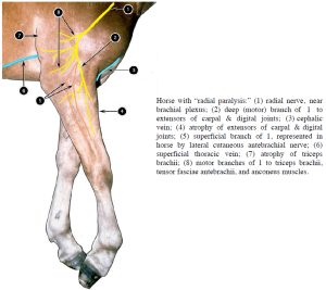
- Horse with “radial paralysis”. 2

-
-
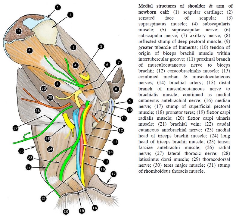
Medial structures of shoulder & arm of
newborn calf. 2
-

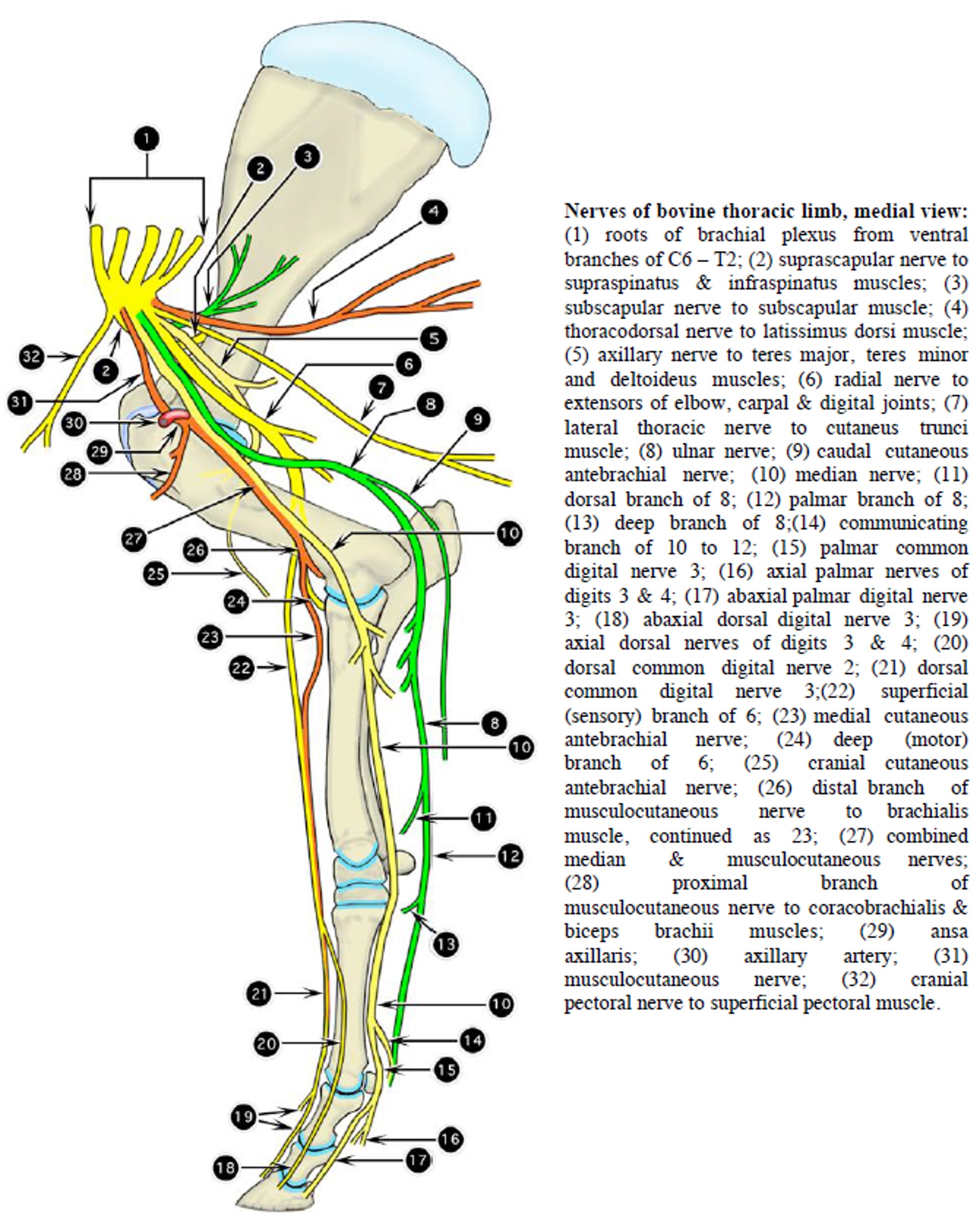
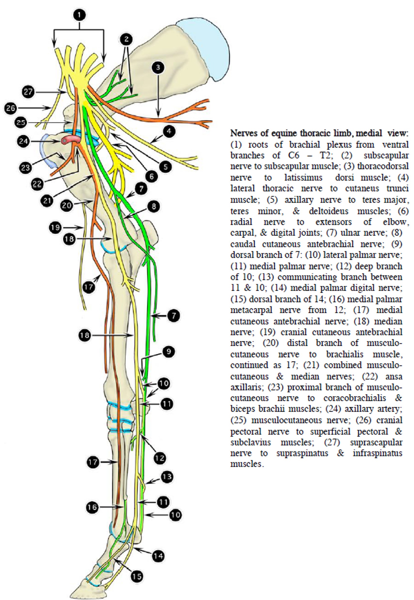
- Nerves of bovine thoracic limb, medial view. 2
-
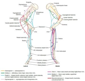
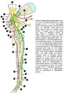
- Muscles of the equine thoracic limb and their innervation. 9

Clinical relevance:
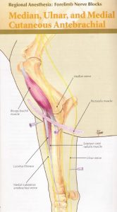
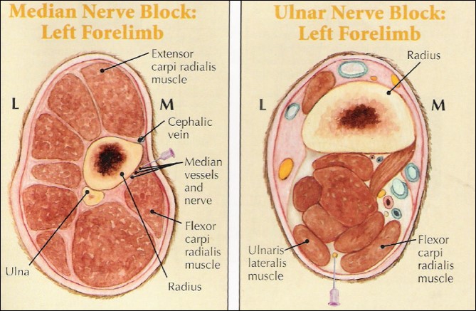
Nerve blocks of ulnar and median nn.; the ulnar n. is blocked 10 cm proximal to the accessory carpal bone, in the groove between the ulnaris lateralis and flexor carpi ulnaris mm. Median n. is blocked about 10 cm distal to the elbow joint on the mid antebrachium, directing the needle in at the caudal border of the flexor carpi radialis m. and toward the caudal surface of the radius. These two blocks are part of the MUM block of the equine thoracic limb to desensitize the entire limb distal to these blocks – it is not commonly performed.
-
- MUM nerve block. 23

-

- Median and Ulnar Nerve Blocks. 23
Equine Distal Limb
Dissect: You have been provided with the distal half of the equine thoracic limb to dissect its nerves. Skin the limb to the mid pastern, being cautious around the palmar surface of the metacarpus where some nerves become more superficial. Use the following paragraphs as a guide to finding and identifying the important nerves.
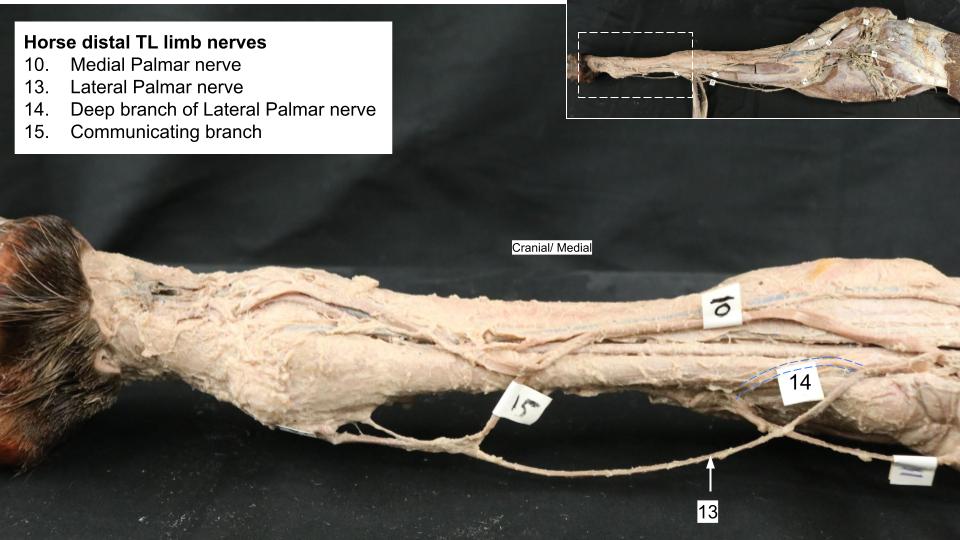
In the distal half of the equine antebrachium, the median n. divides into medial and lateral palmar nn., and an important redistribution of nerve fibers occurs. The medial palmar nerve continues through the carpal canal with the medial palmar a.. A few centimeters to the accessory carpal bone, the ulnar n. divides into dorsal and palmar branches. The dorsal branch immediately penetrates the tendon of the ulnaris lateralis m. and is distributed to the dorsolateral aspect of the carpus and metacarpus. The dorsal branch can be palpated as it crossed the lateral surface of the ulnaris lateralis tendon, just proximal to the accessory carpal bone. The palmar branch of the ulnar n. joins the lateral palmar n. (that arose from the median n.). From this junction, the combined nerve continues as the lateral palmar n. (now a mixture of median and ulnar n. fibers), and it accompanies the lateral palmar a. into the metacarpus.
-
- Nerves of equine thoracic limb, medial view. 2
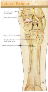
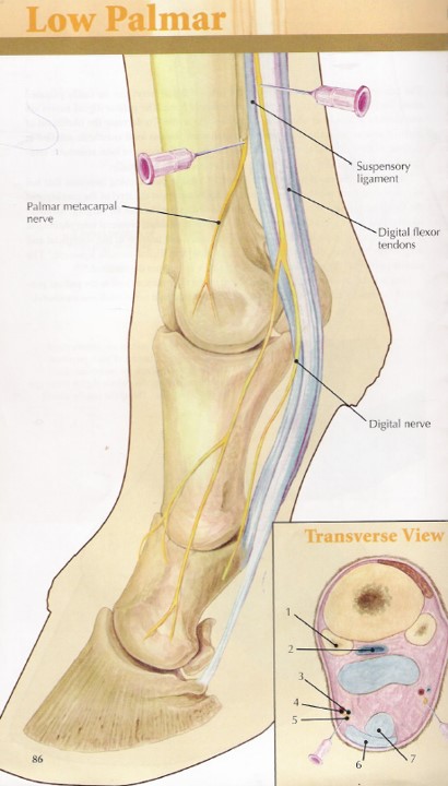
The lateral palmar n. gives off a deep branch in the proximal palmar metacarpus that then divides into the medial and lateral palmar metacarpal nn.. These nerves supply the suspensory ligament (3rd interosseous m.) and continue distally, located deep in metacarpus. The palmar metacarpal nn. may be identified as they branch from the deep branch of the lateral palmar n. proximally, or perhaps as fine nerves emerging just distal to the ‘buttons’ of the splint bones (this is one location they are blocked).
The palmar nerves and vessels descend next to the DDFT in the metacarpal region. Note the communicating branch of medial and lateral palmar nn. at the level of the mid-metacarpus on the palmar surface of the SDFT (this branch may have been disrupted during skin removal).
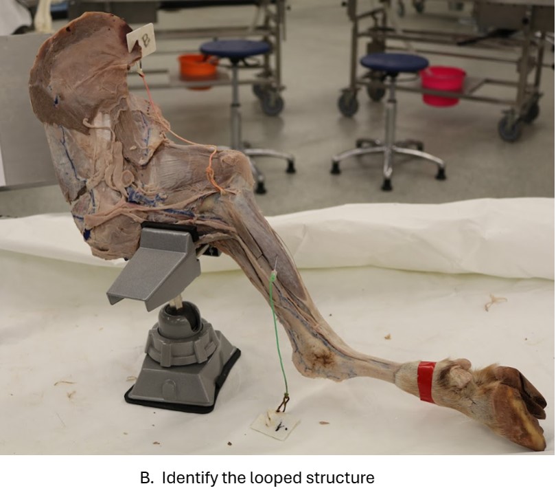
Dissect: Dissect the medial and lateral palmar nn. to the level of the fetlock at which point they continue as the medial and lateral palmar digital nn., passing into the digit alongside the DDFT. The palmar digital nn. provide dorsal branches to the pastern.
Identify the deep branch of the lateral palmar nerve diving deep to the suspensory ligament near the proximal end of the metacarpus.
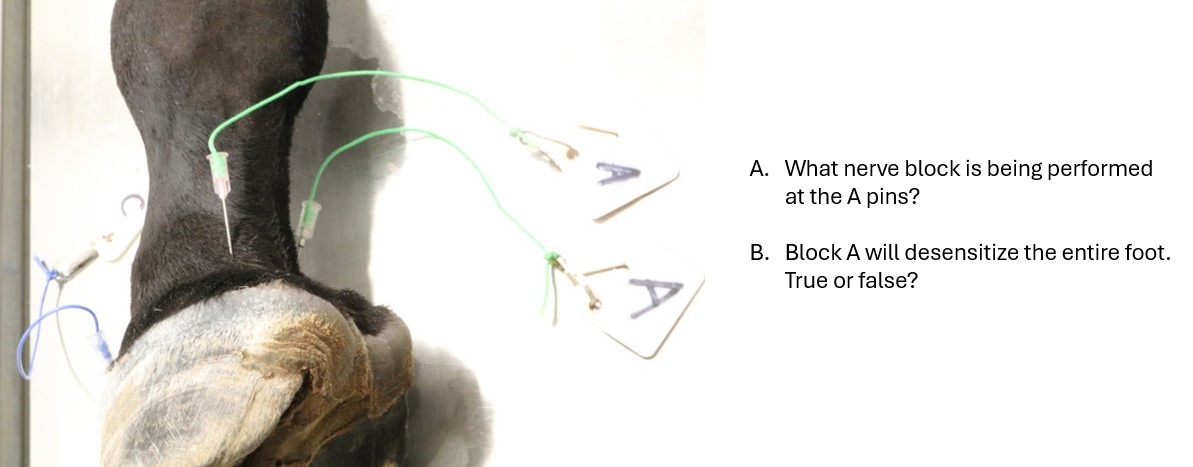
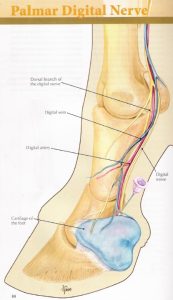
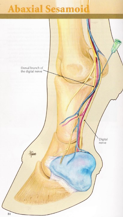
The digital vessels and nerves are palpable in the live horse as they cross the proximal sesamoid bones on their abaxial surface. Here, the digital pulse can be evaluated, and this is also the site of the abaxial sesamoid nerve block. The trio of structures lie closely related in sequence dorsally to palmarly as the vein, artery and nerve i.e. VAN.
-
- Lateral palmar nerve block. 23

-
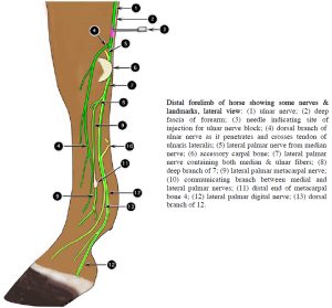
- Distal forelimb of horse showing some nerves & landmarks, lateral view. 2

-
-
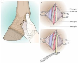
Palmar digital neurectomy, showing path of ligament of ergot over the neurovascular structures. The skin incision is made over the tensed ligament of ergot. From
https://cmapsconverted.ihmc.us

Clinical relevance:
Distal limb nerve blocks for lameness examination, eg low 4 point, abaxial sesamoid block, palmar digital nerve block; knowing the locations of these nerves and vessels helps avoid transecting them during limb surgeries unless indicated to actually transect them! (e.g. palmar digital neurectomy); palpation of digital pulses is common in physical examination.
-
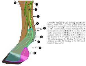
- Left distal forelimb of horse showing sites of nerve blocks, lateral view. 2

-
- Palmar digital nerve block. 23

-

- Abaxial sesamoid nerve block. 23
-

- Low 4 point nerve block. 23
Observe: Refer to prosections and models to study the nerve distribution to the distal thoracic limb of the horse and guide your dissection.
Review Videos
Horse TL nerves (cadaver) – Watch until 9 min
Calf TL nerves (cadaver) – 5 min
Goat TL nerves (cadaver) – Watch until 4:20
Pig TL nerves (cadaver) – 3 min
Horse distal thoracic limb nerve distribution (drawing) – 6 min
Interactive review content:
Terms
| Terms | Species |
| Radial n. | All |
| Musculocutaneous n. | All |
| Median n. | All |
| Ulnar n. | All |
| Medial palmar n. | Horse |
| Carpal canal (i.e., Flexor canal of the carpus) | Horse |
| Lateral palmar n. | Horse |
| Communicating branch | Horse |
| Deep branch of lateral palmar n. | Horse |
| Medial and lateral palmar metacarpal nn. | Horse |
| Medial and lateral palmar digital nn. | Horse |
| Suspensory ligament | Horse; ox |