Lab 6A: Nerves of the Carnivore Pelvic Limb
Learning Objectives
- Recall the roots of the lumbosacral plexus.
- Identify the nerves and their branches in the PL.
- Understand innervation of muscle action groups of the PL.
- Recognize innervation and autonomous zones of the pelvic limb and pes.
Lab Instructions:
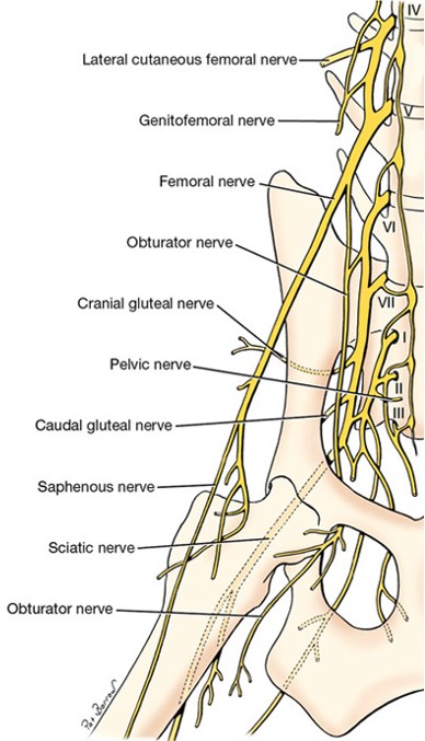
Lumbosacral plexus
The nerves of the pelvic limb originate from the lumbosacral plexus. The lumbosacral plexus is diffuse and consists of the ventral branches of the lumbar and sacral spinal nerves. Unlike the brachial plexus, the spinal nerves that contribute roots to the lumbosacral plexus have variability within and between species. Broadly speaking, spinal nerves of L3-S3 (i.e., third lumbar spinal nerve to the third sacral spinal nerve) contribute to the lumbosacral plexus. We will not identify these roots in the cadaver.
-

- Right lumbosacral nerves and left arteries of the dog, ventral view. 1
-
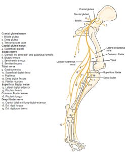
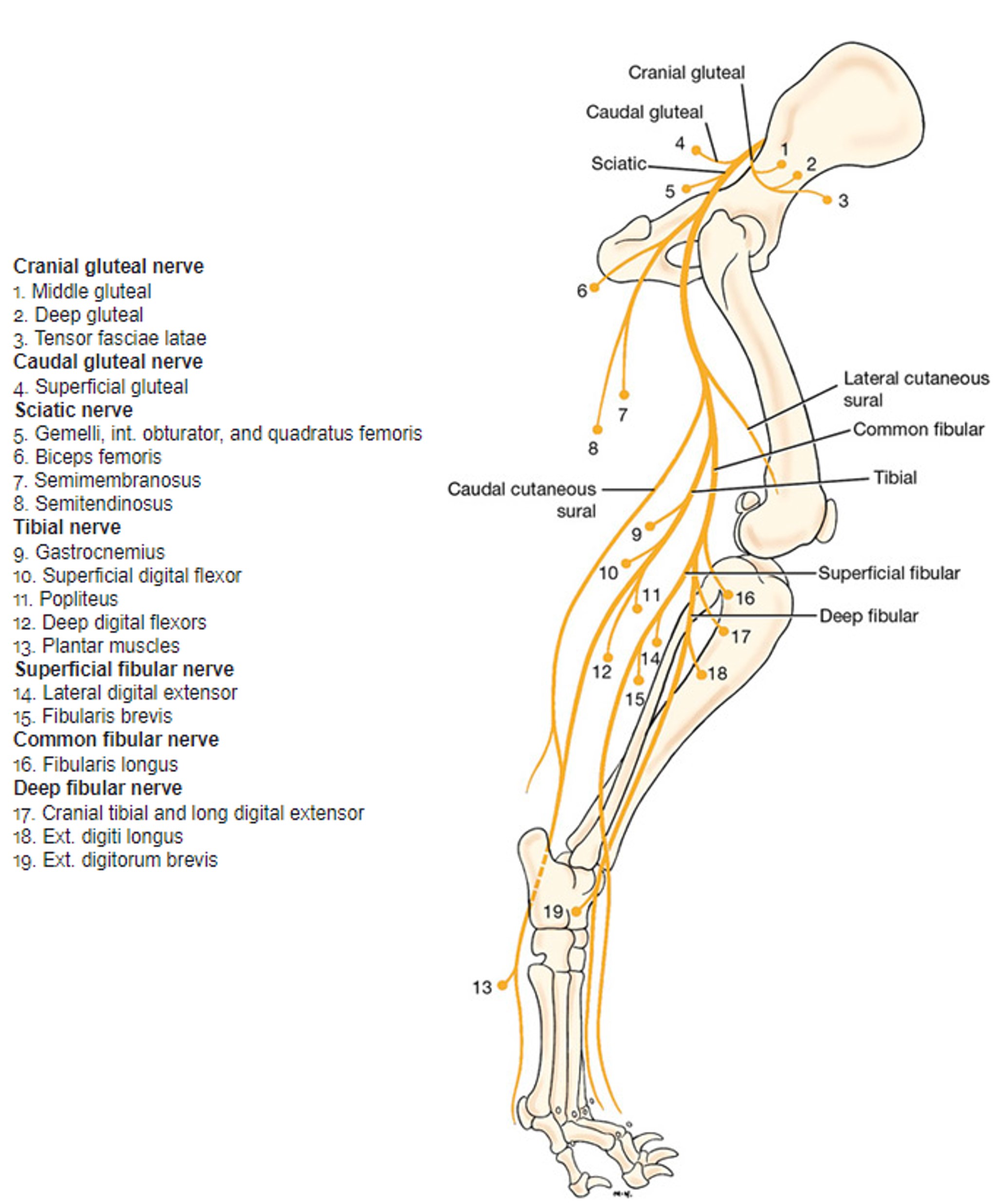
- Distribution of cranial and caudal gluteal nerves and sciatic nerve of right pelvic limb of the dog, schematic lateral view. Muscles innervated by numbered nerves. 1
-

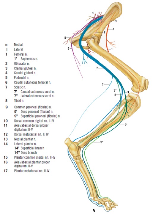
- Schematic illustration of lumbosacral plexus nerves of the cat innervating the pelvic limb. 4
Caudal gluteal
Dissect: The entirety of this lab’s dissection will be done on the right pelvic limb. Expose the caudal insertion of the superficial gluteal m. deep to the proximal edge of the biceps femoris m. Transect the insertion of superficial gluteal m. (and gluteofemoralis if dissecting a cat) at this level and reflect the muscle(s) dorsally. If possible, try not to break the caudal gluteal artery and nerve, and their branches.
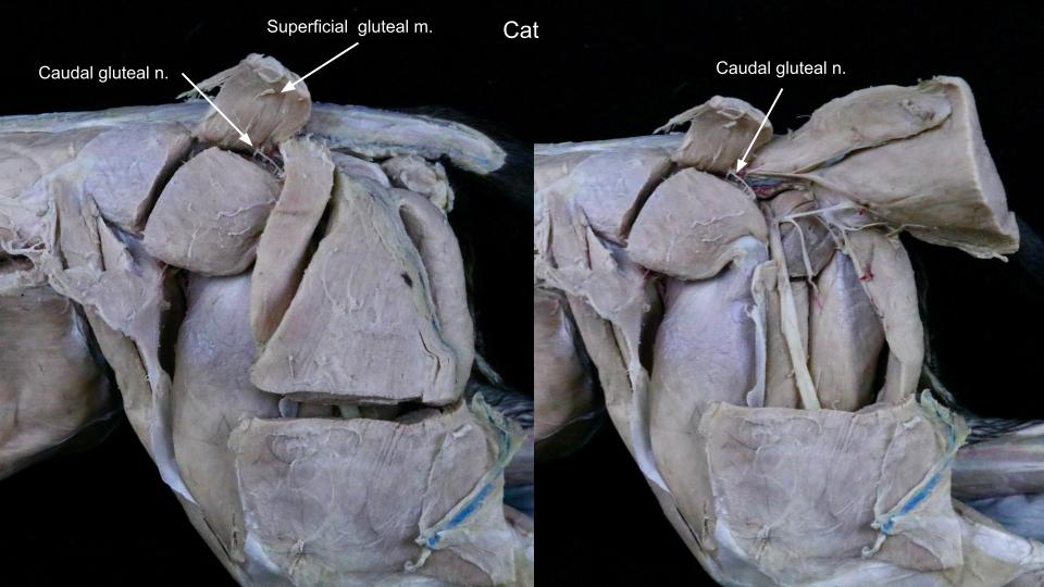
The caudal gluteal nerve passes over the ischiatic notch medial to the middle gluteal muscle and enters the medial surface of the superficial gluteal muscle. It is the sole innervation of the superficial gluteal muscle. It has a variable origin from the seventh lumbar and first two sacral spinal nerves.
Observe: Observe the caudal gluteal nerve passing into the deep surface of the superficial gluteal m.
-

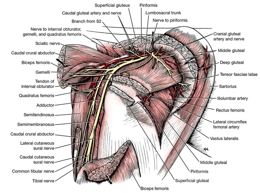
- Nerves, arteries, and muscles of the right hip of the dog, lateral aspect. 1
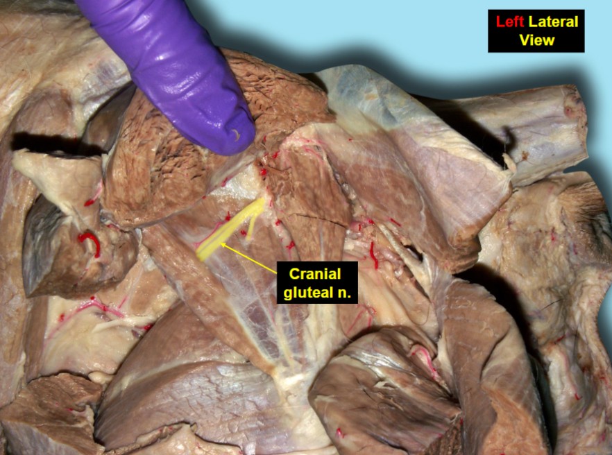
Cranial gluteal
The cranial gluteal nerve passes over the greater ischiatic notch, crosses the lateral surface of the ilium at the origin of the deep gluteal muscle, and innervates the middle and deep gluteal mm., as well as the tensor fasciae latae m. It arises from the ventral branches of the sixth and seventh lumbar nerves, and the first sacral spinal nerve.
-
- Nerves, arteries, and muscles of the right hip of the dog, lateral aspect. 1
-

- Dog cranial gluteal nerve 40
-
- Dog, right lateral
Dissect: Transect the middle gluteal m. midway through its length. Gently reflect the cranial and caudal portions. Try not to break the cranial gluteal artery and nerve, or their branches, in doing so.
Observe short branches of the cranial gluteal n. passing into the deep surface of the middle gluteal m. A larger branch of the cranial gluteal n. is frequently seen passing through the muscle belly of the deep gluteal m. and into the belly of tensor fasciae latae m.
Caudal cutaneous femoral
The caudal cutaneous femoral nerve arises from the sacral plexus and is united to the pudendal for most of its intrapelvic course. The caudal cutaneous femoral nerve follows the caudal gluteal artery to the level of the ischiatic tuberosity, where it becomes superficial near the attachment of the sacrotuberous ligament and terminates in the skin on the proximal caudal half of the thigh.
Dissect: Look for the caudal cutaneous femoral n. on the proximocaudal region of the thigh and/or as a continuation of the caudal gluteal nerve. Typically, this thin cutaneous nerve can be seen along the proximal, caudal portion of biceps femoris (or gluteobiceps) and lateral to semitendinosus. However, it also is very easy to remove this nerve with the skin, so don’t hesitate in moving forward with the dissection if you struggle to find any remnant of the nerve!
-
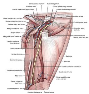
- Vessels and nerves of right thigh and perineum of the dog, lateral view. 1

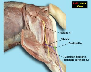
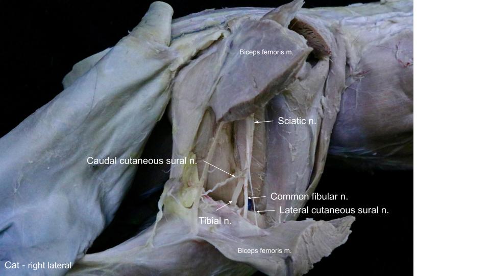
Sciatic
The sciatic n. arises from the last two lumbar and first two sacral spinal nerves. Small branches leave within the pelvis to supply the internal obturator, gemelli, and quadratus femoris mm. Do not dissect these branches. The sciatic nerve passes caudally over the hip medial to the greater trochanter, craniomedial to the ischiatic tuberosity, and then distally caudal to the femur on the lateral side of the adductor muscle. Unnamed branches leave the sciatic nerve at the level of the hip and innervate the biceps femoris, semitendinosus, and semimembranosus mm.
Observe: Observe the large sciatic nerve and note the branches passing into the aforementioned hamstring muscles that it innervates in the caudal thigh.
The sciatic nerve terminates in the thigh as the common fibular and tibial nerves. There are lateral and caudal cutaneous sural nerves that arise from the fibular and tibial components of the sciatic nerve, respectively, and supply the skin on the lateral and caudal surfaces of the crus.
Dissect: Carefully transect the biceps femoris m. midway between its origin and the stifle, and reflect the transected muscle halves. The sciatic nerve and its branches lie directly underneath this muscle. (The sciatic nerve is big and hard to miss!)
Continue distally along the sciatic n. The lateral and caudal cutaneous sural nerves are often broken as the skin is removed. However, it is usually possible to find these transected branches proximal to the common fibular and tibial nerves. Look for the lateral cutaneous sural nerve as it enters the medial (deep) aspect of the biceps femoris muscle. The caudal cutaneous sural nerve follows the tibial nerve to the caudal surface of the gastrocnemius m.
-
- Vessels and nerves of right thigh and perineum of the dog, lateral view. 1
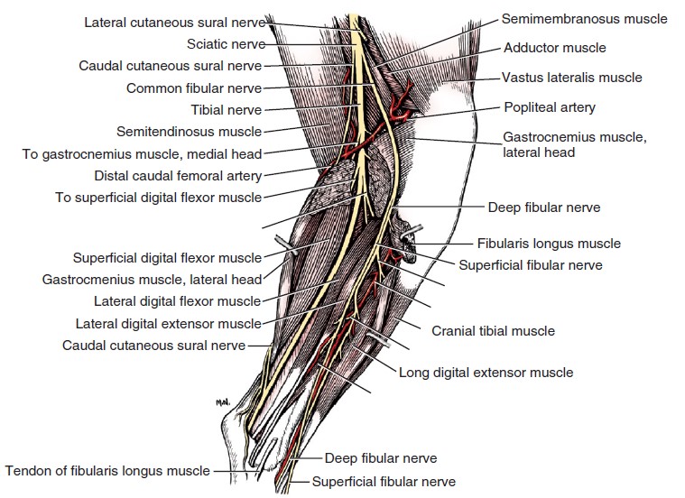
-
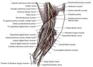
- Nerves, arteries, and muscles of the right leg of the dog, lateral aspect. (The mm. biceps femoris, and portions of the lateral head of the gastrocnemius and the fibularis longus have been removed.) 1
-

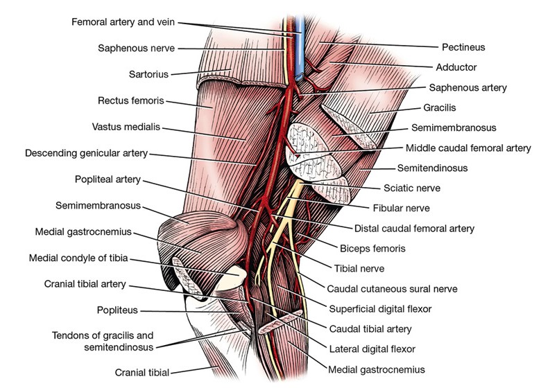
- Arteries and nerves of right popliteal region of the dog, medial view. 1
-
- Dog sciatic nerve 40
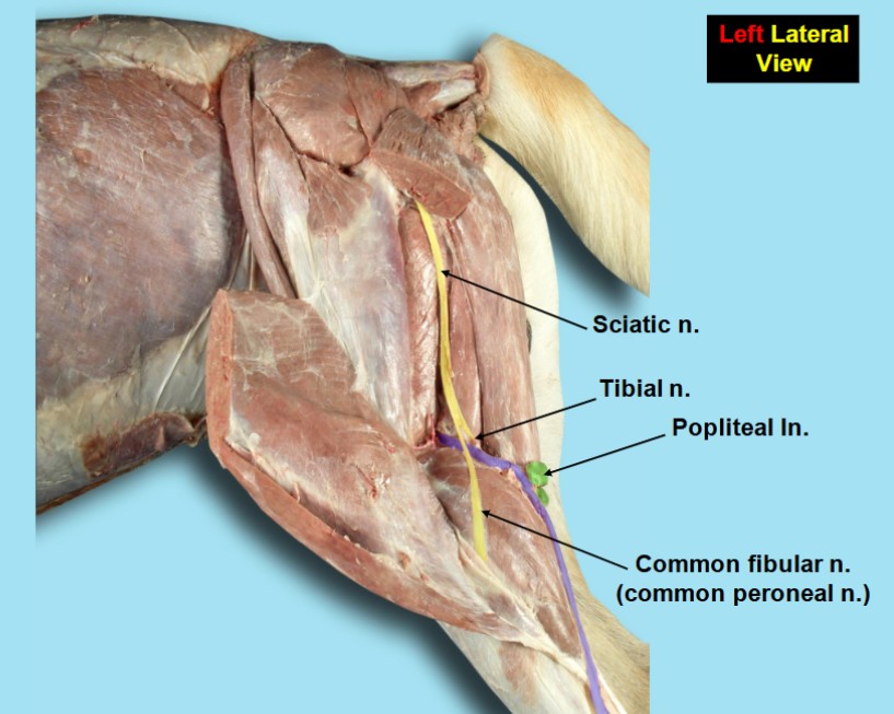
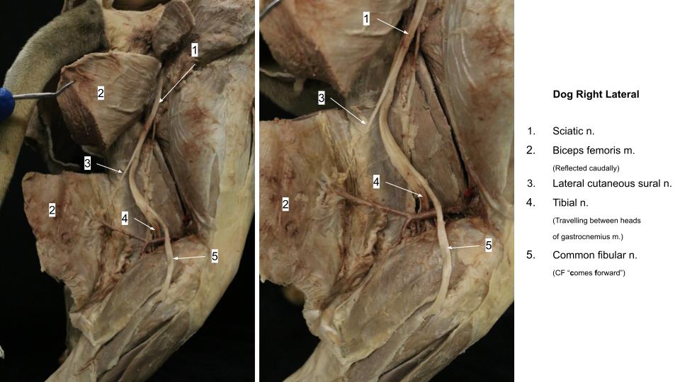
Common Fibular
The common fibular nerve arises primarily from the ventral branches of the sixth and seventh lumbar spinal nerves. It passes laterally, deep to the thin terminal part of the biceps femoris m. and crosses over the superficial surface of the lateral head of the gastrocnemius m. and proximal fibula. It then courses between the lateral digital flexor muscle caudally and the fibularis longus m. cranially to enter the muscles on the cranial side of the crus. Here the common fibular nerve divides into superficial and deep branches (named the superficial and deep fibular nerves, respectively). These nerves innervate the flexor muscles of the tarsus and the extensor muscles of the digits. These include the cranial tibial, fibularis longus, and long digital extensor mm.
Dissect: Clean and follow the common fibular n. as it crosses the lateral head of gastrocnemius m. until it splits into the superficial and deep fibular nn.
The superficial fibular nerve leaves the lateral portion of the parent nerve below the stifle, where it lies between the lateral digital flexor m. (lateral head of the DDF m.) caudally and the fibularis longus m. cranially. At the beginning of the distal third of the crus, it becomes subcutaneous and accompanies the cranial branch of the saphenous artery. Distal to the tarsus the superficial fibular nerve forms dorsal common digital nerves that innervate the paw.
The deep fibular nerve arises from the cranial surface of the parent nerve. It enters the muscles on the cranial part of the crus and courses distally in association with the cranial tibial artery. In the proximal half of the tarsus, they both lie in a groove formed by the tendons of the long digital extensor muscle laterally and by the cranial tibial muscle medially.
At the tarsus the nerve divides into dorsal metatarsal nerves that continue distally to innervate the dorsal aspect of the paw along with the dorsal common digital nerves from the superficial fibular nerve.
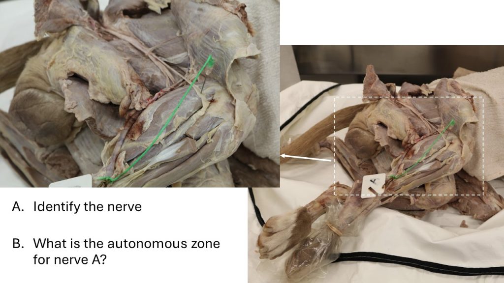
Tibial
The tibial nerve arises primarily from the ventral branches of the seventh lumbar and first sacral spinal nerves and is the caudal portion of the sciatic nerve. It separates from the common fibular nerve in the thigh. At the stifle, it passes between the two heads of the gastrocnemius m. The tibial nerve supplies the caudal muscles of the crus, which include the extensors of the tarsus and flexors of the digits. It innervates both heads of the gastrocnemius m., superficial digital flexor m., popliteus m., and both deep digital flexor mm. The tibial n. emerges from the deep surface of the medial head of the gastrocnemius m. and continues distally along the medial side of the caudal surface of the tibia.
Dissect: Follow the tibial n. from its split from the sciatic n. to where it continues distally along the caudal surface of the tibia. Once again, look for the caudal cutaneous sural nerve following the tibial nerve towards the caudal crus. The tibial nerve will pass between the two heads of the gastrocnemius. The smaller caudal cutaneous sural nerve will stay superficial to the gastrocnemius.
-
- Vessels and nerves of right thigh and perineum of the dog, lateral view. 1
-
- Nerves, arteries, and muscles of the right leg of the dog, lateral aspect. (The mm. biceps femoris, and portions of the lateral head of the gastrocnemius and the fibularis longus have been removed.) 1
-
- Arteries and nerves of right popliteal region of the dog, medial view. 1
-
- Dog sciatic nerve 40
We’ll now shift our attention proximally back up to the medial aspect of the thigh.
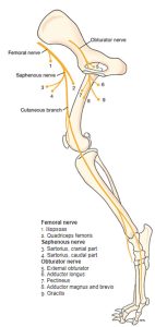
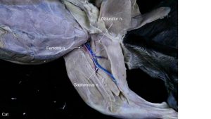
Obturator
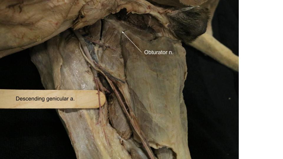
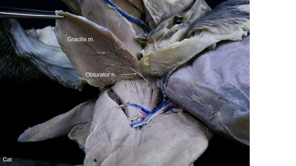
The obturator nerve arises from the fourth, fifth, and sixth lumbar spinal nerves. It is formed within the caudomedial portion of the iliopsoas muscle. It leave the muscle dorsomedially, runs caudoventrally along the body of the ilium, penetrates the medial side of the levator ani muscle and leave the pelvis by passing through the cranial part of the obturator foramen. It supplies the adductors muscles of the limb; i.e., the external obturator, pectineus, gracilis, and adductor mm.
Dissect: Transect the gracilis muscle midway between its proximal and distal attachments, and reflect these two cut portions. Find the obturator n. on the medial limb as it emerges ventrally from the obturator foramen and emerges between the pectineus and adductor muscles. It is most easily appreciated on the lateral (deep) aspect of the transected and reflected gracilis m. From here you can trace it back towards the obturator foramen.
-
- Distribution of saphenous, femoral, and obturator nerves of right pelvic limb, schematic medial aspect. Muscles innervated by numbered nerves. 1
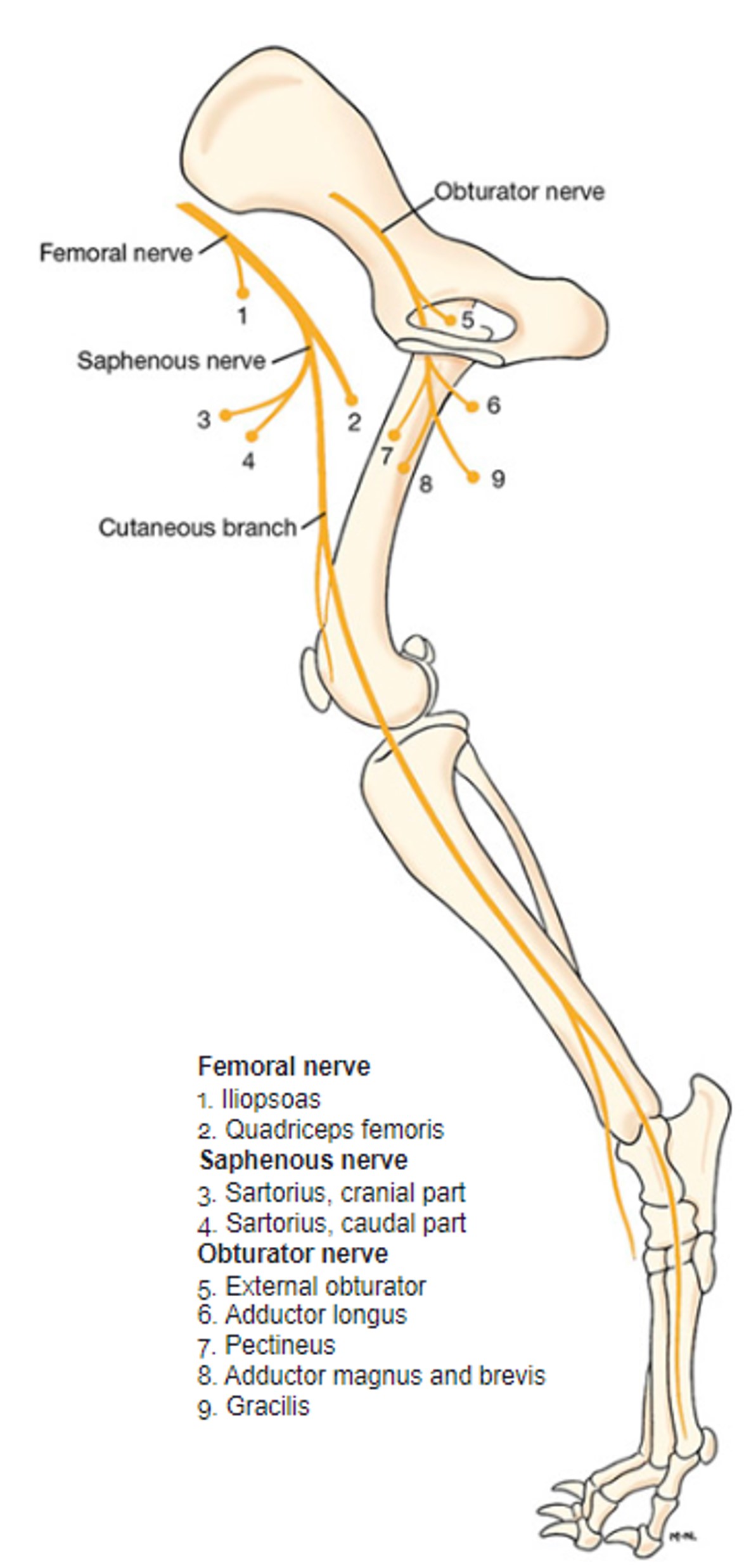
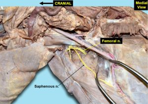
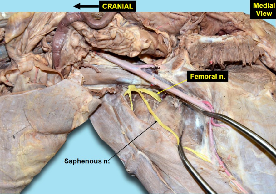
Femoral
The femoral nerve arises primarily from the fourth, fifth, and occasionally sixth lumbar spinal nerves. It supplies branches to the iliopsoas muscle, and enters the quadriceps muscles between the rectus femoris and vastus medialis mm. and supplies all four heads of the quadriceps (rectus femoris m., vastus medialis/lateralis/intermedius mm.).
Observe: Find the femoral n. as it emerges from the iliopsoas muscle just proximal to the origins of the quadriceps. Muscular branches of the femoral n. can often be seen entering the proximal aspects of the muscles of the quadriceps. If necessary, you may transect and reflect the sartorius m. to better visualize the femoral n.
-
- Distribution of saphenous, femoral, and obturator nerves of right pelvic limb, schematic medial aspect. Muscles innervated by numbered nerves. 1
-
- Dog femoral and saphenous nn. 40
-
- Cat medial nerves

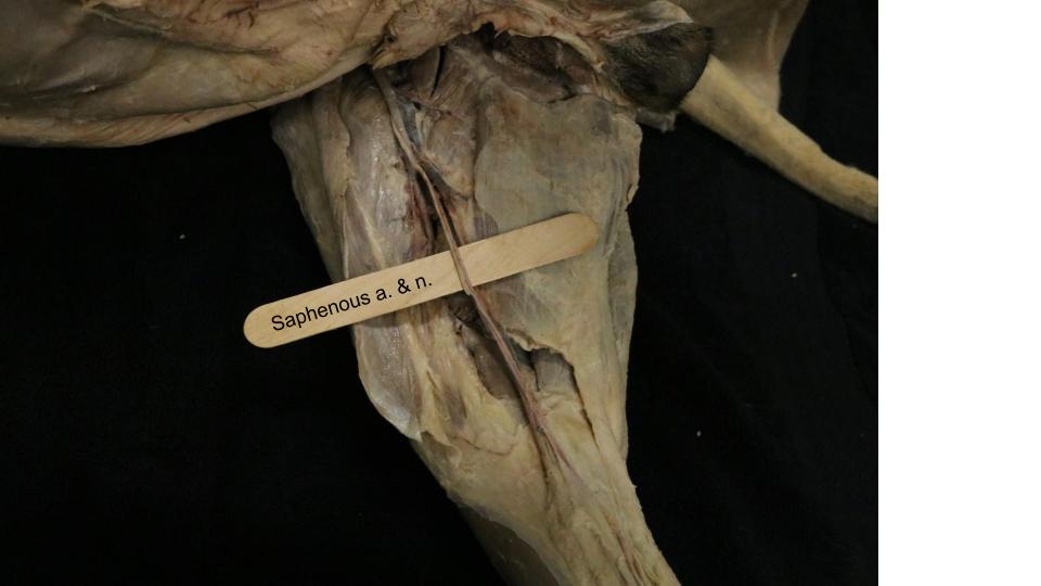
Saphenous
Within the iliopsoas m., the saphenous nerve arises from the cranial side of the femoral n. and passes through the femoral triangle along with the femoral artery and vein. The saphenous n. and/or femoral n. innervates the sartorius m. The cutaneous portion of the saphenous n. supplies the skin on the medial side of the thigh, the stifle, crus, tarsus, and remainder of the paw.
Observe: Follow the saphenous n. from the femoral triangle (notice that it passes with the femoral artery and vein!) and follow it as far distally as the tarsus.
-
- Arteries and nerves of right popliteal region of the dog, medial view. 1
-
- Dog femoral and saphenous nn. 40
-

- Dog saphenous n.
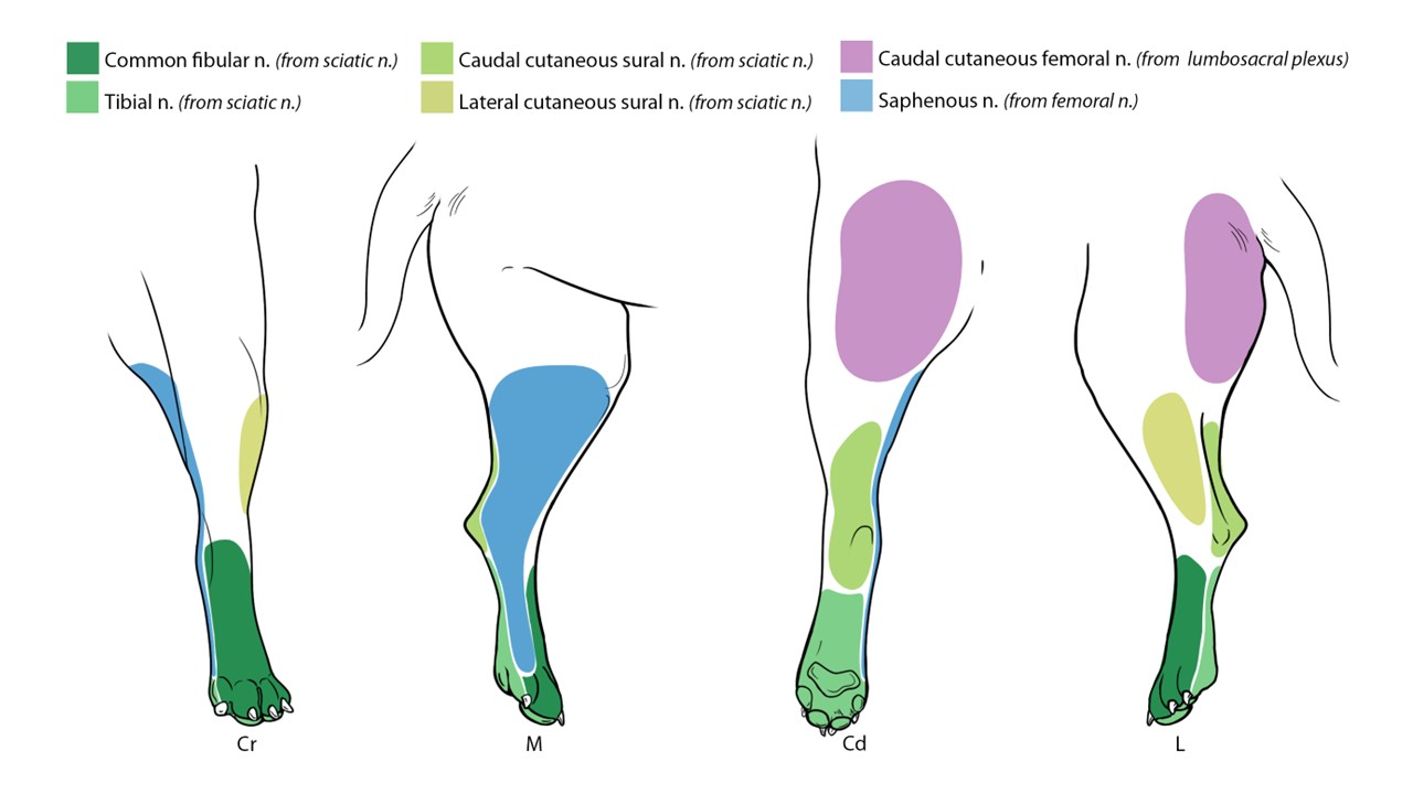
Autonomous Zones of the Pelvic Limb and Pes
You are not required to dissect the nerves of the pes. However, you are responsible for identifying them on models and for naming the autonomous zones of the foot and their corresponding nerves.
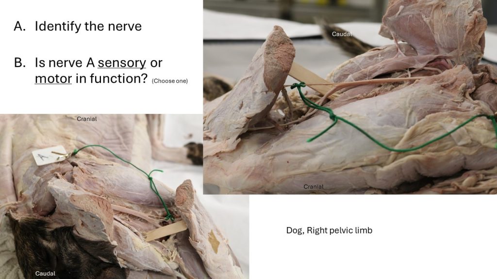
Proximal to the tarsocrural joint, the tibial nerve divides into the medial and lateral plantar nerves. These cross the tarsus medial to the tuber calcanei through the tarsal tunnel and terminate as the plantar common digital and plantar metatarsal nerves, respectively, which are sensory to the plantar surface of the paw. The tarsal tunnel is formed by the sustentaculum tali dorsally, the calcanean tuber laterally, and the flexor retinaculum on the plantar side. It contains the tendon of the lateral head of the deep digital flexor with its synovial sheath, the medial and lateral plantar nerves, and the caudal branch of the saphenous artery. We will come back to these medial and lateral plantar nerves in the equine distal limb. For our purposes in the carnivore, we will focus on the tibial nerve providing cutaneous sensory innervation to the plantar surface of the pes.
The cutaneous innervation of the dorsal surface of the pes (and indeed the tarsus and the cranial crus) is provided by superficial branch of fibular n.
Observe: Review the autonomous zones of the pelvic limb, focusing your attention on the pes (autonomous zones of the proximal limb will not be tested).
-
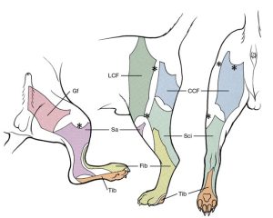
- Autonomous zones of cutaneous innervation of the pelvic limb of the dog. Medial, lateral, and caudal aspects. CCF, Caudal cutaneous femoral; Gf, genitofemoral; LCF, lateral cutaneous femoral; Fib, fibular; Sa, saphenous; Sci, sciatic; Tib, tibial. 1

-

- Pelvic limb autonomous zones L. Hudson
Review Videos
Dog Pelvic Limb Nerves (with some FYI vessels) – Watch until 11:15
Dog pelvic limb nerves – 12 min, after switching to the medial side, jump to 12:10 to hear 1 min about the nerves
Terms
| Terms | Species | Muscle(s) Innervated/Notes |
| Lumbosacral plexus | All | |
| Caudal gluteal n. | All |
|
| Cranial gluteal n. | All |
|
| Caudal cutaneous femoral n. | Carnivore | |
| Sciatic n. | All |
|
|
Caudal cutaneous sural n. |
Carnivore | Innervates skin of the caudal crus |
|
Lateral cutaneous sural n. |
Carnivore | Passes through the deep surface of the biceps femoris m.; innervates skin of lateral crus |
| Common fibular n. | All |
|
| Tibial n. | All |
|
| Obturator n. | All |
|
| Femoral n. | All |
|
| Saphenous n. | All | |
|
Femoral triangle |
All | Borders = abdominal body wall, sartorius m., and pectineus m.; Femoral artery and vein, and saphenous nerve pass through femoral triangle |
| Autonomous zones of pes | Carnivore | Tibial n. and common fibular n. |