Lab 7A: Nerves of the Ungulate Pelvic Limb
Learning Objectives
- Identify the nerves and their branches of the ungulate pelvic limb.
- Understand innervation of muscle action groups of the ungulate pelvic limb.
- Identify the relevant nerves to perform a distal limb nerve block in the equine.
Lab Instructions:
Ungulate Proximal Limb
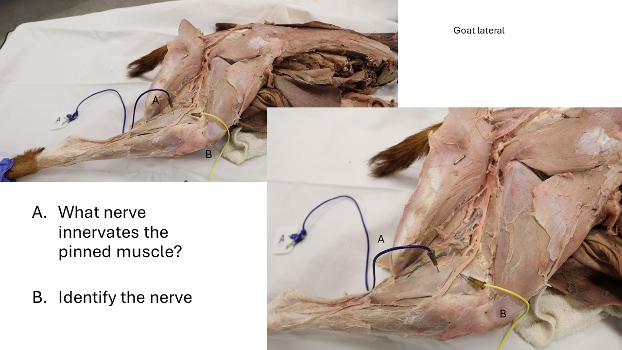
Observe: Identify the following nerves in the instructors’ ungulate prosections. Recall what muscles each of these nerves innervates. While obviously much larger and longer, these nerves generally follow the same path and innervate the same muscles as we observed in the carnivores. The descriptions of the nerves listed below provide context for finding and identifying these nerves in the ungulates.
- Femoral n. (found as stumps entering the proximal ends of the quadriceps muscles)
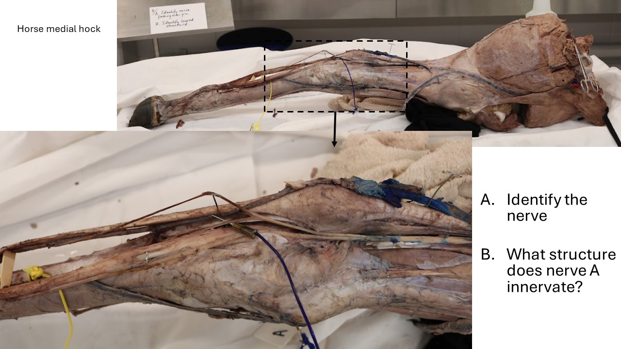
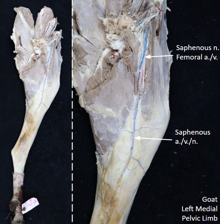
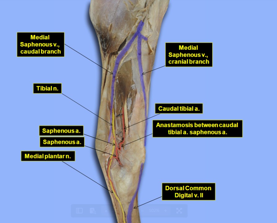
- Saphenous n. (courses with the femoral artery and vein in the femoral triangle, and continues superficially over the medial aspect of the thigh, stifle, and crus)
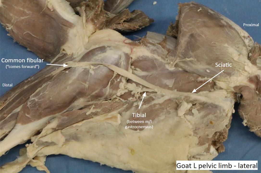
- Sciatic n. (very large nerve in the ungulates. Passes dorsal to the coxofemoral joint across the lateral surface of the sacrosciatic ligament and continues medial to the biceps femoris/gluteobiceps m. in the thigh)
- Tibial n. (passes between the lateral and medial heads of the gastrocnemius m.)
- Common fibular n. (passes over the lateral surface of the lateral head of gastrocnemius m.)
-
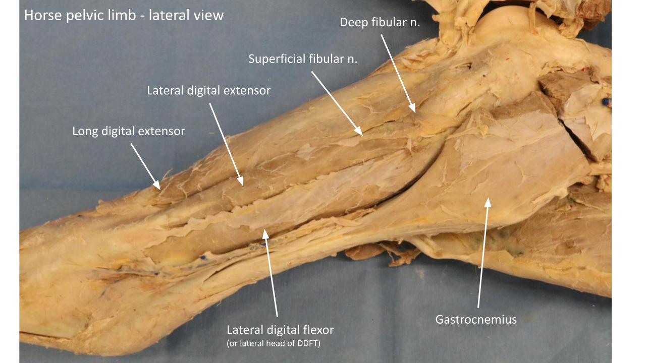
- Superficial and deep fibular nn.
- Superficial and deep fibular nn.
-

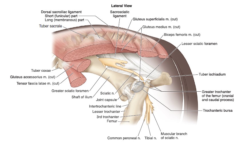
- Lateral view of the equine pelvis depicting the superficial muscles, ligaments, and nerves. 9
-
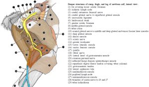
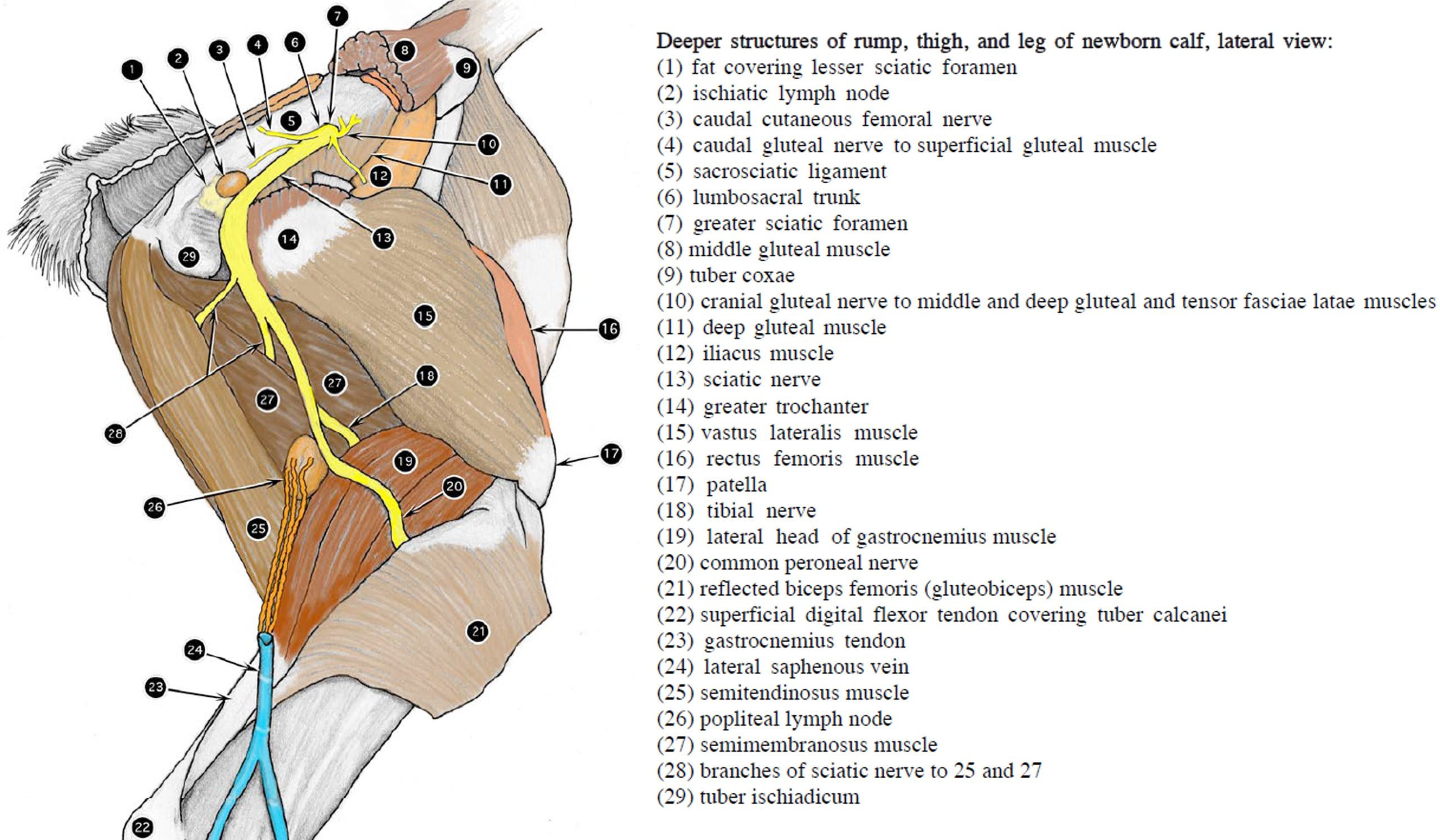
- Deeper structures of rump, thigh, and leg of newborn calf, lateral view. 2
-

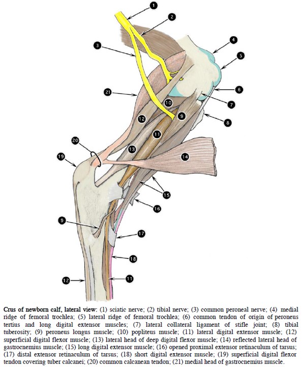
- Crus of newborn calf, lateral view. 2
Clinical application
The following nerves need not be identified, but please be aware of what muscles they innervate:
- Obturator n. – innervates external obturator, pectineus, gracilis, and adductor mm.
- Cranial gluteal n. – innervates middle gluteal m., deep gluteal m., and tensor fasciae latae m.
- Caudal gluteal n. – innervates superficial gluteal m.
-
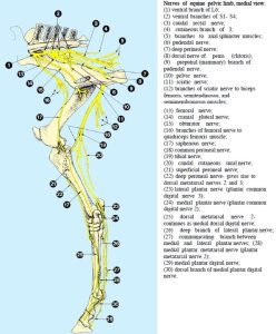
- Nerves of equine pelvic limb, medial view. 2

-
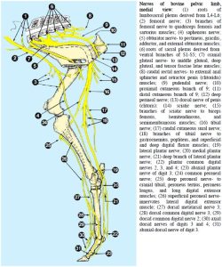
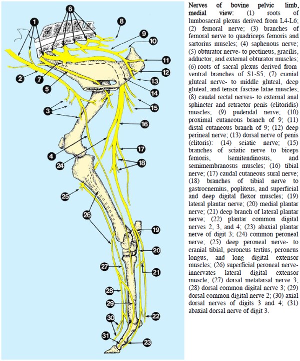
- Nerves of bovine pelvic limb, medial view. 2
Equine Distal Limb
Dissect: Each dissection team has been given the distal pelvic limb of a horse. Remove the skin from the limb past the fetlock and bluntly dissect the nerves described below. Note that we have a very similar distribution of nerves to what we saw in the thoracic limb (and a very similar naming convention, just substitute “plantar” for “palmar”!).
Also, take extra care while skinning the metatarsus as there is a superficial branching crossing over the plantar surface of the superficial digital flexor tendon approximately half way down the metatarsus.
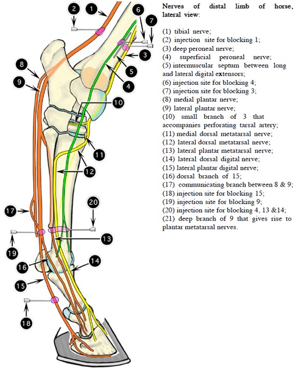
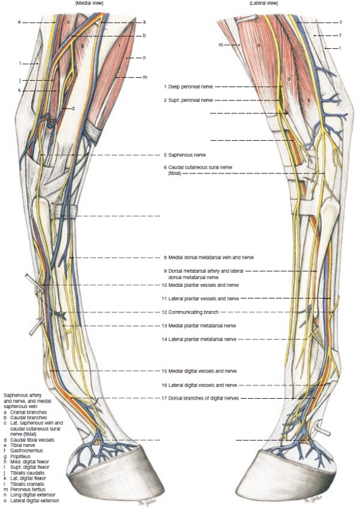
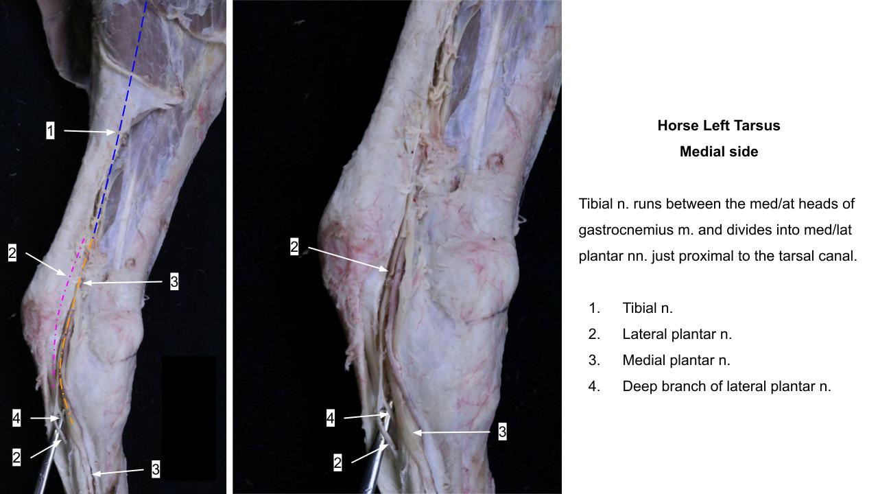
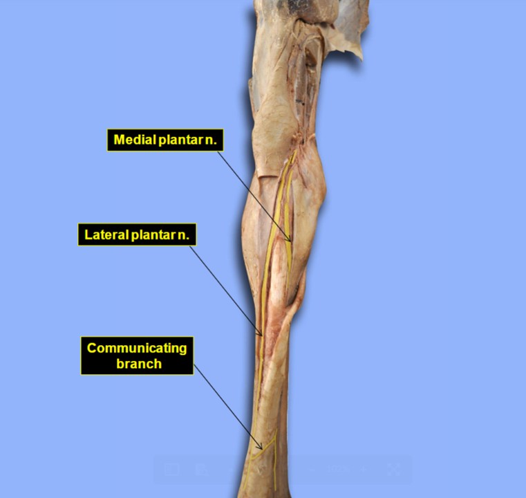
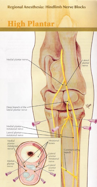
The tibial n. divides into medial and lateral (M/L) plantar nn. at the tarsus. A deep branch of the lateral plantar n. is found in the proximal metatarsus and it divides into M/L plantar metatarsal nn. to supply the suspensory ligament. The M/L plantar nerves continue in the metatarsus on each side of the deep digital flexor tendon. A communicating branch may still be intact at the mid/distal third of the metatarsus.
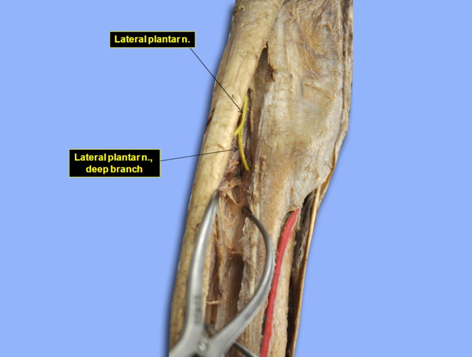
Dissect: Identify the communicating branch crossing over from the medial to lateral plantar nerves, approximately midway down the metatarsus. This branch may have been removed during skinning. Also identify the medial and lateral plantar nerves. Trace the deep branch of the lateral plantar nerve diving deep to the suspensory ligament, near the proximal end of the metatarsus.
-

- Nerves of distal limb of horse, lateral view. 2
-

- Nerves of the Distal Hindlimb. 14
-

- Horse tibial nerve 40
-

- Horse plantar nn. 40
-
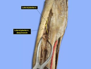
- Horse deep branch 40
Clinical relevance:
-
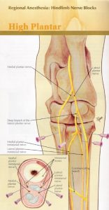
- High Plantar Nerve Block. 23
The name change is at the fetlock from ‘plantar’ to ‘plantar digital’ structures. Locate the M/L plantar digital nn. at the level of the fetlock. We should understand the distal distribution of the superficial and deep fibular nn. in the horse, but do not worry about identifying their continuations in the metatarsus, they are very small nerves.
Dissect: Follow the medial and lateral plantar nerves as they approach the fetlock joint. As they cross this joint, their names change to the medial and lateral plantar digital nerves, respectively.
Clinical relevance:
Distal limb nerve blocks for lameness examination, eg low 4 point, abaxial sesamoid block, plantar digital nerve block; knowing the locations of these nerves and vessels helps avoid transecting them during limb surgeries unless indicated to actually transect them! (e.g. plantar digital neurectomy); palpation of digital pulses is common in physical examination.
-
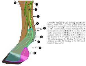
- Left distal forelimb of horse showing sites of nerve blocks, lateral view. 2

-
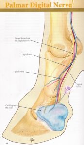
- Palmar digital nerve block. 23

-

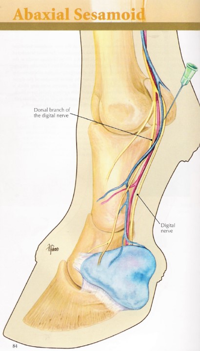
- Abaxial sesamoid nerve block. 23
-

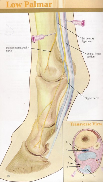
- Low 4 point nerve block. 23
Review Videos
Horse pelvic limb nerves, wire model – 15 minutes
Horse pelvic limb nerves, cadaver – 17 minutes
Horse distal pelvic limb nerve distribution and nerve blocks – 25 min
Goat pelvic limb nerves – 5 min
Calf pelvic limb nerves – 4 min, watch until 24 min
Pig pelvic limb nerves – 4 min (mostly discusses arteries)
Interactive Practice Content:
Terms
| Terms | Species |
| Caudal gluteal n. | All |
| Cranial gluteal n. | All |
| Caudal cutaneous femoral n. | All |
| Sciatic n. | All |
| Common fibular n. | All |
| Superficial and deep fibular nn. | All |
| Tibial n. | All |
| Obturator n. | All |
| Femoral n. | All |
| Saphenous n. | All |
| Medial and lateral plantar nn. | Horse |
| Communicating branch | Horse |
| Deep branch of lateral plantar n. | Horse |
| Medial and lateral plantar metatarsal nn. | Horse |
| Medial and lateral plantar digital nn. | Horse |