Lab 1A: Nerves of the Neck and Thoracic Wall
Learning Objectives
- Identify ventral branches of cervical spinal nn, the accessory n, and the vagosympathetic trunk.
- Relate the condition of “cribbing” to the innervation of the strap muscles, and describe how this condition is treated.
- Describe the location of the intercostal a, v, n, and the clinical importance of this information.
Carnivore Cervical Nerves
Dissect: Alright, time to incise and reflect skin much as we did all the way back in Unit 1. The objective is to remove the skin from the right side of the cadaver (except for the head). Examine the images and follow the instructions below.
Ensure the cadaver is lying in left lateral recumbency (i.e. the right side of the animal is facing up towards you). We wish to reflect the skin from the right side of the body. Ensure that the skin of the neck is removed to its most cranial extent. Once reflected the skin will be removed and discarded.
- Palpate the dorsal midline and perform a skin incision from cranial to caudal, starting at the back of the head (just behind the level of the ears) and extending to the butt of the tail.
- Roll the cadaver into dorsal recumbency and extend an incision along ventral midline, starting at the cranial end of the neck at a point just behind the palpable jaw line.
- Continue this ventral midline incision caudally (careful! don’t cut too deep or you may open the abdominal cavity), skirting around external genitalia, between the thighs, and up and around the left margin of the anus to connect to the dorsal midline incision at the butt of the tail.
- With the cadaver still in dorsal recumbency, abduct the left thoracic and pelvic limbs. From the point on the ventral midline incision directly opposite the left thoracic limb, extend a skin incision onto the medial surface of the limb and continue it to the level of the paw. Repeat this incision for the left pelvic limb.
- Make a complete circular incision through the skin at the paws, just above the level of the digits.
- Roll the cadaver back to right lateral recumbency as needed. Returning to the cranial end of the dorsal midline incision, extend a transverse skin incision from dorsal midline around the left side of the cranial neck to end at the ventral midline incision.
- Now, with all skin incisions made, carefully reflect the skin of the left side of the body. More skin incisions can be made to create smaller sections of skin for removal, as preferred.
- The skin will be intimately ‘fused’ with the thin underlying cutaneous muscles over the neck, thorax, and abdomen. These muscles may be left on the specimen as is practical, otherwise they may be reflected with the skin.
Example incisions from the skin incision on left side of the body (these will obviously be mirrored to the right side):


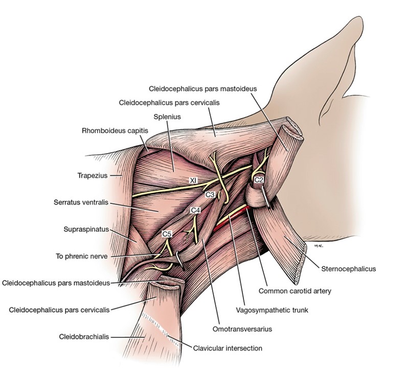
With the skin removed, let’s return to the neck. There are eight pairs of cervical spinal nerves in the dog. The first cervical spinal nerve passes through the lateral vertebral foramen of the atlas. The remaining nerves pass through the succeeding intervertebral foramina. The eighth cervical spinal nerve emerges from the intervertebral foramen between the seventh cervical and first thoracic vertebrae. Immediately upon leaving the foramina, the nerves divide into large ventral and small dorsal branches. The dorsal branches supply structures dorsal (such as the epaxial mm.) to the vertebrae and are not dissected.
When nerves are being dissected, it is helpful to separate the tissue bluntly by inserting scissors and opening it to spread the tissue. Fascial strands of connective tissue will break with minimal effort, but nerves usually stretch.
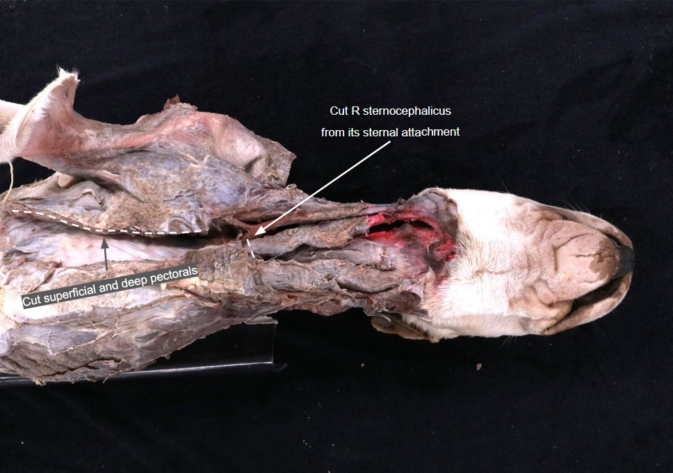
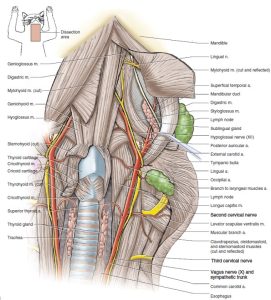
Dissect: We will dissect the cervical spinal nerves from a ventral view, so turn the cadaver to dorsal recumbency. Transect the superficial and deep pectoral mm. two centimeters parallel to their ventral midline attachment. This will allow greater abduction of the limb. On the right side of the neck, transect the sternocephalicus m. at its sternal attachment, then reflect it laterally to access the deeper fascia of the neck. Taking care to not transect any of the large vessels or nerves passing caudally next to the trachea, proceed to clear this fascia until you can clearly identify the right longus capitis m. At this point you should be able to find nerves passing laterally from the neck. These are the ventral branches of cervical spinal nerves.Palpate the wing of the atlas (C1), and working away fascia at the caudoventral border of the atlas, uncover the ventral branch of the second cervical spinal nerve (C2). It emerges between the mastoid part of the cleidocephalicus and the omotransversarius mm.
This dissection can also be attempted on the left side if you feel that the amputated limb on that side makes it easier to access the neck.
-

- Ventral branches of cervical spinal nerves emerging through the lateral musculature. 1
-

- Cervical nerves and formation of the brachial plexus of the cat, ventral view. 4
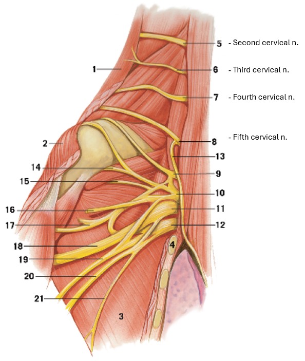
The second cervical spinal nerve lies along or deep to the middle of the caudoventral border of the platysma m. (a cutaneous muscle of the head and neck) and emerges between the mastoid part of the cleidocephalicus m. and the omotransversarius m..
The ventral branch of the second cervical spinal nerve divides into two cutaneous branches:
(1) The great auricular nerve extends toward the ear. It branches and supplies the skin of the neck, the ear, and the back of the head with sensory branches.
(2) The transverse cervical nerve branches to the skin of the cranioventral part of the neck. These will not be dissected.
Dissect: Trace the great auricular nerve as it passes through the cleidocephalicus m. (cranial portion of brachiocephalicus m.) and courses to the caudal aspect of the ear. It can be helpful to start with the C2 spinal nerve and trace it laterally to approximate where the great auricular nerve should be piercing through the cleidocephalicus m.
-
- Ventral branches of cervical spinal nerves emerging through the lateral musculature. 1
-
- Cervical nerves and formation of the brachial plexus of the cat, ventral view. 4
-
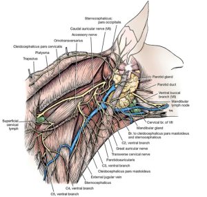
- Superficial nerves of the neck of the dog, lateral aspect. 1

-

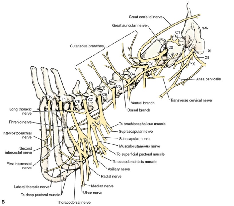
- Schema of the cervical nerves and brachial plexus. The numbers C-1 through C-8 and T-1 refer to spinal nerves, not vertebrae.1
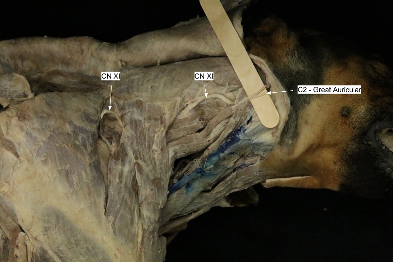
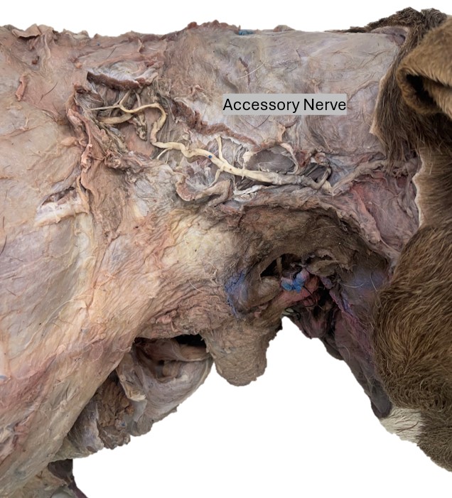
The accessory nerve, or eleventh cranial nerve (CN XI) is a large nerve found deep to the occipital part of the sternocephalicus m. and the cervical part of cleidocephalicus m.. As it emerges from the neck, it crosses the second cervical spinal nerve, runs along the dorsal border of the omotransversarius, and terminates in the thoracic part of the trapezius. The accessory nerve is the only motor nerve to the trapezius m.. In addition, it supplies in part the omotransversarius, the cleidocephalicus, and the sternocephalicus mm.
Dissect: Continuing on the right side of your cadaver, dissect between the trapezius and cleidocephalicus mm. and identify the accessory nerve coursing caudally to the trapezius m. You should also be able to identify the accessory nerve on the deep surface of the trapezius m. It can also be helpful to find the accessory n. as it passes at a right angle near the second cervical spinal nerve, close to the wing of the atlas. Refer to the images below for an idea as to where to find this nerve.
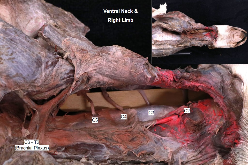
Find and clean the ventral branches of the third, fourth, and fifth cervical spinal nerves (C3-5) deep to the omotransversarius m. It can be helpful to use the brachial plexus as reference (the brachial plexus is the large bundle of nerves running to the forelimb. It consists of nerve roots from C6-T2; so if we identify the big bundle then move one vertebral level cranially, we should be looking at C5 spinal nerve!).
-
- Ventral branches of cervical spinal nerves emerging through the lateral musculature. 1
-
- Cervical nerves and formation of the brachial plexus of the cat, ventral view. 4
-
- Superficial nerves of the neck of the dog, lateral aspect. 1
-
- Schema of the cervical nerves and brachial plexus. The numbers C-1 through C-8 and T-1 refer to spinal nerves, not vertebrae.1

-

- Dog accessory nerve
They are distributed in a segmental manner to the muscles and skin of the neck. The third and fourth nerves, after emerging from the intervertebral foramina, pass through the deep fascia and the omotransversarius m.
The esophagus, the first part of the alimentary canal, is the connecting tube between the laryngeal part of the pharynx and the stomach. In medium-sized dogs it is approximately 30 cm long and 2 cm in diameter when it is collapsed. Because this passage traverses most of the neck and all of the thorax, and ends on entering the abdomen, it is divided into cervical, thoracic, and abdominal portions. We’ll explore and learn more about the esophagus in a later unit.
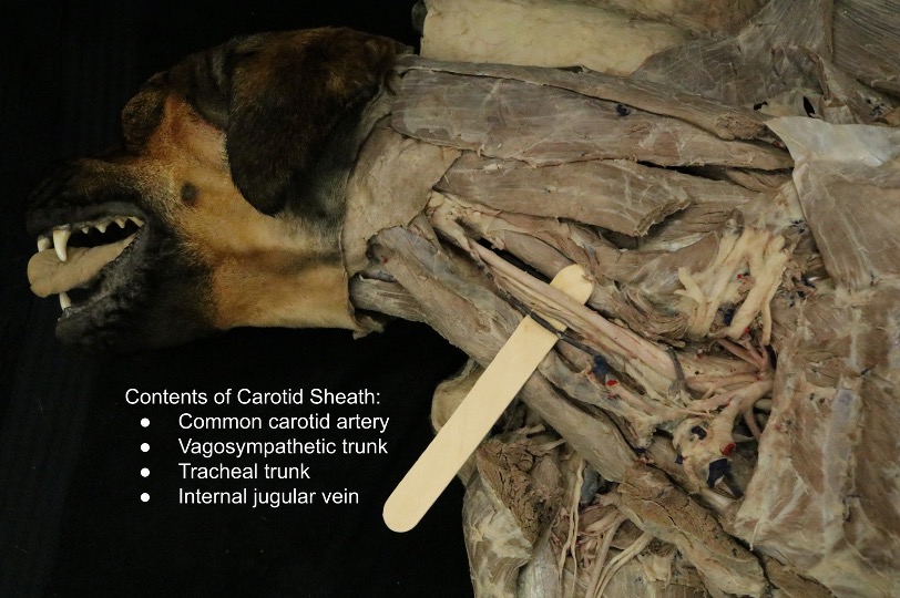
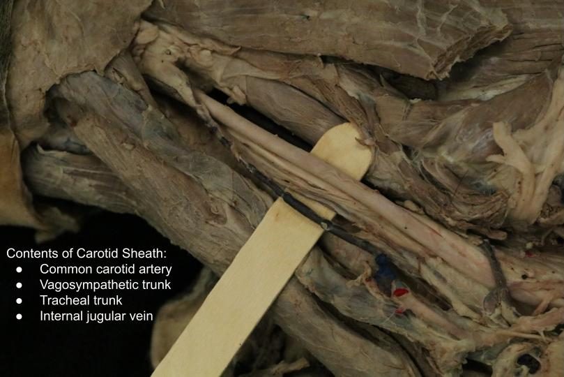
The cervical portion (pars cervicalis) of the esophagus is related mainly to the left longus colli and longus capitis muscles dorsally, and to the trachea ventrally and to the right. At its origin it starts to incline to the left, so that at the thoracic inlet it usually lies left lateral to the trachea. Its position here varies; in some specimens it is left dorsal and in others it is left ventral to the trachea. On the left side, the contents of the left carotid sheath (common carotid artery, vagosympathetic trunk, internal jugular vein, and tracheal trunk) run in the angle between the esophagus and the longus capitis muscle. The corresponding structures of the carotid sheath on the right side are located lateral to the trachea.
Dissect: Carefully open the carotid sheath. Identify the common carotid artery, vagosympathetic trunk, internal jugular vein, and tracheal trunk. Note that the tracheal trunk and internal jugular v. can often be difficult to visualize and/or distinguish from one another, so be aware of their inclusion within the carotid sheath but don’t spend too much time trying to clearly identify them.
-
- Ventral branches of cervical spinal nerves emerging through the lateral musculature. 1
-
- Branches of the common carotid artery, and muscles and nerves of the throat of the cat in ventral view. 5
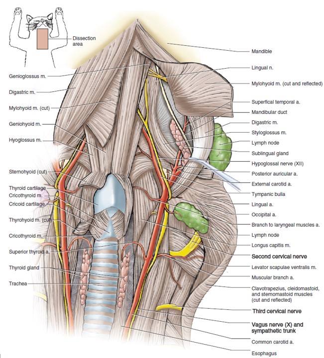
The vagosympathetic trunk is a large bundle of nerve fibers representing tracts of both the vagus nerve (CN X) and the sympathetic trunk. The vagus nerve originates from the brainstem and is an important nerve for the innervation of structures in the head and neck, as well as providing parasympathetic innervation to the viscera of the thorax and abdomen. The sympathetic trunk, as its name suggests, is responsible for sympathetic innervation of many different tissues. Here in the vagosympathetic trunk, we are seeing sympathetic fibers ascending cranially towards the head and neck from their origin in the thoracolumbar regions of the spinal cord. We’ll see more of the sympathetic trunk (separate from the vagus n.) when we open up the thorax.
Ungulate Cervical Nerves
Using the instructor prosections of the ungulates available to you in the lab, recall that there is a left and right sternocephalicus m. Identify the ventral branch of the accessory n. (CN XI) entering the belly of the sternocephalicus m. at its cranial myotendinous junction, by rotating the muscle to look at its media (inner surface).
-
-
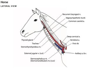
Cervical structures in the neck of the horse.
6

-
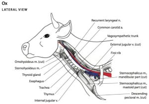
- Cervical structures in the neck of the ox. 6
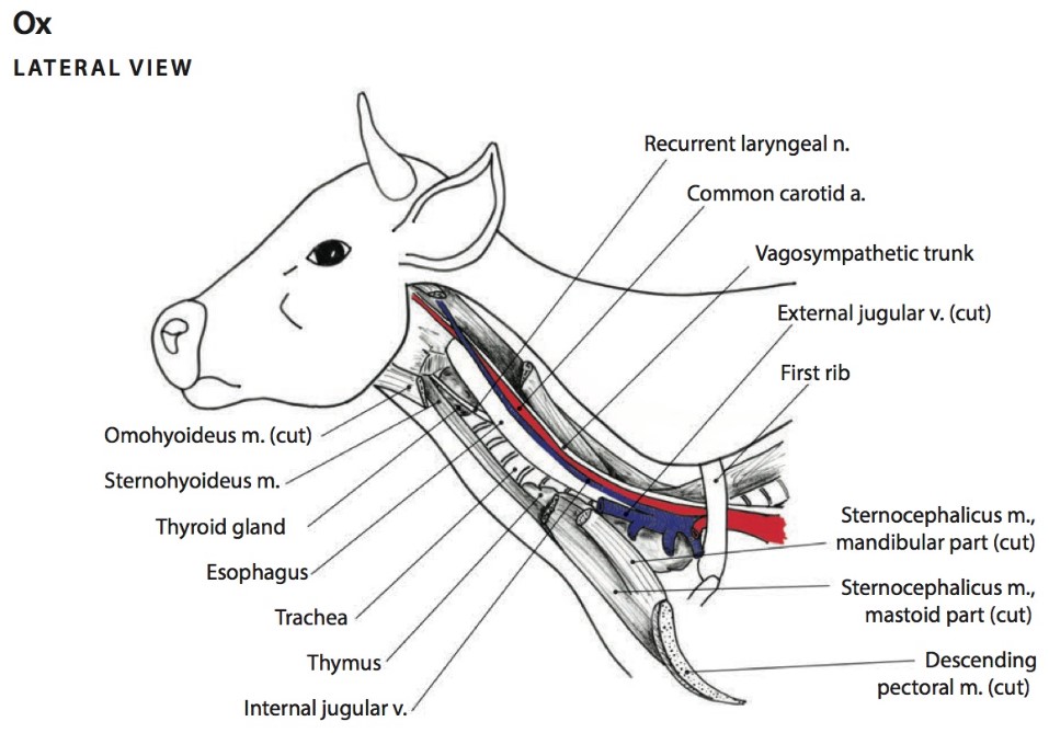
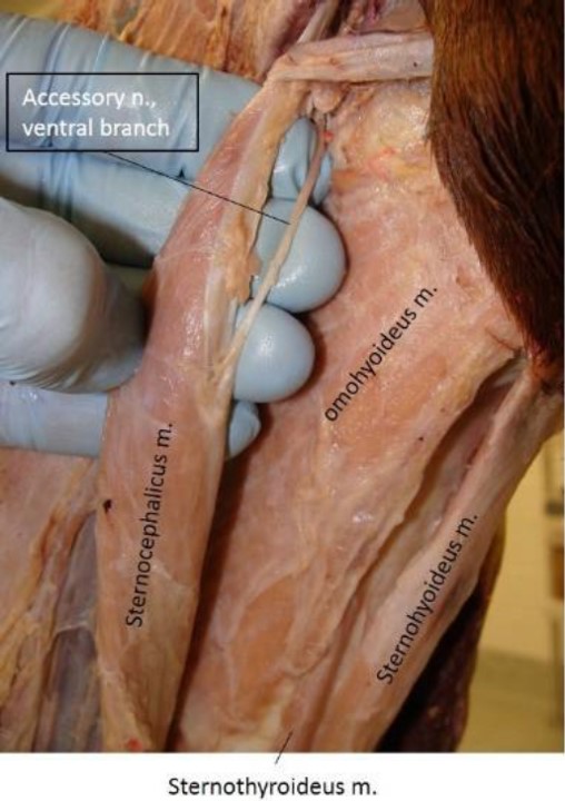

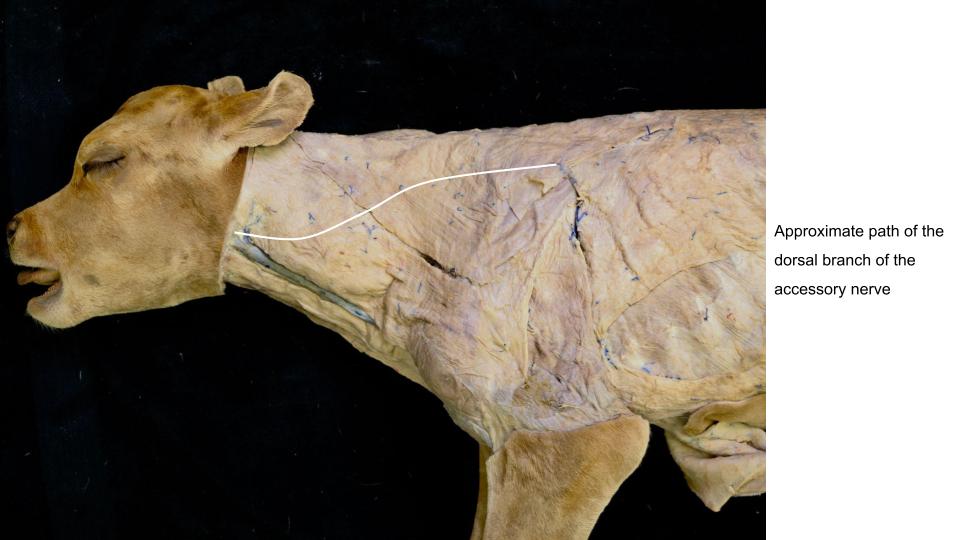
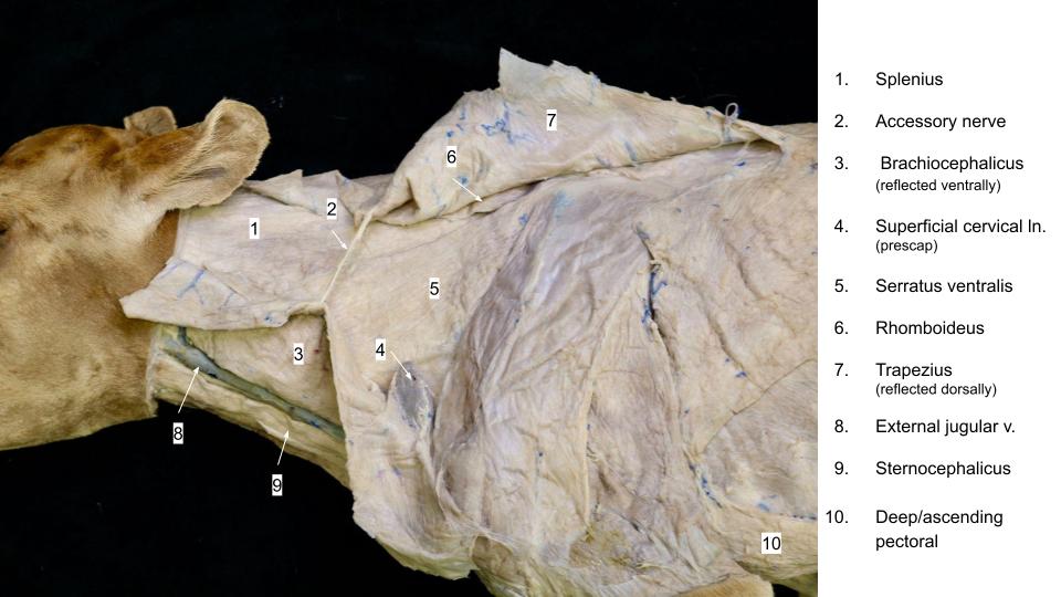
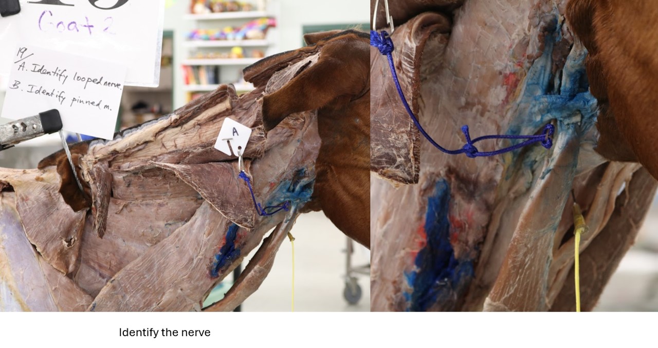
For the ruminants, locate the dorsal branch of the accessory n. as it passes along the dorsal border of the omotransversarius m. and at the point where the omotransversarius, trapezius, and brachiocephalicus mm. meet. The nerve is relatively thin and often hard to find in the horse and goat, whereas it is large and readily identifiable in the calf. If possible, follow the nerve to the deep surface of the reflected trapezius m.
-
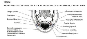
- Transverse section of the neck of the stallion at the level of cervical vertebra 5 (C5), caudal view. Located inside the carotid sheath are the vagosympathetic trunk and common carotid artery. The horse does not have an internal jugular vein as in the ox. 6
-

- Comparative diagrams of the neck in transverse section of the ox and sheep, caudal views. The drawing of the ox neck is of an adult animal so the cervical thymus has regressed and is no longer visible in the neck. The drawing of the sheep neck is of a young animal so the cervical lobes of thymus are prominent. 6
-
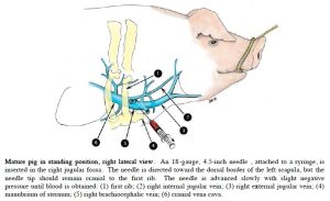
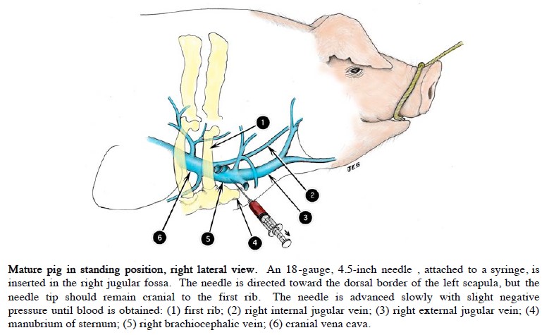
- Pig internal jugular vein, right lateral view. 2
Clinical relevance:
Hypertrophy of the ventral neck muscles occurs in horses that crib (crib-biting or “wind- sucking”). The Modified Forssell’s procedure involves excising a segment of the “strap muscles” (combined name is the sternothyrohyoideus mm. at their caudal fused portion) and omohyoideus m., and transecting the innervation of the sternocephalicus m. Partial myectomy of the strap muscles or a sternothyroideus tenectomy may also be performed to help manage dorsal displacement of the soft palate (DDSP) in horses.
Carnivore Thorax Nerves
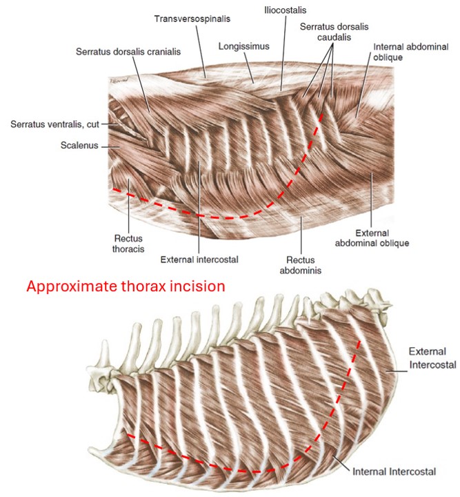
Dissect: We will enter the thorax by following these directions, using the provided diagrams as guides:
- On the left side (the left limb should already have been amputated), incise through the costal (rib) cartilage about two centimeters lateral to the sternum at ribs 2-8.
- Reach inside the thorax and pass your hand caudally until you feel the line of pleural reflection (your fingers should reach a point where they cannot pass any further). Use care when doing so, as the incised ribs may be sharp. The line of pleural reflection is where the tissue covering the deep surface of the ribs (the costal parietal pleura) sharply turns and is continuous with the tissue that lines the cranial surface of the diaphragm (diaphragmatic parietal pleura). We will review this landmark later.
- Cut through the thoracic wall from ventral to caudodorsal, staying about an inch cranial to the line of pleural reflection, so as not to enter the abdomen.
- Reach inside the thorax and snip each rib with bone cutters (provided by the anatomy team) as far dorsally as possible. These snips will provide hinge points on the ribs so as to allow the thoracic wall to be reflected dorsally as a unit.
- Leave rib 1 intact for now in order to preserve the nerves and arteries of the cranial mediastinum.
- Here’s a video of Dr. Gerard opening up the left side of the thorax to help guide you through the process.
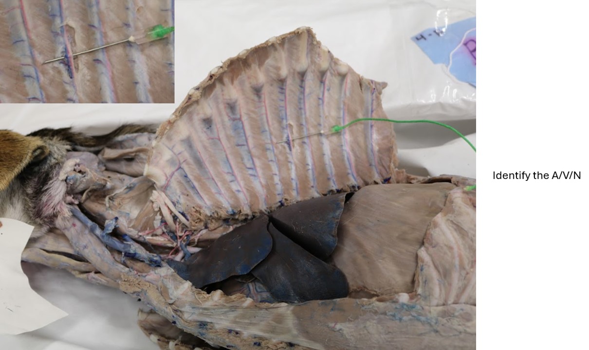
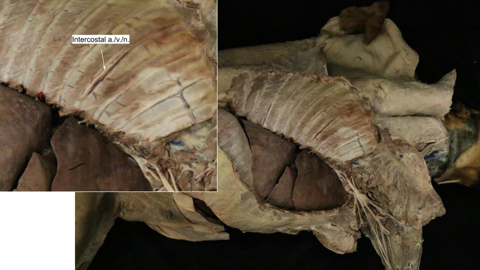
Okay, we’re in the thorax! Take a look at the deep surface of the thoracic wall, between the ribs. On the caudal edge of each rib, notice that there is a small neurovascular bundle. These are the intercostal artery, vein, and nerve. Notice that the artery and vein of each intercostal space divide into dorsal and ventral branches.
Each intercostal n. is derived from a spinal nerve as it emerges from the intervertebral foramen. The ventral branches of the first 12 thoracic spinal nerves are intercostal nerves and have lateral and ventral cutaneous branches (see next paragraph) and muscular branches supplying the intercostal muscles.
Dorsal and lateral rows of lateral cutaneous branches of intercostal nerves, and dorsal intercostal arteries, and veins emerge at regular intervals between the ribs and supply the cutaneous muscle, subcutaneous tissue, skin and thoracic mammae. The nerves of the dorsal row arise from the dorsal branches of the thoracic spinal nerves. A row of ventral cutaneous nerve branches emerges through the origin of the deep pectoral muscle after having penetrated the ventral ends of the intercostal spaces. These ventral nerves are the terminal branches of the intercostal nerves.
-
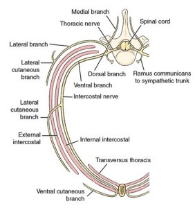
- Schema of a thoracic spinal nerve. 1

Clinical Application:
Thoracocentesis is performed on the cranial aspect of the eight or ninth rib, avoiding the neurovascular bundles which are located on the caudal aspect of each rib.
Review Videos
Terms
| Term | Species |
| Cervical spinal nerve 2 (C2) | Carnivore |
| Great auricular n. | Carnivore; innervates skin on caudal surface of ear |
| Cervical spinal nerves 3-5 (C3-5) | Carnivore |
| Accessory n. (CN XI) | All; innervates trapezius, omotransversarius, sternocephalicus, and cleidocephalicus mm. |
| Ventral br. of accessory n. | Horse; innervates sternocephalicus m. |
| Carotid sheath | All; know contents |
| Vagosympathetic trunk | All |
| Vagus n. (CN X) | Carries parasympathetic fibers from brain |
| Sympathetic trunk | Carries sympathetic fibers |
| Common carotid a. | All |
| Internal jugular v. | Identify in the Pig; Otherwise, know that it is included in the contents of the carotid sheath in other species, but don’t identify in species other than pig |
| Tracheal trunk | Lymphatic vessel; know that it is included in the contents of the carotid sheath, but don’t identify |
| Intercostal a./v./n. | All |