Labs 13A & 14A: Neuroanatomy
Learning Objectives
- Identify and list functions of structures seen on the surface of the cerebrum, cerebellum, and brainstem.
- Identify the parts of the ventricular system and describe the flow pathway of cerebrospinal fluid.
- Identify and list functions of structures seen on the surface of the cerebrum, cerebellum, and brainstem.
- Identify and list functions of structures seen in transverse cross sections throughout the brain.
Lab 13: Surface Neuroanatomy
Lab Instructions
In order to study neuroanatomy, we will not be doing any active dissection with our cadavers. Instead, you will be provided with plastic casts of canine brains (what this guide will refer to as the “blue brains”), as well as transverse slices of canine brains. Other specimens around the lab may be referenced, particularly for blood supply to the brain.
Meninges
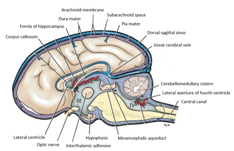
Just like the spinal cord, the brain is covered by three membranes of connective, collectively known as the meninges. Let’s have a quick review of these meninges before we describe some important structures associated with them around the brain. From most superficial to deep, these meninges are: the dura mater, the arachnoid membrane, and the pia mater.
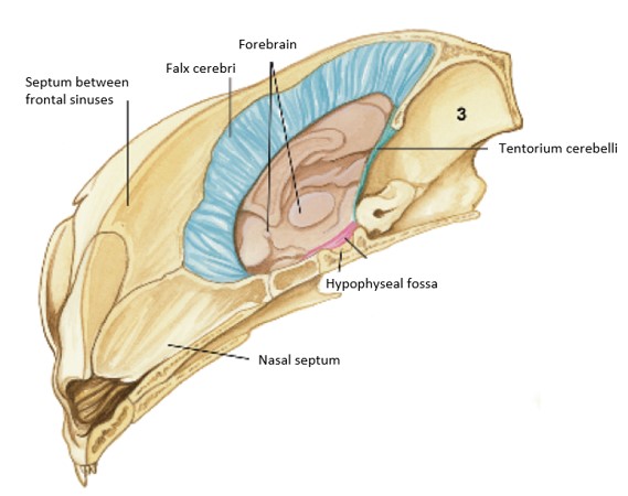
Throughout most of the vertebral canal, the dura is separated from the periosteum of the bony canal by the loose connective tissue of the epidural space, which often contains fat. As the spinal cord approaches the brain stem, the dura adheres to the periosteum in the first one or two cervical vertebrae and to the atlanto-occipital membrane. Inside the cranial cavity, the dura and periosteum are fused, which means we do not have an epidural space in a normal head. On one half of the head, a fold of dura will be found extending ventrally from the midline in the longitudinal cerebral fissure between the two cerebral hemispheres. This is the falx cerebri (falx [L] = sickle), which contains the dorsal sagittal sinus dorsally.
Observe: Identify the meninges, the falx cerebri, and the dorsal sagittal sinus in a bisected head. The falx cerebri will appear as a sickle-shaped extension of the dura mater descending from the dorsal roof of the neurocranium.
-
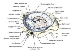
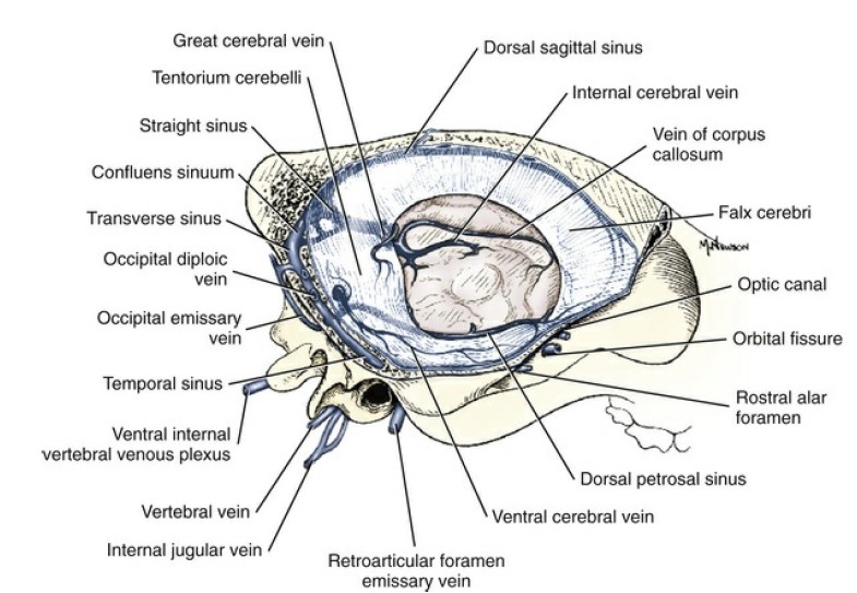
- Cranial venous sinuses of the dog, lateral aspect. (Right cerebral hemisphere removed.) 1
-
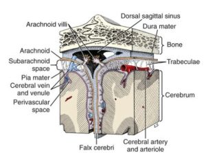
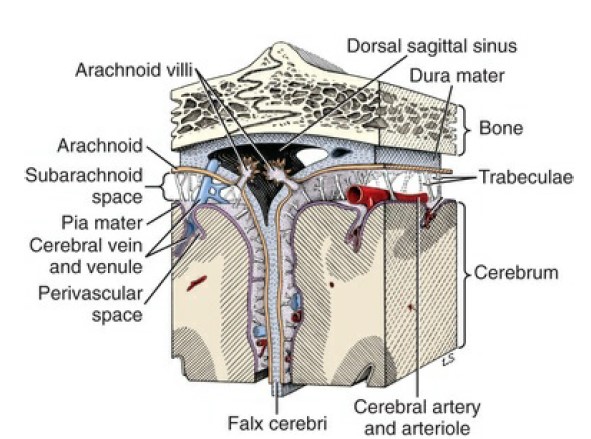
- Schematic view of arachnoid villi projecting into the dorsal sagittal venous sinus. The sinus is located where dura mater splits into a periosteal layer and a partition, the falx cerebri. 1
-

- Special areas of dura mater of the cat associated with the brain, median section with hindbrain removed, craniomedial view. 4

-
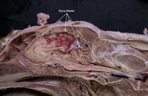
- Dog dura mater

-
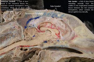
- Dog falx cerebri

The pia mater and arachnoid membrane are the other two connective tissue coverings of the central nervous system. The pia mater adheres to the external surface of the nervous tissue. The arachnoid in the live animal is attached to the dura and sends delicate trabeculae to the pia. These trabeculae closely invest the blood vessels that course on the surface of the pia. The space between the pia and the arachnoid is the subarachnoid space, which is filled with cerebrospinal fluid that surrounds the entire central nervous system. After death, the arachnoid membrane breaks free from the dura and collapses onto the pia of the central nervous system.
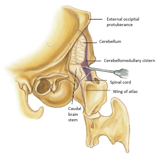
Subarachnoid cisterns occur in areas where the arachnoid and pia are more widely separated. The largest cistern is the cerebellomedullary cistern, located in the angle between the cerebellum and medulla. Cerebrospinal fluid may be obtained from this cistern by means of a needle puncture through the atlanto-occipital membrane, dura, and arachnoid.
-

- Schema of meninges and ventricles of the dog. Arrows indicate the flow of cerebrospinal fluid. 1
-

- Position of spinal needle in subarachnoid space of cerebellomedullary cistern of the cat. 4
-

- Dog head with a probe inserted into the subarachnoid space
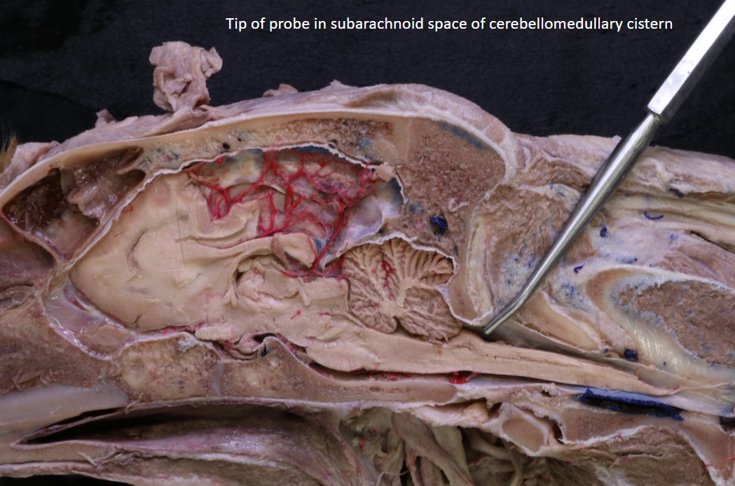
Observe: Identify the cerebellomedullary cistern in a bisected head.
Ventricular System
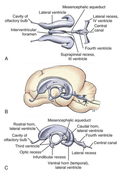
The ventricular system is the CSF filled passageway within the CNS that communicates with the subarachnoid space and is the remainder of the lumen of the neural tube. The lateral ventricles are located in each cerebral hemisphere and are normally separated from each other by a middle membrane (the septum pellucidum). Because of their lateral location in the brain, the lateral ventricles can only be observed in lab on transverse sections.
The lateral ventricles connect to the third ventricle caudally. The third ventricle is located in the diencephalon, and roughly resembles a squashed doughnut turned on its edge, with the doughnut hole occupied by the interthalamic adhesion.
Observe: Observe the third ventricle and the interthalamic adhesion on the blue brain models.
The third ventricle connects to the fourth ventricle via the mesencephalic aqueduct, located, as indicated, in the mesenencephalon. The mesencephalic aqueduct is a small passageway within the midbrain. It is a frequent site of inflammatory blockage, resulting in hydrocephalus.
Observe: Observe the mesencephalic aqueduct and fourth ventricle on the blue brain models.
-
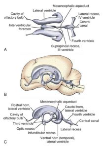
- Ventricular system of the dog brain. A, Dorsal view; B and C, lateral views. The arrows indicate the direction of flow through the ventricles and, through the lateral recess and aperture, to the subarachnoid space. 1
-

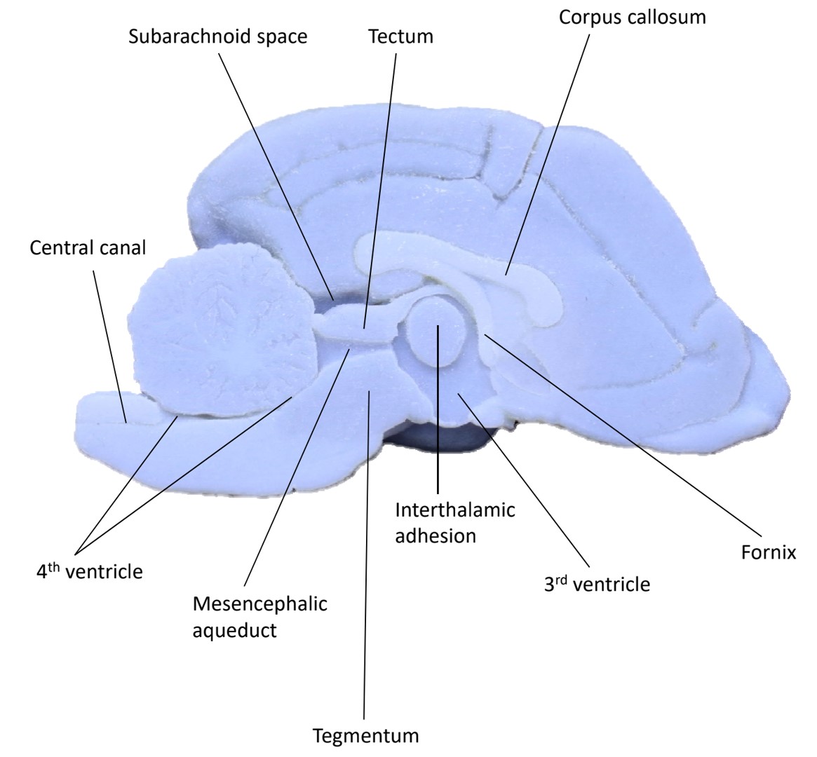
- Medial view of sagittal 3D printed brain
-

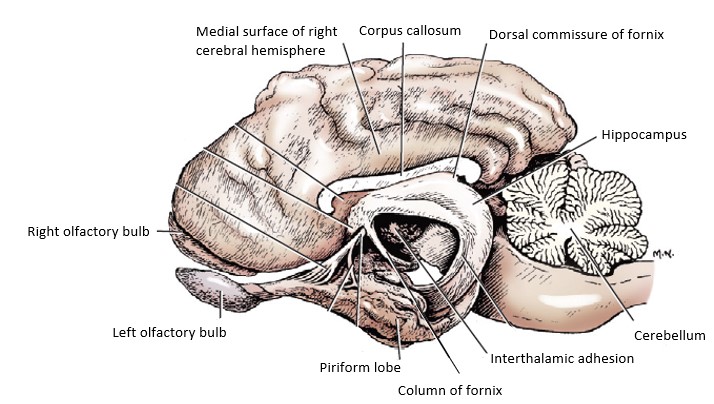
- Brain with the left half removed except for most of the left rhinencephalon. 1
Surface Structures of the Brain
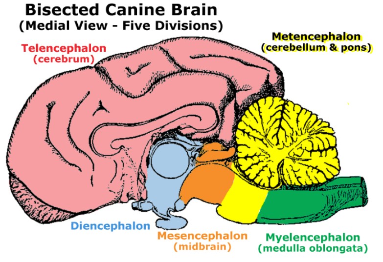
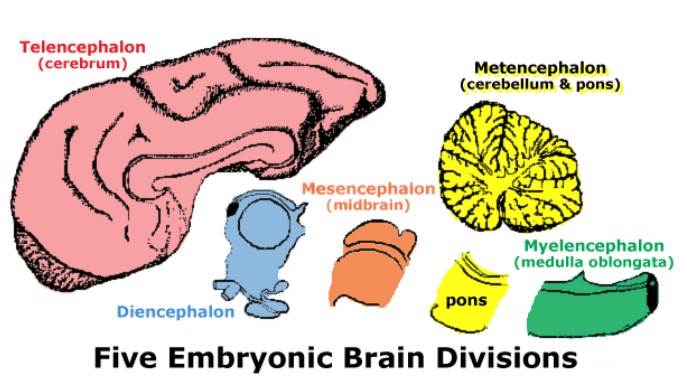
The brain is composed of the embryologically segmented brain stem and two suprasegmental portions, the cerebrum (telencephalon) and the cerebellum (dorsal metencephalon). The brain stem includes the myelencephalon (medulla), the ventral metencephalon (pons), the mesencephalon (midbrain), and the diencephalon (epithalamus, thalamus, and hypothalamus).
-
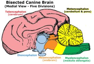
- Canine brain showing the Five Embryonic Divisions: Telencephalon, Diencephalon, Mesencephalon, Metencephalon, and Myelencephalon.
-
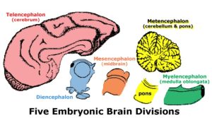
- Five embryonic brain divisions: Telencephalon, Diencephalon, Mesencephalon, Metencephalon, and Myelencephalon.
-
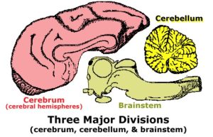
- Three Major Divisions of the brain: Cerebrum, Cerebellum, and Brainstem.

Cerebrum
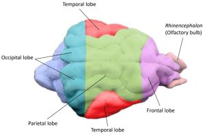
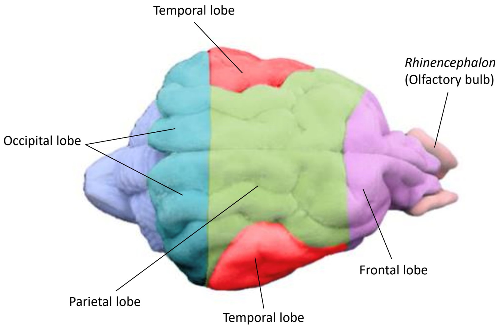
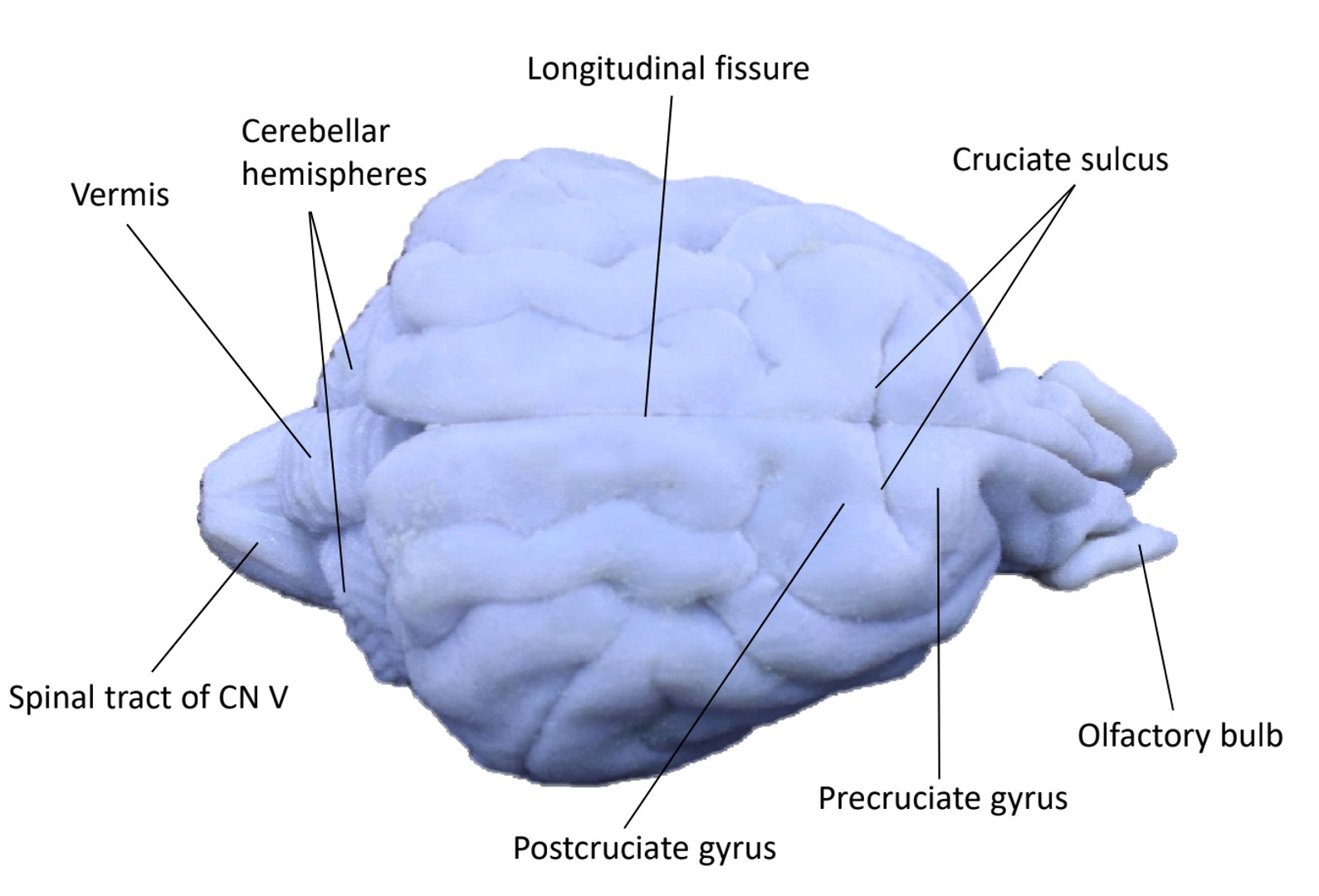
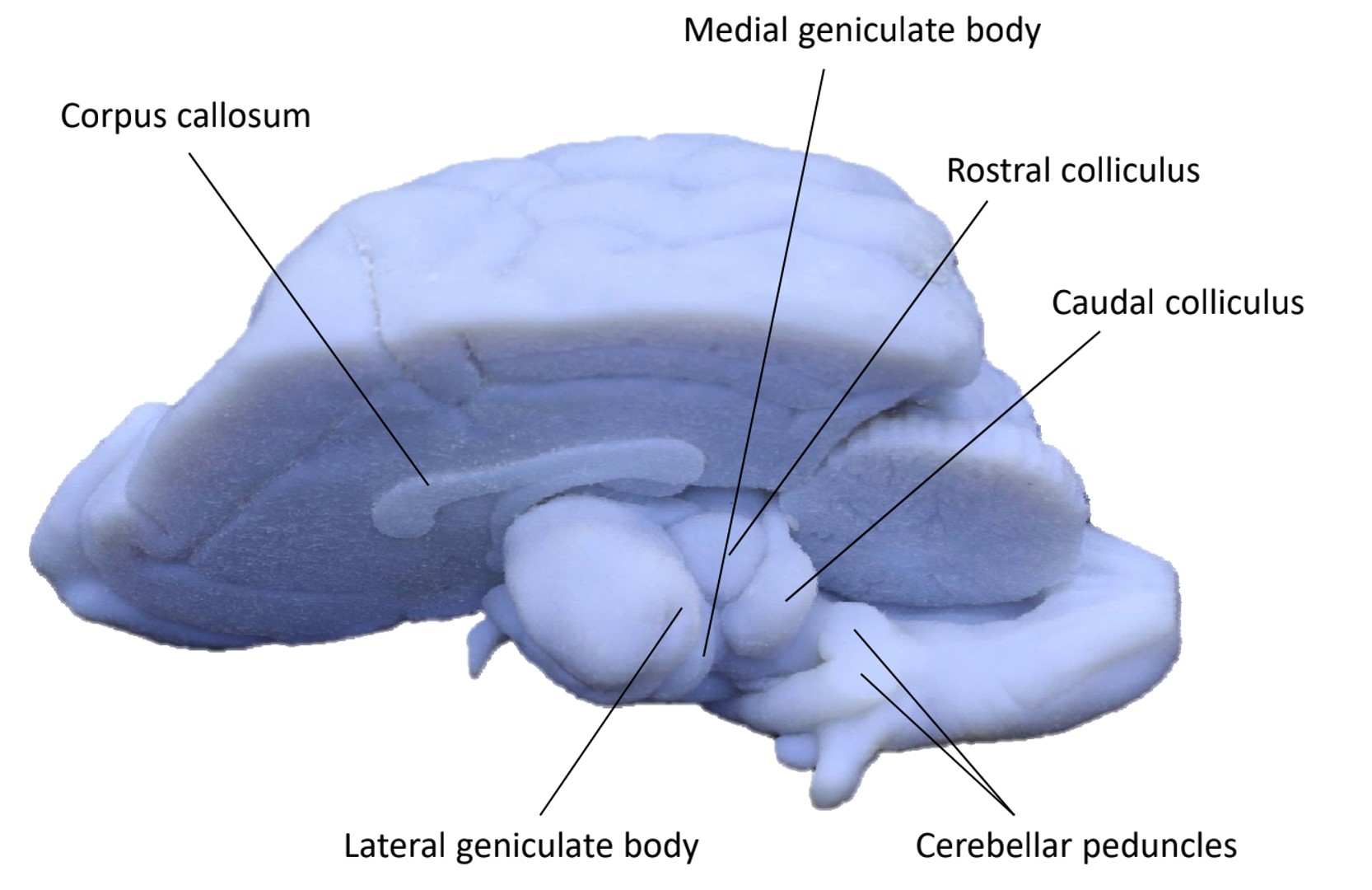
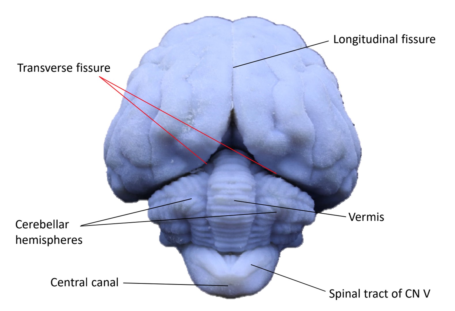
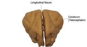
The cerebrum is divided into two cerebral hemispheres by the longitudinal fissure. Each cerebral hemisphere has outward folds (convolutions) called gyri separated by inward folds called sulci.
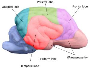
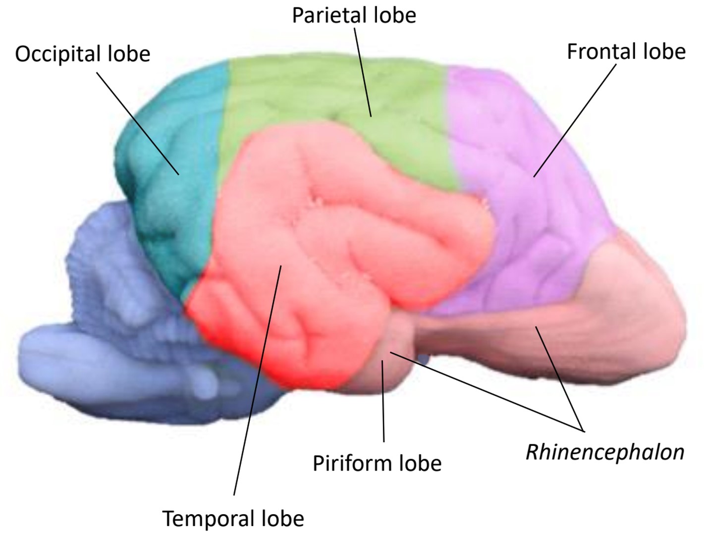
Each cerebral hemisphere may be divided into lobes named for that portion of the calvaria that covers them. The relationship is not precise and varies among species. The frontal lobe is that portion of each cerebral hemisphere rostral to the cruciate sulcus. The parietal lobe is caudal to the cruciate sulcus. It extends caudally to approximately the caudal third of the cerebral hemisphere. The occipital lobe includes the caudal third of the cerebral hemisphere. Caudal portions of this lobe on both medial and lateral sides function as the visual cortex. The temporal lobe is composed of the gyri and sulci on the ventrolateral aspect of the cerebral hemisphere.
Observe: Identify the cruciate sulcus on the dorsal surface of the cerebrum. You can find it close to the rostral end of the cerebrum, running perpendicular to the large longitudinal fissure which separates the left and right hemispheres of the cerebrum. See images below to help you find this sulcus.
Portions of the more primitive cerebrum (i.e., the “paleopallium”) that are visible are the olfactory bulb, which rests on the cribriform plate, and the olfactory peduncle, which joins the bulb to the cerebral hemisphere. The olfactory peduncle courses caudally with a band of fibers on its ventral surface. Caudally, this band divides into lateral and medial olfactory tracts, which course their way to the piriform lobe.
Observe: Identify the frontal, parietal, occipital, temporal, and piriform lobes of the cerebrum.
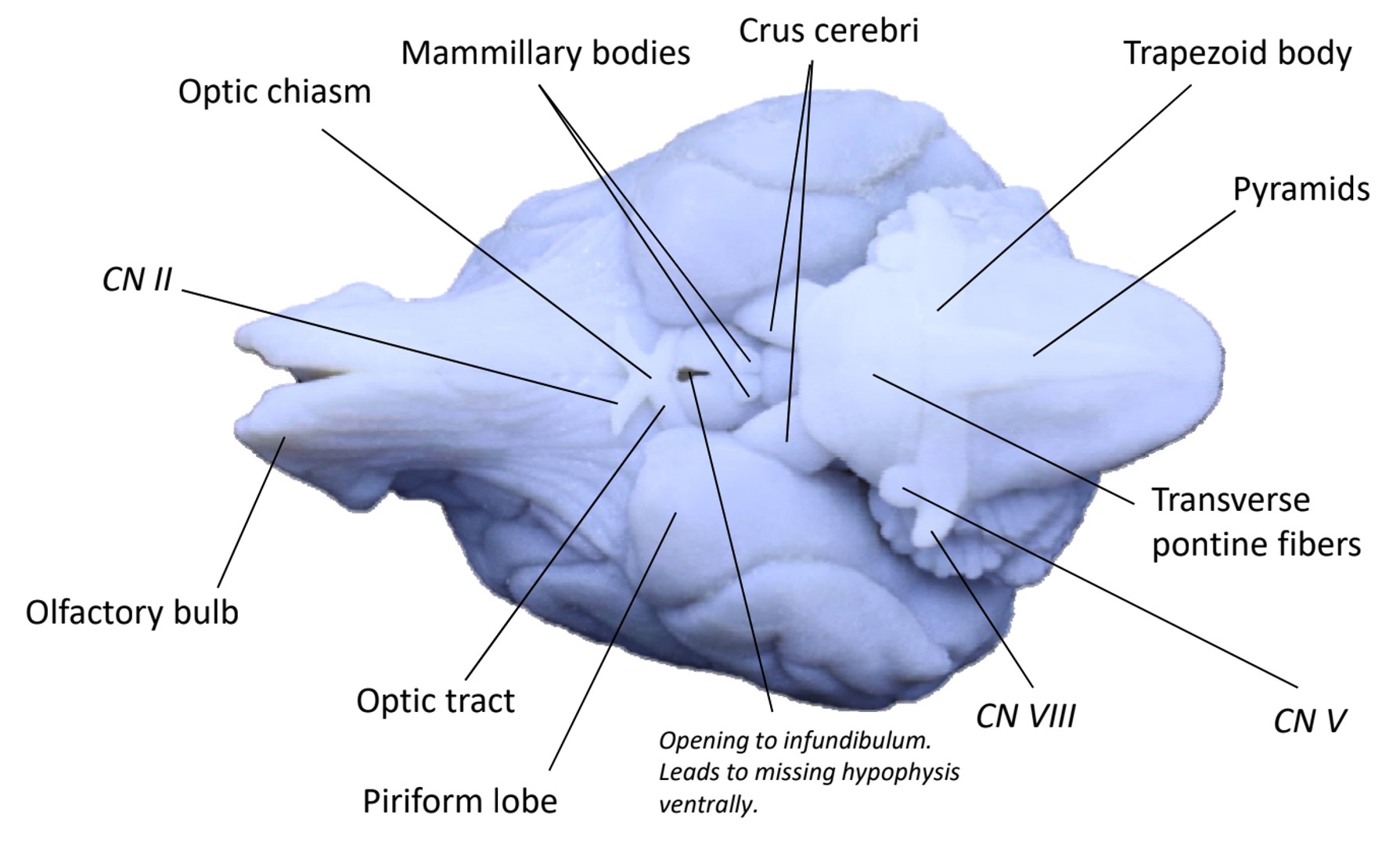
On the bisected blue brain, notice a conspicuous horn-like, or antenna-like structure on the medial surface; this is the major white matter commissural tract carrying axons from one cerebral hemisphere to the other, the corpus callosum. Ventral to the corpus callosum, we will find the brainstem (see below).
Observe: Identify the corpus callosum on the medial surface of a bisected blue brain.
-
- Color-coded dog brain model illustrating the major cerebral lobes
-
- Color-coded dog brain model illustrating the major cerebral lobes
-

- Dorsal view of a 3D printed dog brain showing key surface features.
-

- Midsagittal view of a 3D printed dog brain showing deep structures

-
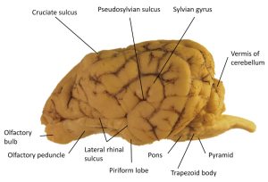
- Lateral view of a dog brain showing surface structures

-
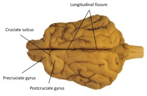
- Dorsal view of a dog brain showing surface structures

Cerebellum
The cerebellum is derived from the dorsal portion of the metencephalon and lies caudal to the cerebrum and dorsal to the fourth ventricle. The transverse cerebral fissure separates it from the cerebrum. The dural and osseous tentorium cerebelli is located in this fissure. The cerebellum is connected to the brain stem by three cerebellar peduncles on each side of the fourth ventricle and by portions of the roof of the fourth ventricle.
Observe: Identify the tentorium cerebelli in the transverse cerebral fissure in a bisected head.
-

- Medial section of a cat skull showing the specialized dural folds like the Falx cerebri and Tentorium cerebelli. 4
-
- Dog falx cerebri
The cerebellar peduncles connect the medulla and cerebellum. The rostral cerebellar peduncle contains mainly efferent axons from the cerebellum to the brain stem. Afferent axons to the cerebellum from the brain stem and spinal cord pass primarily through the middle and caudal cerebellar peduncles.
Observe: Identify the transverse fibers of the pons on the ventral surface of the brain stem. Follow these fibers laterally as they course dorsocaudally into the cerebellum on each side as the middle cerebellar peduncles.
-
- Ventral view of a 3D printed dog brain model
-

- Medial view of a 3D printed dog brain with the left cerebrum resected, exposing the deep diencephalic and midbrain structures
-

- Ventral view of a dog brain

The cerebellum is composed of lateral cerebellar hemispheres and a middle portion, the vermis. The convolutions of the cerebellum are known as folia (in place of “gyri”, as we see in the cerebrum). These are grouped into three lobes and numerous cerebellar lobules that have specific names. The vermis comprises the entire middle portion of the cerebellum directly above the fourth ventricle. Some of its lobules are found on the ventral surface of the cerebellum facing the roof plate of the fourth ventricle. Each hemisphere projects over the cerebellar peduncles and the adjacent brain stem. A lateral component lies in the cerebellar fossa of the petrosal part of the temporal bone.
Observe: Identify the vermis running dorsoventrally at the midline of the cerebellum.
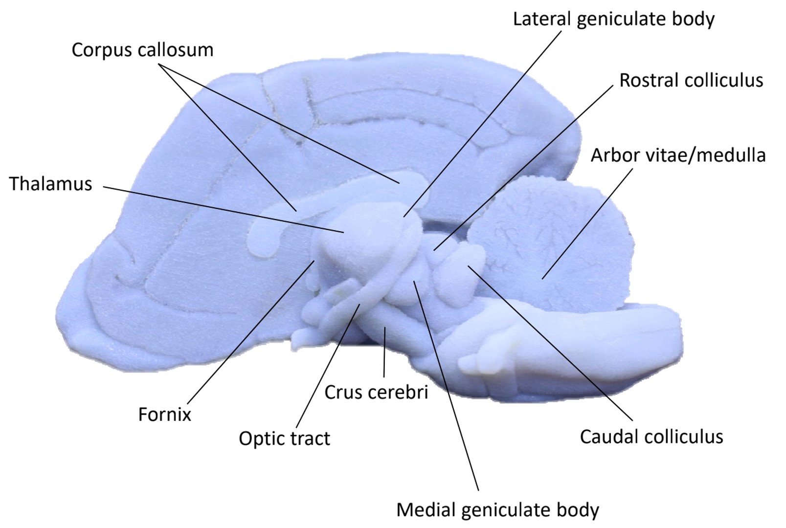
On a model with the cerebellum longitudinally hemisected through the vermis, examine the medial cut surface of the cerebellum. The medulla of the cerebellum is the white matter in its central portion that contains the cerebellar nuclei and connects with all the folia and the cerebellar peduncles. Note the pattern of white matter as it branches and arborizes from the medulla of the cerebellum into the folia. The medulla is often referred to as the arbor vitae ([L] “tree of life”).
-

- Caudal view of a 3D printed dog brain showing the cerebrum and cerebellum
-

- Caudal view of a dog brain showing the cerebrum and cerebellum

-

- Medial view of a 3D printed dog brain with resected left cerebrum
Brainstem
While the most evolutionarily “primitive” portion of the brain, the brainstem nevertheless performs an array of complex and vital functions for the animal. The brainstem includes, from rostral to caudal: the diencephalon, the mesencephalon, the ventral metencephalon, and the myelencephalon.
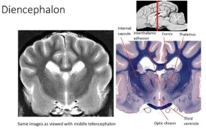
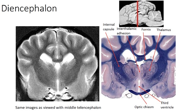
Diencephalon
The diencephalon consists of a large, centrally located thalamus; a smaller hypothalamus below; and a very small epithalamus on the dorsal midline. It is the most rostral part of the brainstem. Recall that while the diencephalon is part of the brainstem anatomically, functionally it is grouped with the telencephalon. In the laboratory setting we will consider the diencephalon from an anatomical perspective. However, it’s important to recognize that as the relay system to and from the telencephalon, deficits here will cause clinical signs which localize to the forebrain.
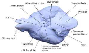
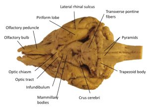
The prechiasmatic optic tract (CN II) forms the optic chiasm of the diencephalon rostral to the hypophysi (i.e., the pituitary gland). The postchiasmatic optic tracts course laterally and dorsocaudally from the chiasm, pass over the lateral surface of the diencephalon, and enter the lateral geniculate nuclei of the thalamus.
Caudal to the optic chiasm on the median plane is the hypophysis (pituitary gland), which is attached by the infundibulum to the hypothalamus. If the gland is missing, the lumen of the infundibulum will be evident. This lumen communicates with the overlying third ventricle of the diencephalon.
The mammillary bodies of the hypothalamus bulge ventrally caudal to the lumen of the infundibulum. They demarcate the most caudal extent of the hypothalamus on the ventral surface of the diencephalon.
Observe: Identify the optic chiasm and mammillary bodies of the diencephalon.
-
- Ventral view of a 3D printed dog brain model
-
- Ventral view of a dog brain

The internal capsule bounds the diencephalon laterally and was cut when the left cerebral hemisphere was removed. The thalamus and epithalamus can be seen on the dorsal aspect of the diencephalon. The thalamus lies between the internal capsule on each side and dorsal to the hypothalamus. It is covered by pia, arachnoid trabeculations, and the subarachnoid space. It consists of two bilaterally symmetrical collections of nuclei that primarily project axons to the ipsilateral cerebral hemisphere. These two collections are separated by the third ventricle. Two of these nuclei are prominent and readily recognized on the caudal surface. These are the geniculate nuclei, which comprise the metathalamus. A lateral eminence on the caudodorsal surface of the thalamus is the lateral geniculate nucleus, which receives fibers of the optic tract and functions in the visual system. The lateral geniculate nucleus is connected with the rostral colliculus of the midbrain. Caudoventral to the lateral geniculate nucleus is the medial geniculate nucleus of the thalamus. This nucleus functions in the auditory system and is connected to the caudal colliculus of the midbrain by the brachium of the caudal colliculus.
Observe: In the third ventricle, observe the interthalamic adhesion between the right and left sides of the thalamus. Identify the lateral and medial geniculate nucleus on the doral surface of the diencephalon.
-
- Midsagittal view of a 3D printed dog brain showing deep structures
-
- Medial view of a 3D printed dog brain with resected left cerebrum
-
- Medial view of a 3D printed dog brain with the left cerebrum resected, exposing the deep diencephalic and midbrain structures
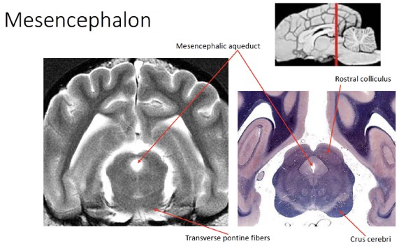
Mesencephalon
The mesencephalon (midbrain) is relatively short. It is nearly round on the transverse section with a canal, the mesencephalic aqueduct, passing through it. The mesencephalon consists of a tectum, or roof, dorsal to the aqueduct, which is composed of four groups of neuronal cell bodies: the colliculi. That portion of the mesencephalon ventral to the aqueduct is the cerebral peduncle, which consists of a tegmentum, substantia nigra, and crus cerebri, from dorsal to ventral. For our purposes, it is important that you recall that the tegmentum is that part of the mesencephalon ventral to the mesencephalic aqueduct.
Observe: Identify the mesencephalic aqueduct on the medial surface of a blue brain. Note that dorsal to this aqueduct is the tectum, and ventral to is the tegmentum.
We’ll return to the mesencephalon to see the structures described above when we look at brain transverse sections; for now let’s review what we can see of the mesencephalon on the blue brains.
Caudally projecting tracts of projection processes that connect portions of the cerebral cortex with brain stem centers and the spinal cord course on the ventral surface of the midbrain. These are grouped together on each side as the crus cerebri. The left and right oculomotor nn. (CN III) leaves the midbrain medial to the crura.
Observe: Identify the crus cerebri on the ventral surface of the brain, just caudal to the hypothalamus.
The mesencephalic structures dorsal to the mesencephalic aqueduct compose the tectum of the midbrain. The mesencephalic aqueduct is a short, narrow tube, derived from the neural canal in the midbrain that connects the third ventricle rostrally with the fourth ventricle caudally. Four dorsal bulges, collectively known as the corpora quadrigemina, are evident on the dorsal side. The rostral pair are the rostral colliculi, which function with the visual system. The smaller caudal pair are the caudal colliculi, which function in the auditory system.
The trochlear nerve (CN IV) courses laterally out of the roof of the fourth ventricle adjacent to the caudal colliculus. It continues rostroventrally on the lateral surface of the midbrain.
Observe: Identify the rostral and caudal colliculi. Recognize that these are parts of the tectum of the mesencephalon.
-
- Midsagittal view of a 3D printed dog brain showing deep structures
-
- Medial view of a 3D printed dog brain with resected left cerebrum
-
- Medial view of a 3D printed dog brain with the left cerebrum resected, exposing the deep diencephalic and midbrain structures
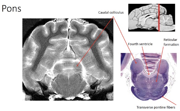
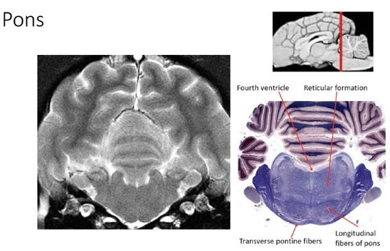
Metencephalon
The metencephalon includes a segment of the brain stem, called the pons, and the dorsal development, the cerebellum. The ventral surface of the pons includes the transverse fibers of the pons, which course laterally into the middle cerebellar peduncles. This large band of transverse fibers borders the trapezoid body of the medulla caudally. Its rostral border covers part of the ventral surface of the midbrain. The trigeminal nerve (CN V) is associated with the pons and can be found entering the pons along the caudolateral aspect of the transverse fibers.
Observe: Identify the pons, and specifically note the transverse pontine fibers on the ventral surface. Note that the dorsal metencephalon (i.e, the cerebellum) was discussed earlier.
-
- Ventral view of a 3D printed dog brain model
-
- Ventral view of a dog brain
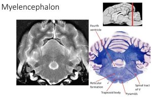
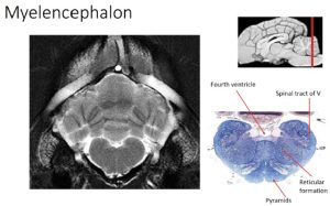
Myelencephalon
The myelencephalon, or medulla, extends from the transverse fibers of the pons to the level of the ventral rootlets of the first cervical spinal nerve. The trapezoid body is the transverse band of fibers rostrally that course parallel but caudal to the transverse pontine fibers. It is continuous with the vestibulocochlear nerve and cochlear nuclei laterally on the side of the medulla and functions in the auditory system.
Observe: On the ventral surface of the brain stem, just caudal to the transverse fibers of the pons, note the presence of the trapezoid body. Do not concern yourself with identifying the trapezoid body, but rather use it as a convenient landmark for separating the pons (ventral metencephalon) from the more caudal myelencephalon.
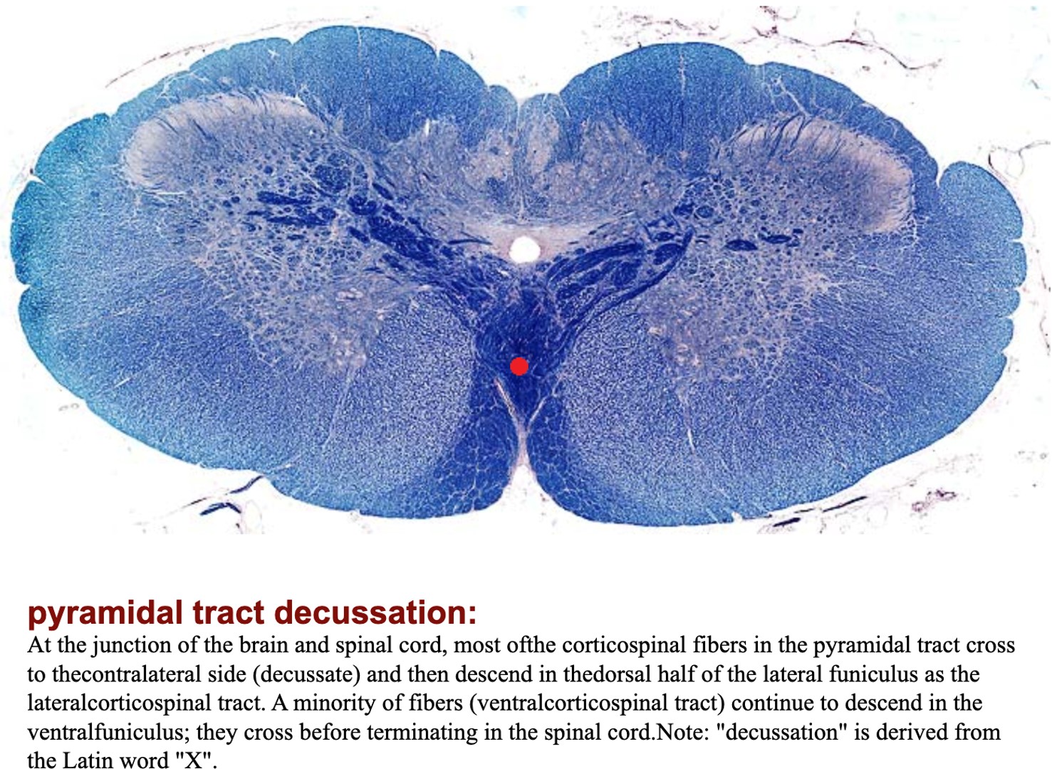
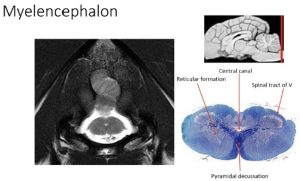
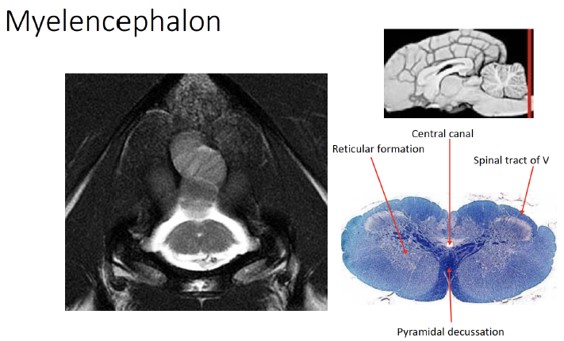
The pyramids are a pair of longitudinally coursing fiber bundles on either side of the ventral median plane. They emerge from the transverse fibers as the caudal continuations of axons from the longitudinal fibers of the pons that did not terminate in pontine nuclei. They course caudally across the trapezoid body to continue on the ventral surface of the medulla. They are separated by the ventral median fissure. This fissure can be followed caudally until it is obliterated over a short distance by the decussation of the pyramids located at the level of the emerging hypoglossal nerve fibers. The decussation itself is difficult to see because it occurs as the pyramidal fibers are passing dorsally into the parenchyma of the medulla. Pyramidal axons continue in the spinal cord as the corticospinal tracts. These are cerebral projection processes that project to the spinal cord.
Observe: Identify the pyramids on the ventral surface of the medulla.
-
- Ventral view of a 3D printed dog brain model
-
- Ventral view of a dog brain
-

- Histological transverse section of the brainstem/spinal cord junction showing the decussation of the pyramidal tracts
Lab 14: Transverse Cross-section Neuroanatomy
Lab Instructions
You have been given a jar with a canine brain transversely cut into a number of slices. Understanding the cross sectional anatomy of the brain is a challenging task; first and foremost it requires a solid understanding of the surface anatomy that was covered in the previous lab. While you move your way caudally through the brain slices, be sure you are using the blue brains as references to better conceptualize the way the cross sectional anatomy relates to what you are seeing on the surface.
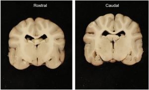
In order to facilitate your understanding of this material, we will present the brain slices from rostral to caudal and cover the important anatomy that you see in each one. Please note: The slices presented in this lab guide are not guaranteed to be precisely like your own. These brains are sliced by your instructors, and therefore subject to degrees of variation. This is why it’s important to consider consistent landmarks to help your identification.
Additionally, we will provide you with images of the cranial and caudal surfaces of each slice; be sure to look at both sides of your own slices to get a complete picture of the anatomy.
Also note: There are a number of important nuclei, particularly in the brainstem, of which you should be aware. These are italicized in the key terms listed above. We will not be directly identifying these nuclei on the slices, but you should know which anatomic division of the brain is home to which nuclei. Please refer to the Brain lecture notes for more information regarding these nuclei.
With all of that in mind, let’s jump in!
Slice #1
Anatomic divisions present:
- Telencephalon
The rostral surface of slice #1 is also the most rostral part of the brain. Here we have sliced the brain at the cruciate sulcus. Rostral to the cruciate sulcus is the frontal lobe of the cerebrum (i.e., telencephalon). Separating the left and right half hemispheres of the cerebrum from one another is the deep longitudinal fissure. While not seen in this image, you may have olfactory bulbs still present on the ventral surface of your brain.
Observe: Identify the longitudinal fissure and olfactory bulbs in your specimen (if present). Recognize that the cruciate sulcus separates the frontal lobe rostrally from the parietal lobes.
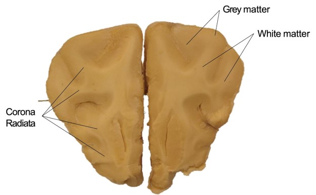
Flipping to the caudal surface of slice #1, we now can appreciate the internal anatomy of the telencephalon. Along the margins, you will notice the darker grey matter of the cerebral cortex. This grey matter contains the neuronal cell bodies of the telencephalon. Deep to the grey matter, you can identify tracts of white matter, which contain the axons of the neurons whose bodies are found in the cerebral cortex. Together these white matter projection tracts are known as the corona radiata ([L], “radiated crown”). Projection tracts are white matter tracts in the brain that connect the cerebral cortex to subcortical structures, such as the thalamus.
Observe: Identify the grey matter and corona radiata.
-
- Rostral surface Slice #1

-

- Caudal surface Slice #1
-
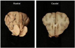
- Slice 1: Rostral and caudal surface

-

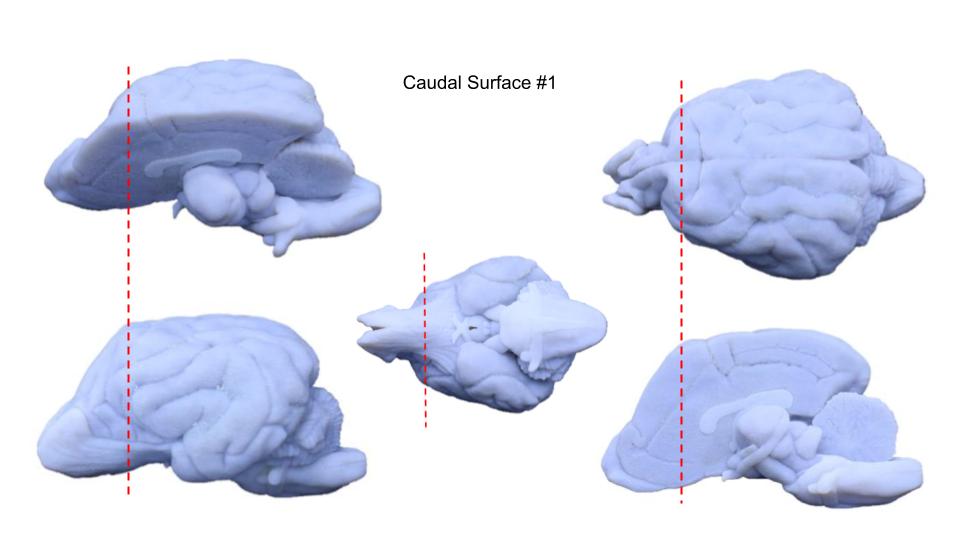
- Approximate location of slice #1
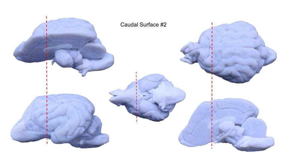
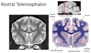
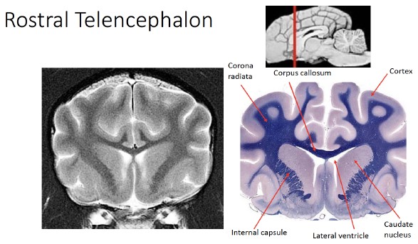
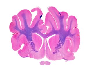
Slice #2
Anatomic divisions present:
- Telencephalon
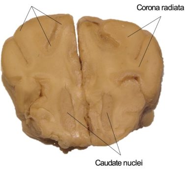
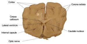
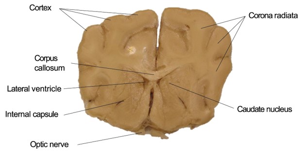
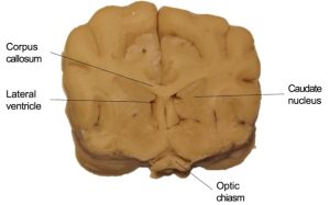
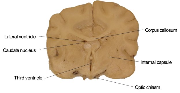
On the rostral surface of slice #2, we can one again identify the corona radiata and surrounding grey matter of the cortex. On the ventral aspect of this slice, you may notice distinct clumps of grey matter close to midline; these are the caudate nuclei, examples of the subcortical basal UMN nuclei of the telencephalon.
On the caudal side, you can now see a larger variety of structures within the transverse section. Ventral to the corpus callosum, you will notice two spaces just lateral to the midline; these are the rostral-most extents of the lateral ventricles. Lateral to the caudate nuclei on the left and ride sides, there are white matter tracts that descend ventrally from the corona radiata known as the internal capsule. The internal capsule contains projection fibers that course between the telencephalon and the diencephalon and course caudally from the telencephalon to the brain stem and the spinal cord.
Observe: Identify the lateral ventricles and internal capsule.
-

- Slice 2: Rostral surface
-
- Slice 2: Caudal surface
-
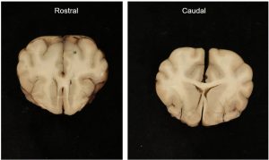
- Slice 2: Rostral and caudal surface

-
- Approximate location of slice #2
-
- Slice 2: Radiographic and stained
-
- Slice 2: Stained
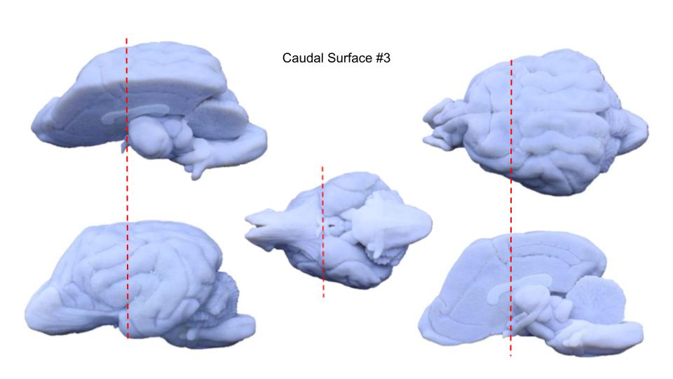
Slice #3
Anatomic divisions present:
- Telencephalon
- Diencephalon
On the rostral surface of slice #3, we see the same structures from the caudal surface of slice #2 extending caudally. Notice the caudate nuclei shifting dorsally as they reach their caudal extent, and the lateral ventricles expanding laterally as we move caudally in the brain.
The caudal surface of slice #3 likely represents the first available view of the third ventricle in cross-section. Compare this to the view that we get of the circular third ventricle that we see in the longitudinal cross-section on the medial surface of the bisected blue brain. Recall that the lateral ventricles are connected to the third ventricle via the interventricular foramina (not seen, but important to understand flow of CSF). The third ventricle is also the ventricular cavity associated with the diencephalon, giving us our first view of an anatomic region besides the telencephalon.
-
- Slice 3: Rostral surface

-

- Slice 3: Caudal surface
-

- Slice 3: Rostral and caudal surface

-
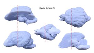
- Approximate location of slice #3
-
- Slice 3: Radiographic and stained

-

- Slice 3: Stained
-
- Slice 3: Stained

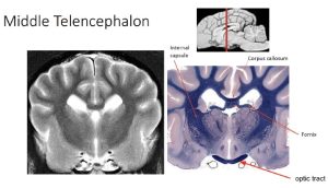
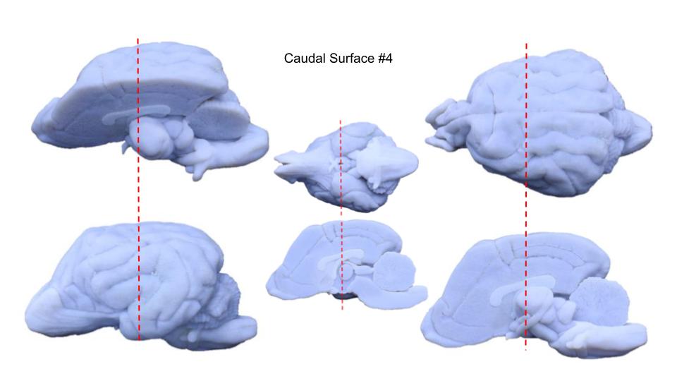
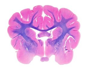
Slice #4
Anatomic divisions present:
- Telencephalon
- Diencephalon
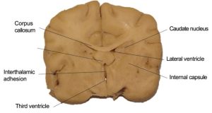
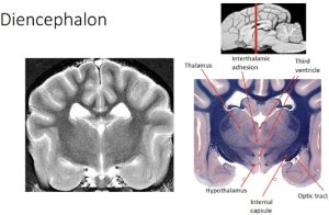
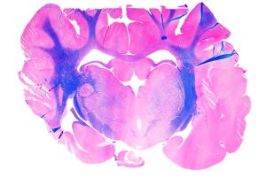
On the rostral and caudal surfaces of slice #4, we see a continuation of the structures of the telencephalon that we identified in more rostral slices; including the corpus callosum, internal capsule, caudate nuclei, and lateral ventricles. We also see structures of the diencephalon, including the third ventricle, which in your specimen may now be seen in two sections, dorsal and ventral. Separating these dorsal and ventral portions of the third ventricle is the interthalamic adhesion. The third ventricle roughly resembles a squashed doughnut turned on its edge, with the doughnut hole occupied by the interthalamic adhesion. The interthalamic adhesion is the medial connection of the left and right halves of the thalamus. The lateral and ventral walls of the third ventricle are formed by the hypothalamus. The hypothalamic nuclei are symmetrically organized on both sides of the ventral portion of the third ventricle bordered laterally by the ventral portion of the internal capsule and the post-chiasmatic optic tract.
Observe: Identify the interthalamic adhesion, third ventricle, and hypothalamus.
-
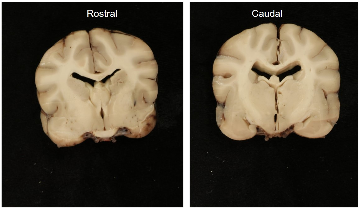
- Slice 4: Rostral surface

-

- Slice 4: Caudal surface

-
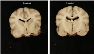
- Slice 4: Rostral and caudal surface
-
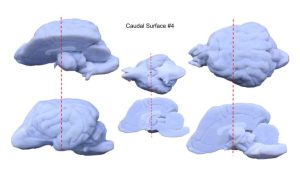
- Approximate location of slice #4
-
- Midsagittal view of a 3D printed dog brain showing deep structures
-
- Slice 4: Radiographic and stained
-
- Slice 4: Stained

-
- Slice 5: Stained

-
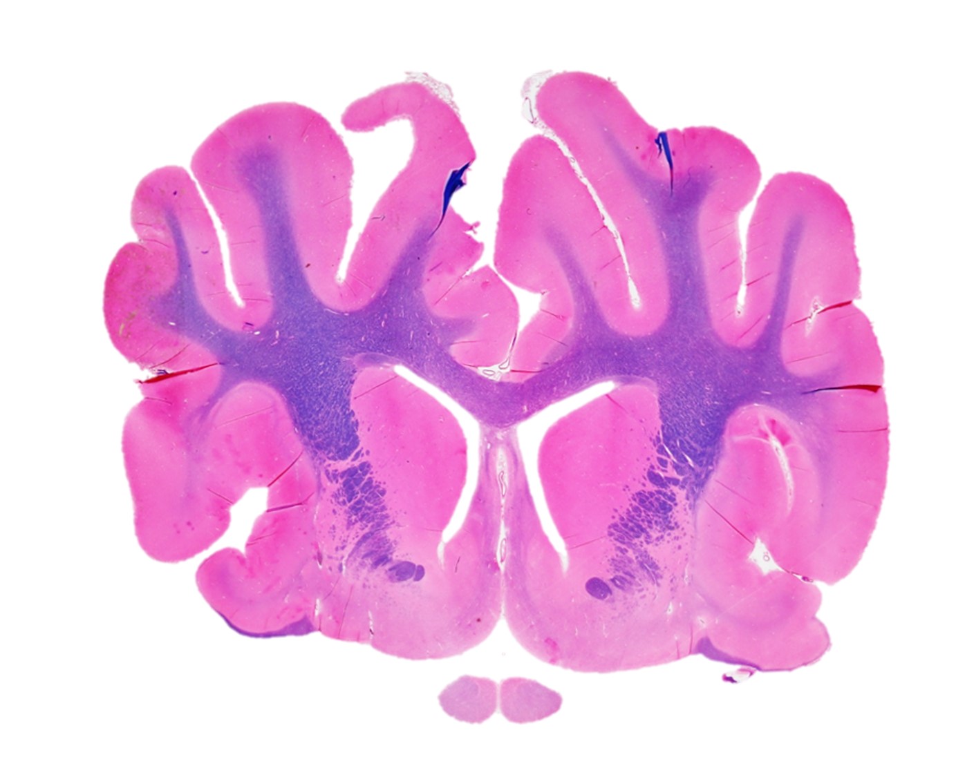
- Prosencephalon, transverse section. Note that the red arrow is pointing to a very thin membrane called the tela choroidea ventriculi tertii (FYI) which separates the subarachnoid space from the third ventricle. Choroid plexus is present on the ventral surface of this structure and produces cerebrospinal fluid in the third ventricle. It is not present in any of the cadaveric brains in the laboratory, nor will it appear in photographs in this guide as it is invariably broken in handling the specimens. It’s important to know that it is located here in life to understand where the subarachnoid space exists dorsal to the third ventricle. 27
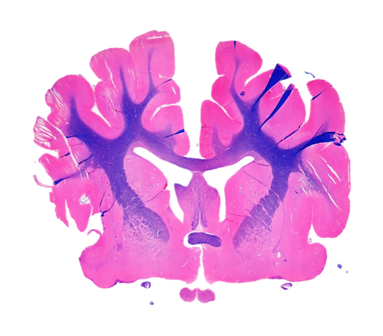
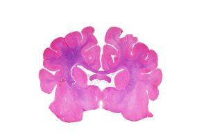
Slice #5
Anatomic divisions present:
- Telencephalon
- Diencephalon
- Mesencephalon
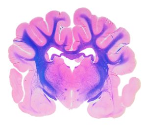
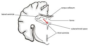
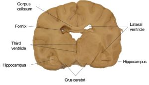
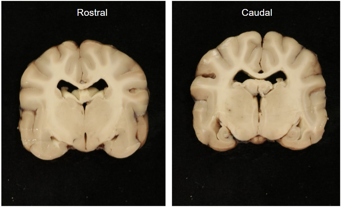
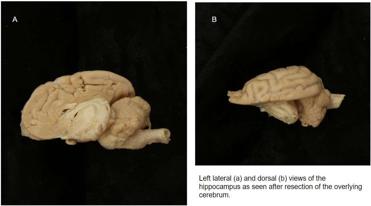
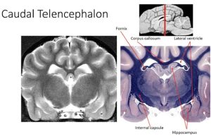
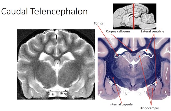
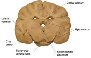
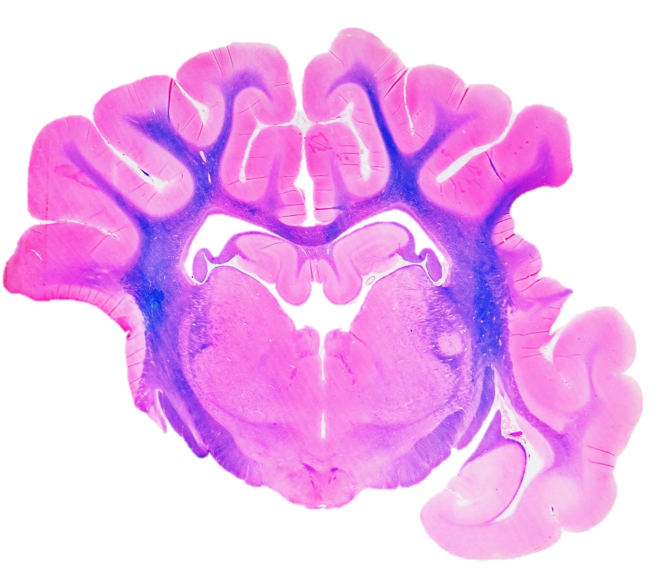
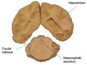
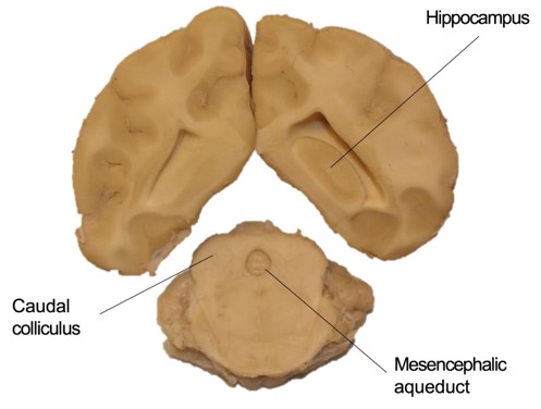
Looking at slice #5, we notice that the lateral ventricles are now visible both dorsomedially and ventrolaterally. Closely associated with the lateral ventricles in this slice we can also visualize the grey matter of the hippocampus. The hippocampus is a specialized region of the cerebral cortex. The white matter closely associated with the hippocampus is the fornix. The fornix begins caudally by the accumulation of fibers on the lateral side of the hippocampus. These form the crus of the fornix. The crura join rostral to the hippocampus and dorsal to the thalamus to form the body of the fornix.
Ventral to the third ventricle, you will notice a pair of large “mounds” close to the ventral midline of the brain slice. Caudally projecting tracts of projection processes that connect portions of the cerebral cortex with mesencephalic centers and the spinal cord course on the ventral surface of the midbrain. These are grouped together on each side as the crus cerebri.
Observe: Identify the hippocampus, fornix, and crus cerebri. Note the appearance dorsomedially and ventrolaterally of the lateral ventricles in cross section.
-

- Slice 5: Rostral surface

-
- Caudal surface

-
- Slice 5: Rostral and caudal surface
-
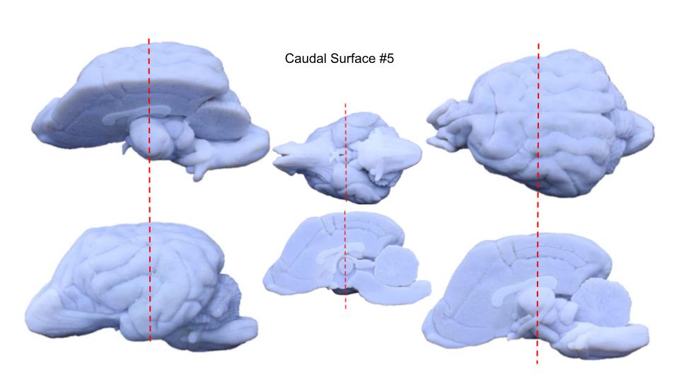
- Approximate location of slice #5
-
- Midsagittal view of a 3D printed dog brain showing deep structures
-
- Slice 5a: Radiographic and stained
-
- Slice 5b: Radiographic and stained

-
- Slice 5: Stained
-
- Slice 6: Stained

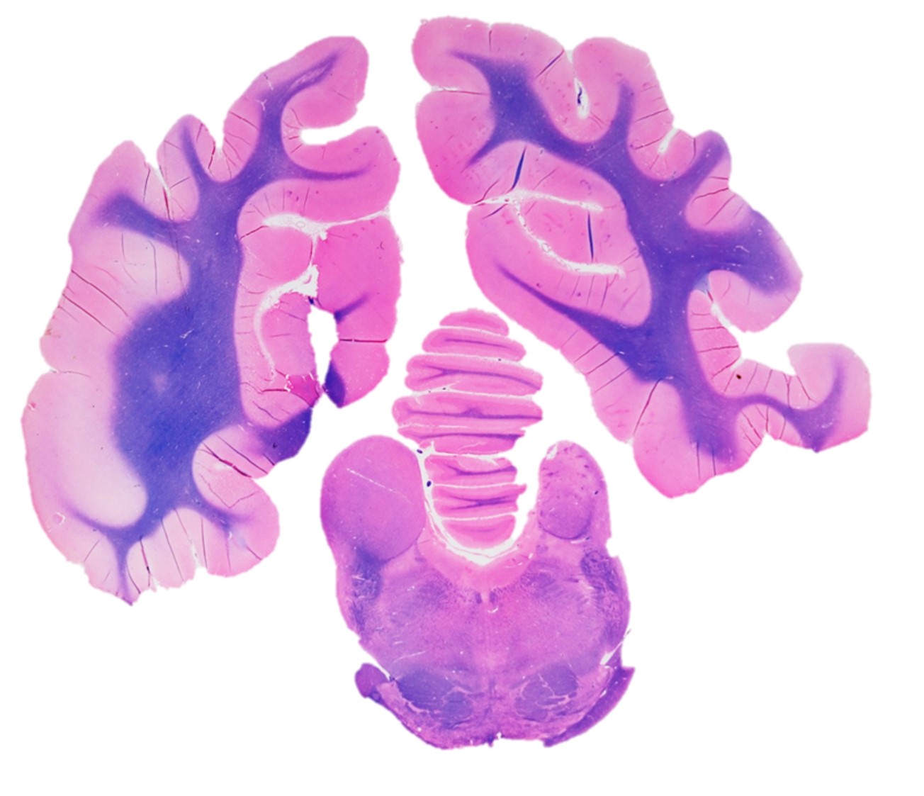
Slice #6
Anatomic divisions present:
- Telencephalon
- Diencephalon
- Mesencephalon
- Metencephalon (ventral)
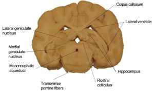
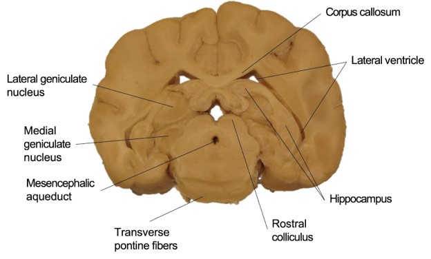
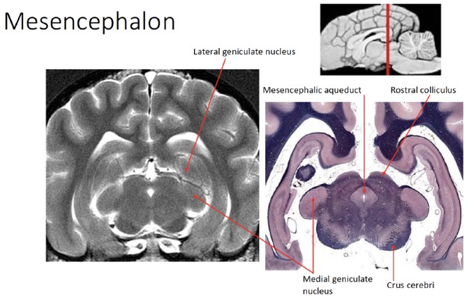
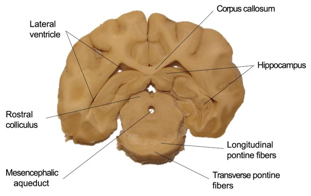
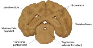
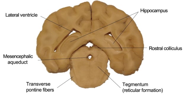
In slice #6, we can observe a number of notable structures associated with the diencephalon, mesencephalon and metencephalon. Recall that the diencephalon includes the thalamus, which consists of two bilaterally symmetrical collections of nuclei that primarily project axons to the ipsilateral cerebral hemisphere. These two collections are separated by the third ventricle. Two of these nuclei are prominent and readily recognized on the caudal surface. These are the geniculate nuclei, which comprise the metathalamus. A lateral eminence on the caudodorsal surface of the thalamus is the lateral geniculate nucleus, which receives fibers of the optic tract and functions in the visual system. The lateral geniculate nucleus is connected with the rostral colliculus of the midbrain. Caudoventral to the lateral geniculate nucleus is the medial geniculate nucleus of the thalamus. This nucleus functions in the auditory system and is connected to the caudal colliculus of the midbrain by the brachium of the caudal colliculus.
Observe: Identify the lateral and medial geniculate nuclei just medial to the lateral ventricles and dorsolateral to the mesencephalon.
The majority of the ventromedial aspect of this slice is the mesencephalon (midbrain). The mesencephalon is readily identifiable by the presence of the mesencephalic aqueduct, which connects the third ventricle to the fourth ventricle caudally. The mesencephalon consists of a tectum (or roof) dorsal to the aqueduct, which is composed of four groups of neuronal cell bodies: the colliculi. The rostral colliculi (sing. “colliculus”) sit dorsomedial relative to the caudal colliculi, and both sets of colliculi sit medial to the geniculate nuclei of the diencephalon. Ventral to the mesencephalic aqueduct is the tegmentum of the mesencephalon, as well as the crus cerebri. The tegmentum is that portion of the mesencephalon which includes the reticular formation.
The reticular formation is a complex network of nuclei and neurons in the brainstem that serve as a major integration and relay center for many vital brain systems to coordinate functions necessary for survival. The transverse pontine fibers cover part of the caudal mesencephalon ventrally. Stated another way, the portion of the reticular formation ventral to the mesencephalic aqueduct and dorsal to the transverse pontine fibers (which is part of the metencephalon) is considered part of the mesencephalic reticular formation (tegmentum). Caudal to the mesencephalic aqueduct, in between the fourth ventricle and the transverse pontine fibers, the reticular formation is considered to be part of the pons (ventral metencephalon). Caudal to that still, the reticular formation is part of the myelencephalon where it exists dorsal to the trapezoid body and pyramids.
Observe: Observe the tectum (including the rostral and caudal colliculi) and tegmentum of the mesencephalon, just dorsal and ventral, respectively, to the mesencephalic aqueduct. Your specimen likely has transverse pontine fibers that are also identifiable ventral to the tegmentum, and will have distinct, transversely-coursing fibers.
-
- Slice 6: Rostral surface

-
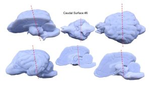
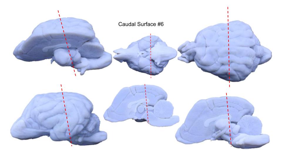
- Slice 6: Caudal surface
-
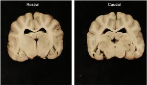
- Slice 6: Rostral and caudal surface

-
- Approximate location of slice 6
-

- Slice 6: Radiographic and stained
-
- Slice 6: Stained
-
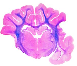
- Slice 6: Stained
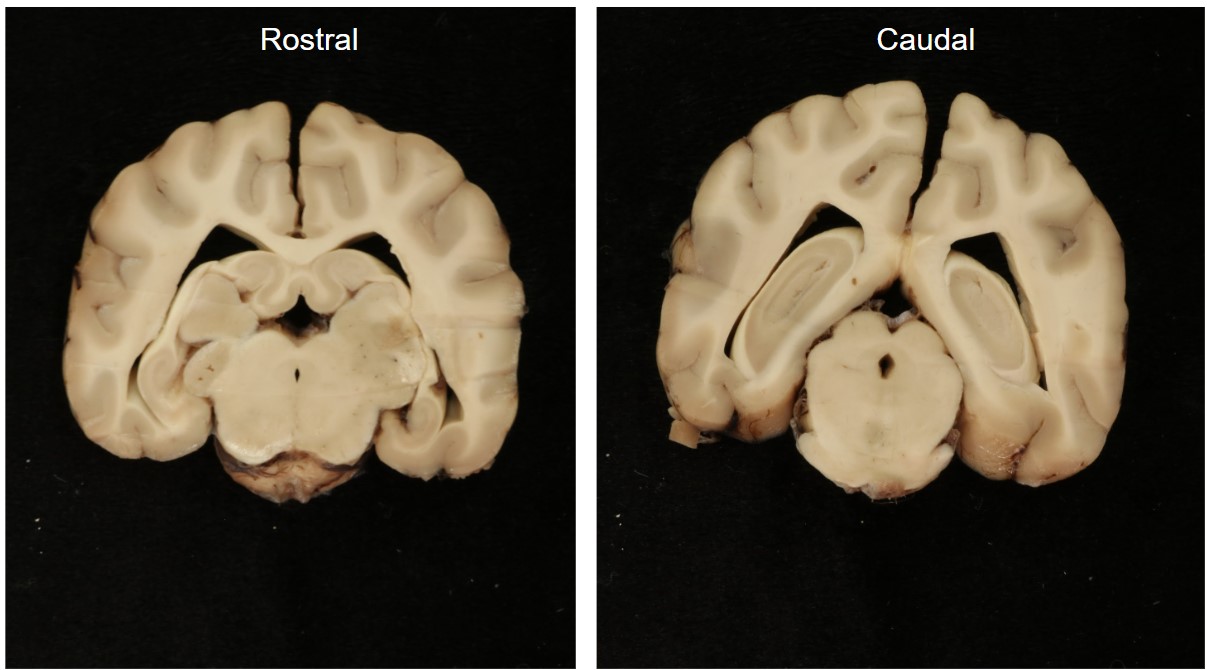
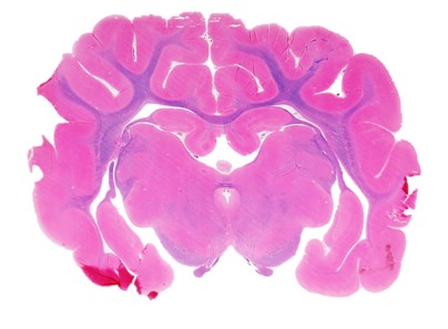
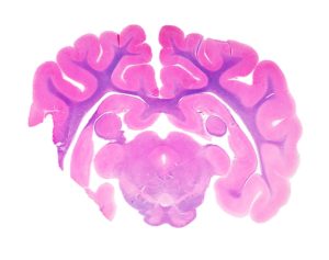
Slice #7
Anatomic divisions present:
- Telencephalon
- Mesencephalon
- Metencephalon (ventral)
-

- Slice 7: Rostral surface
-
- Slice 7: Caudal surface
-
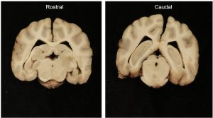
- Slice 7: Rostral and caudal surface
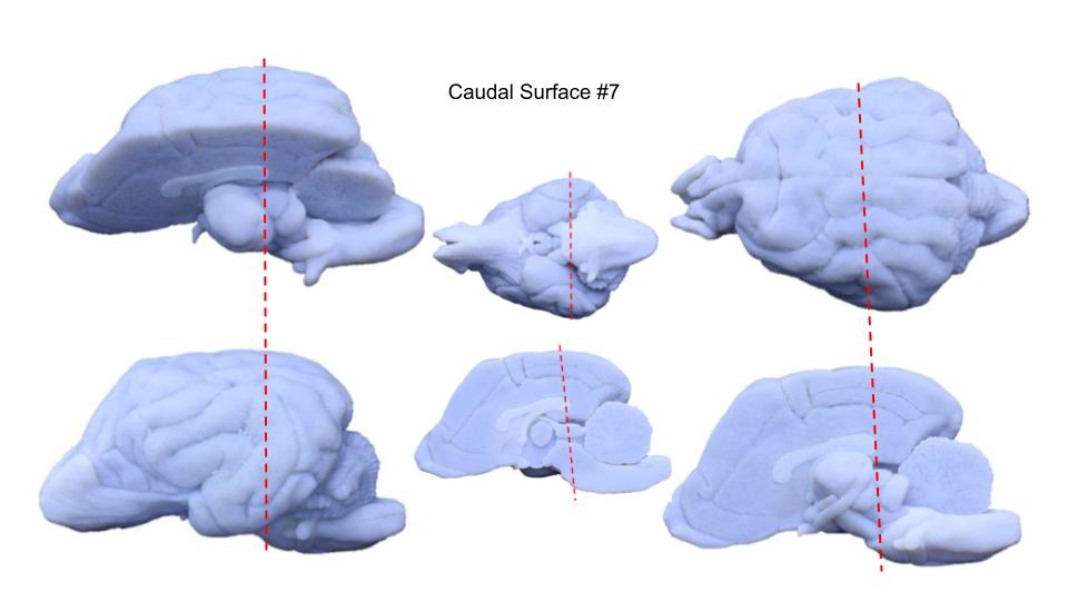
-
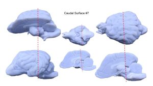
- Approximate location of slice 7
-

- Slice 7: Radiographic and stained
-
- Slice 7a: Stained
-
- Slice 7b: Stained
-
- Slice 7c: Stained

-
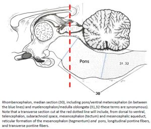
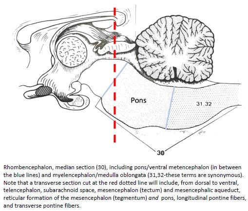
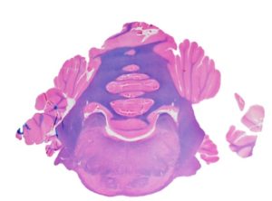
- Median section of rhombencephalon. 27
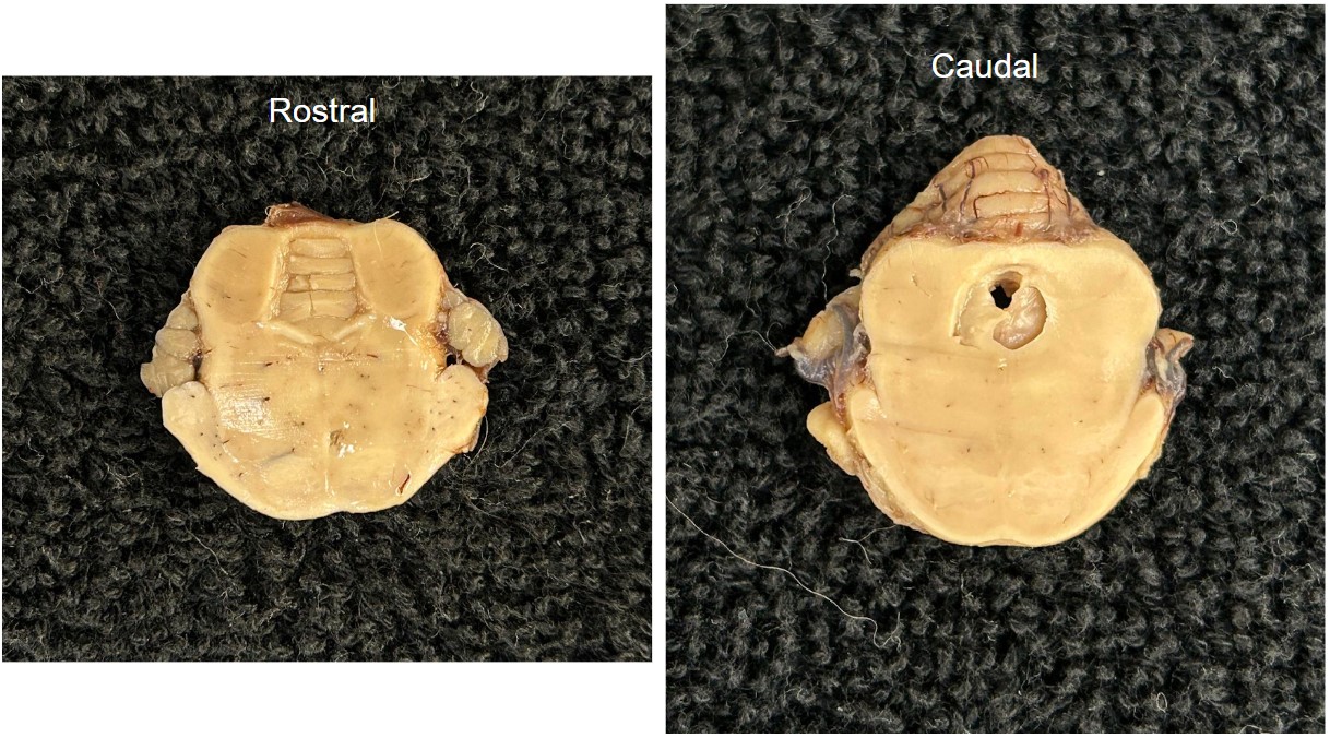
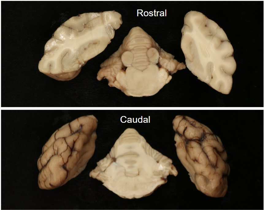
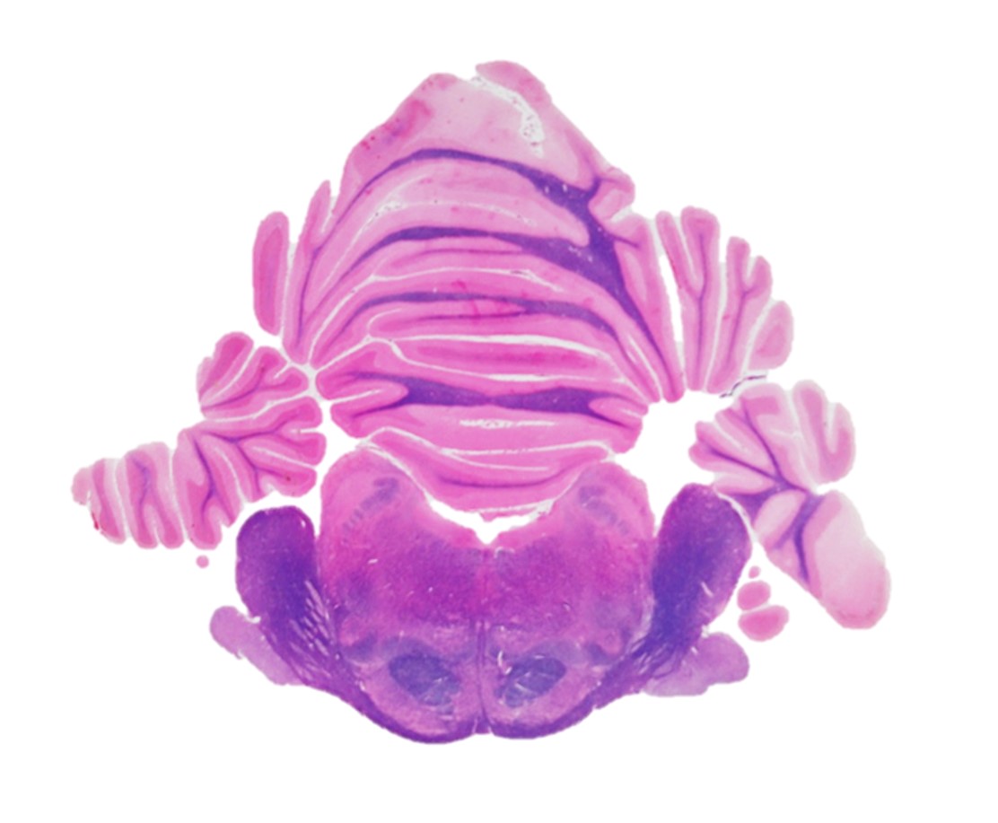
Slice #8
Anatomic divisions present:
- Telencephalon
- Mesencephalon
- Metencephalon (dorsal)
- Myelencephalon
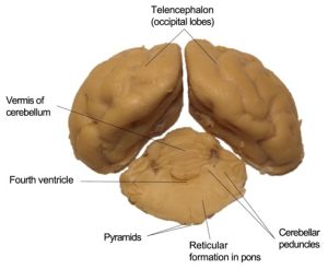
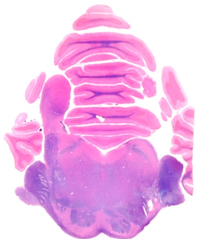
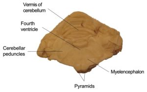
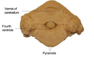
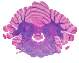
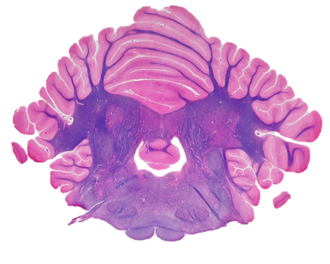
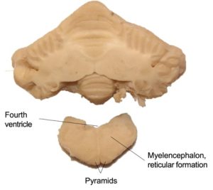
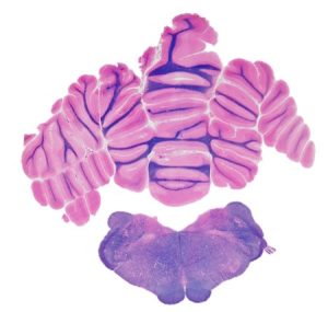
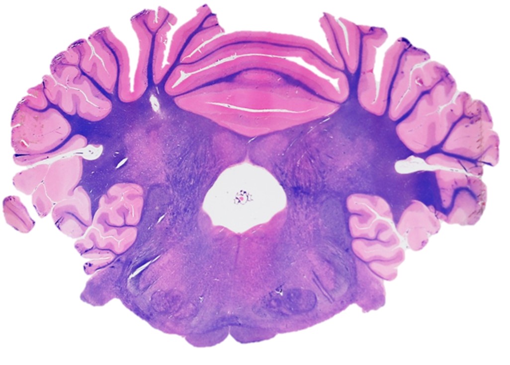
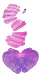
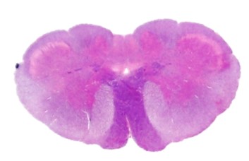
On the caudal surface, we can observe the caudal-most region of the brainstem: the myelencephalon. The myelencephalon, or medulla, extends from the transverse fibers of the pons to the level of the ventral rootlets of the first cervical spinal nerve. In the transverse cross section, the myelencephalon can be readily identifiable by the presence of the pyramids. The pyramids are a pair of longitudinally coursing fiber bundles on either side of the ventral median plane.
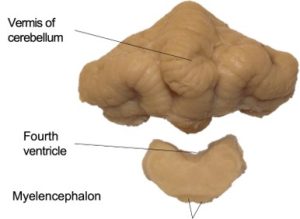
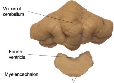
Also present is the cerebellum, or dorsal metencephalon. Note the vermis of the cerebellum in the midline. Between the vermis and the myelencephalon, we can see a space known as the fourth ventricle. This is the caudal-most space of the ventricular system, from which CSF exits via the lateral apertures or into the central canal of the spinal cord.
On either side of the fourth ventricle are the cut ends of the three cerebellar peduncles. The rostral cerebellar peduncle is medial and courses rostrally into the mesencephalon; the middle cerebellar peduncle is lateral and arises from the transverse fibers on the lateral side of the pons; and the caudal cerebellar peduncle is in the middle, entering from the myelencephalon after passing beneath the acoustic stria.
Observe: Observe the myelencephalon, fourth ventricle, and cerebellar peduncles.
-
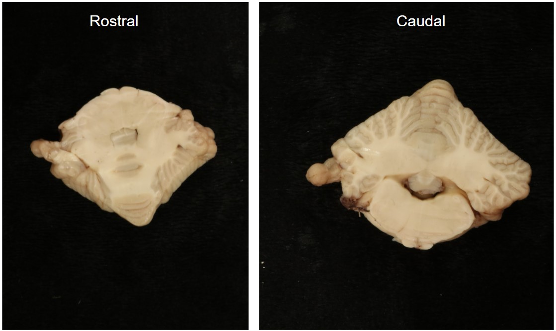
- Slice 8: Rostral surface
-
- Slice 8: Caudal surface

-
- Slice 8: Rostral and caudal surface
-

- Slice 8: Rostral and caudal surface
-
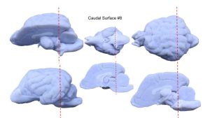
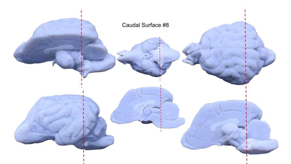
- Approximate location of slice #8
-
- Slice 8a: Radiographic and stained

-

- Slice 8b: Radiographic and stained
-

- Slice 8a: Stained
-

- Slice 8b: Stained
-

- Slice 8c: Stained
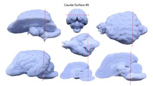
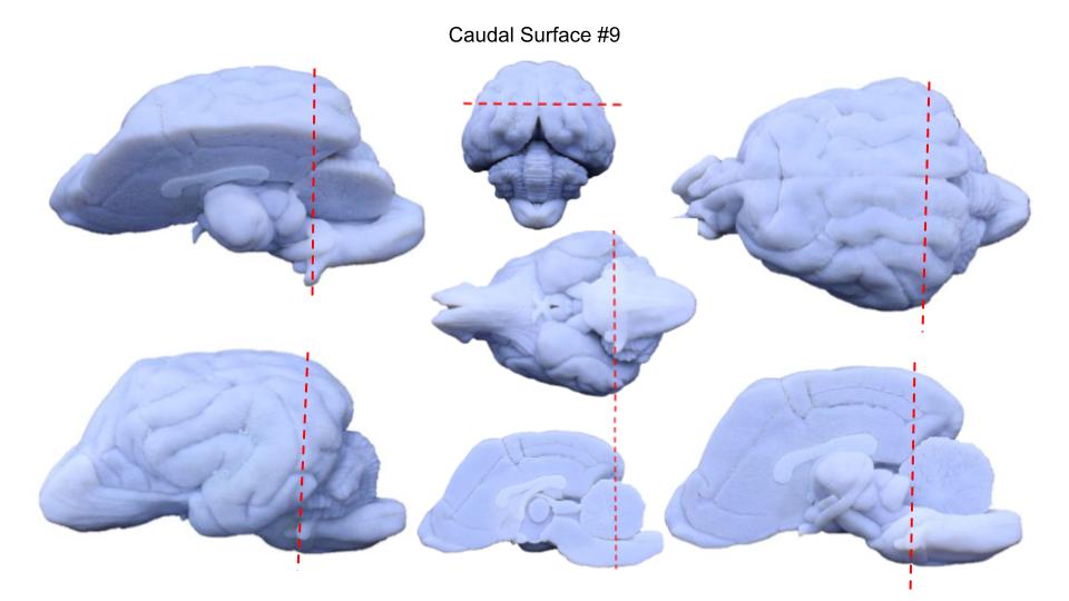
Slice #9
Anatomic divisions present:
- Metencephalon (dorsal)
- Myelencephalon
-
- Slice 9: Rostral surface

-
- Slice 9: Caudal surface

-

- Slice 9: Rostral and caudal surface
-
- Approximate location of slice #9
-

- Slice 9: Radiographic and stained
-
- Slice 9a: Stained

-
- Slice 9b: Stained
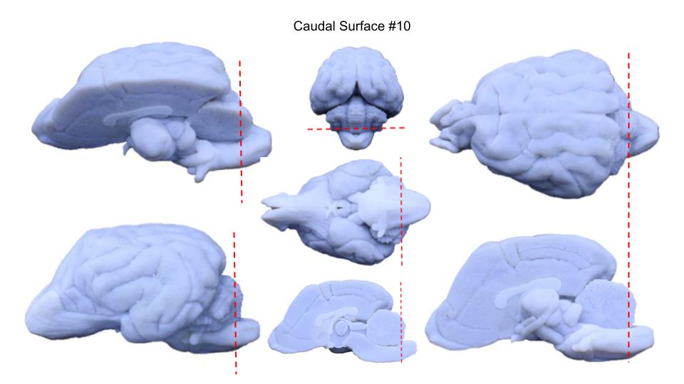
Slice #10
Anatomic divisions present:
- Metencephalon (dorsal)
- Myelencephalon
-
- Slice 10: Rostral surface
-
- Slice 10: Caudal surface

-
- Slice 9: Rostral and caudal surface
-
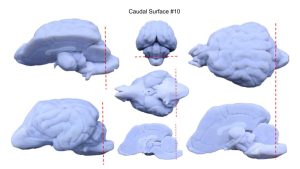
- Approximate location of slice #10
-
- Slice 10: Radiographic and stained

-
- Slice 10a: Stained

-

- Slice 10b: Stained
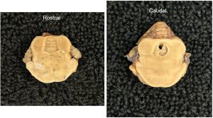
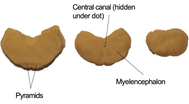
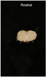
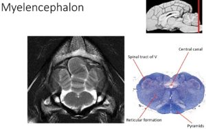
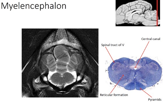
Remaining slices
The remaining slices that you have in your jar represent the caudal extent of the brainstem and the transition to the spinal cord. Note the presence of the pyramids with the myelencephalon, and the presence of the central canal (a lumen filled with CSF) indicating the spinal cord.
-

- Slice 10, 11, 12: Caudal surface
-

- Slice 10: Rostral and caudal surface

-

- Slice 11: Rostral and caudal surface

-
- Slice 12: Rostral and caudal surface

-
- Slice 11: Radiographic and stained
-
- Slice 12: Radiographic and stained
-
- Slice 11: Stained

-

- Slice 12: Stained
Finally, let’s review the cranial nerves (CNs), upper motor neuron nuclei (UMN), and lower motor neuron nuclei (LMN) present in each anatomic division of the brain. Reference the table below for this information:
| Anatomic Division | Cranial nerves | Important UMN Nuclei | LMN Nuclei |
| Telencephalon | CN I |
|
None |
| Diencephalon | CN II | Hypothalamus | None |
| Mesencephalon |
|
|
|
| Metencephalon | CN V |
|
Trigeminal nucleus |
| Myelencephalon |
|
|
|
Review Videos
Surface brain structures – 31 min
Surface brain structures – 21 min
Transverse brain slices – 43 min
Transverse brain slices – 30 min
Interactive Review Content:
Lab 13 Terms (Know the following on the blue brains)
| Terms | Notes |
| Meninges | |
| Dura mater | |
| Arachnoid membrane | |
| Pia mater | |
| Falx cerebri | |
| Dorsal sagittal sinus | |
| Subarachnoid space | |
| Cerebellomedullary cistern | |
| Gyrus | |
| Sulcus | |
| Cerebral cortex | |
| Ventricular system | Ventricular system best identified in midsagittal plane |
| Lateral ventricles | |
| Third ventricle | |
| Mesencephalic aqueduct | |
| Fourth ventricle | |
| Cerebrum | |
| Longitudinal fissure | Separates left and right cerebral hemispheres |
| Cerebral hemispheres | |
| Transverse fissure | Separates cerebrum from cerebellum |
| Frontal lobe | |
| Cruciate sulcus | Separates frontal lobe from parietal lobes |
| Parietal lobe | |
| Temporal lobe | |
| Occipital lobe | |
| Olfactory bulb | |
| Piriform lobe | |
| Cerebellum | |
| Vermis | |
| Folia | |
| Cerebellar peduncles | |
| Arbor vitae | Best seen in midsagittal plane |
| Tentorium cerebelli | |
| Brain stem | |
| Mammillary bodies | |
| Optic chiasm | |
| Optic tract | |
| Lateral geniculate body | |
| Medial geniculate body | |
| Rostral colliculus | |
| Caudal colliculus | |
| Crus cerebri | |
| Pons (ventral metencephalon) | |
| Transverse pontine fibers | |
| Pyramids | |
| Decussation of pyramids | Understand, don’t identify |
Lab 14 Terms (Know the following on the brain cross-sections)
| Terms | Notes |
| Telencephalon | |
| Longitudinal fissure | Separates left and right hemispheres of cerebrum |
| Olfactory bulb | |
| Hippocampus | |
| Fornix | White matter associated with the hippocampus |
| Corpus callosum | |
| Internal capsule | |
| Cerebral cortex | |
| Corona radiata | |
| Caudate nucleus | |
| Lateral ventricles | |
| Diencephalon | |
| Thalamus | |
| Interthalamic adhesion | |
| Lateral geniculate body | |
| Medial geniculate body | |
| Mammillary bodies | |
| Optic chiasm | |
| Optic tract | |
| Hypothalamus | |
| Third ventricle | |
| Mesencephalon | |
| Tectum | |
| Rostral colliculus | |
| Caudal colliculus | |
| Tegmentum | |
| Red nucleus | UMN; Know region in which it’s found, don’t identify directly |
| Oculomotor nucleus | LMN; Know region in which it’s found, don’t identify directly |
| Parasympathetic nucleus of CN III | LMN; Know region in which it’s found, don’t identify directly |
| Trochlear nucleus | LMN; Know region in which it’s found, don’t identify directly |
| Reticular formation | UMN; Know region in which it’s found, don’t identify directly |
| Ascending reticular activating system (ARAS) | UMN; Know region in which it’s found, don’t identify directly |
| Fourth ventricle | |
| Mesencephalic aqueduct | |
| Metencephalon | |
| Ventral metencephalon (i.e., pons) | |
| Transverse pontine fibers | |
| Reticular formation | UMN; Understand region in which it’s found, don’t identify directly |
| Dorsal metencephalon | |
| Vermis | |
| Folia | |
| Arbor vitae | |
| Cerebellar peduncles | |
| Myelencephalon | |
| Pyramids | |
| Vestibular nuclei | UMN; Know region in which it’s found, don’t identify directly |
| Facial nucleus | LMN; Know region in which it’s found, don’t identify directly |
| Nucleus ambiguus (Nuclei of CNs IX, X, and XI) | LMN; Know region in which it’s found, don’t identify directly |
| Hypoglossal nucleus | LMN; Know region in which it’s found, don’t identify directly |
| Reticular formation | UMN; Know region in which it’s found, don’t identify directly |
| Parasympathetic nuclei of CNs VII, IX, and X | LMN; Know region in which it’s found, don’t identify directly |