Lab 4A: Nerves of the Carnivore Thoracic Limb
Learning Objectives
- Name and identify roots of the brachial plexus.
- Name and identify nerves and their branches in the extrinsic and intrinsic mm. of the TL.
- Understand innervation of muscle action groups.
- Recognize innervation and autonomous zones of the manus.
Lab Instructions:
Brachial plexus
The brachial plexus (plexus [L]: “braid”) is formed by the ventral branches of the sixth, seventh, and eighth cervical and the first and second thoracic spinal nerves. In some dogs there is a small contribution from the ventral branch of the fifth cervical spinal nerve. These branches arise from their spinal nerve just lateral to their respective intervertebral foramen. They emerge along the ventral border of the scalenus and extend across the axillary space to the thoracic limb. In the axilla numerous branches of these spinal nerves communicate to form the brachial plexus. From the plexus arise nerves of mixed origin that supply the structures of the thoracic limb and adjacent muscles and skin.
The pattern of interchange in the brachial plexus is variable, but the specific spinal nerve composition of the named nerves that continue into the thoracic limb is consistent. These nerves include the suprascapular, subscapular, axillary, musculocutaneous, radial, median, ulnar, thoracodorsal, lateral thoracic, and pectoral nerves.
-

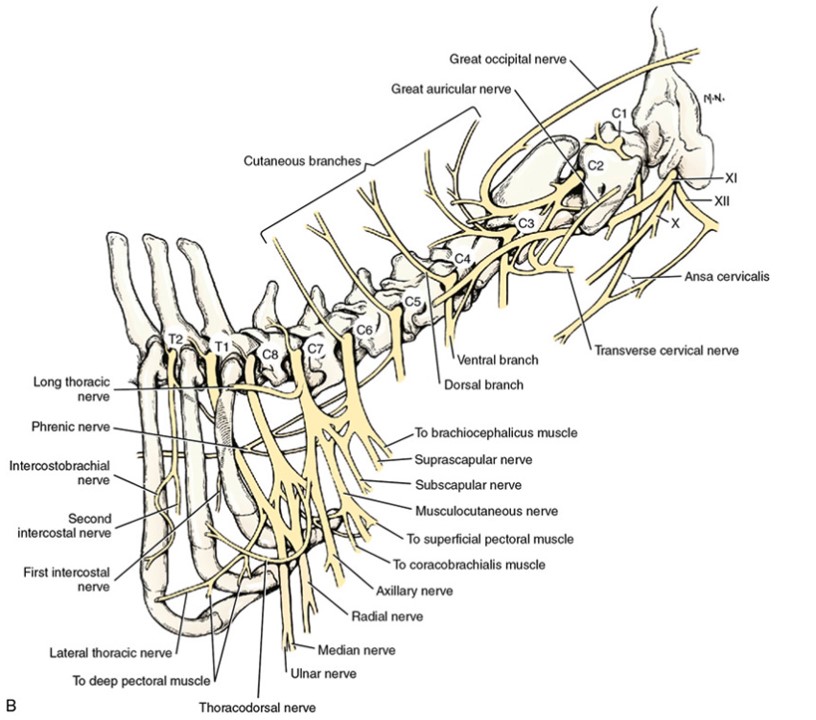
- Schema of the cervical nerves and brachial plexus. The numbers C-1 through C-8 and T-1 refer to spinal nerves, not vertebrae.1
-
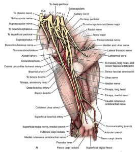
- Dog brachial plexus, right thoracic limb, medial aspect. 1

-

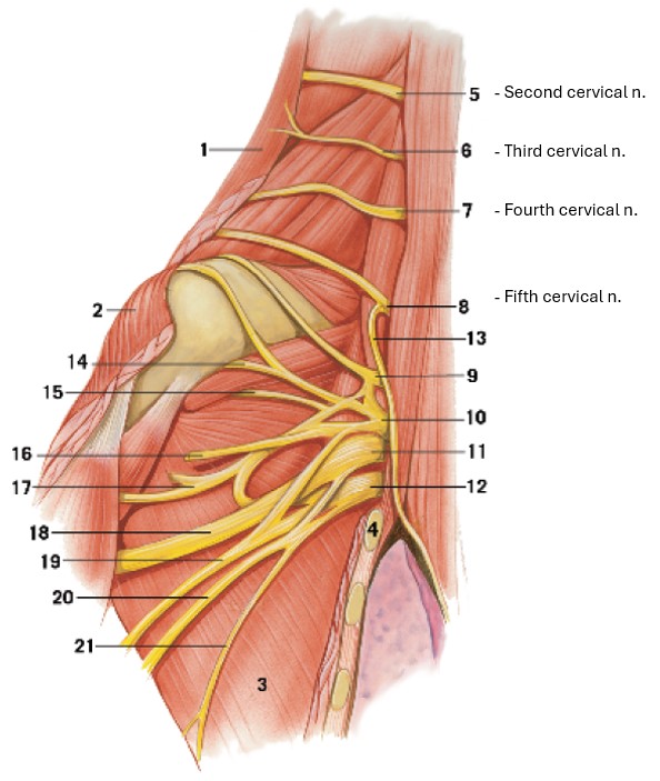
- Cervical nerves and formation of the brachial plexus of the cat, ventral view. 4
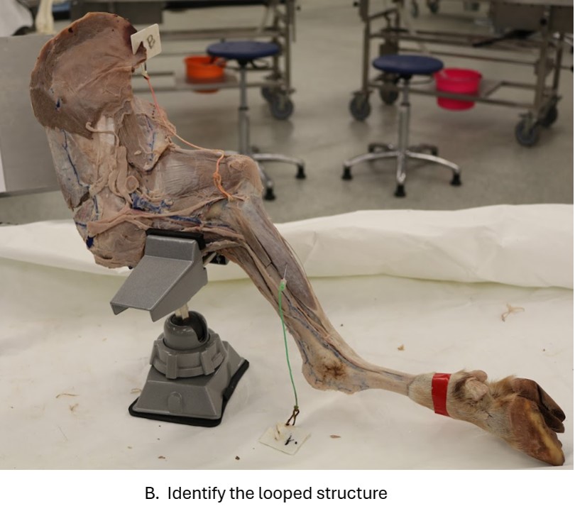
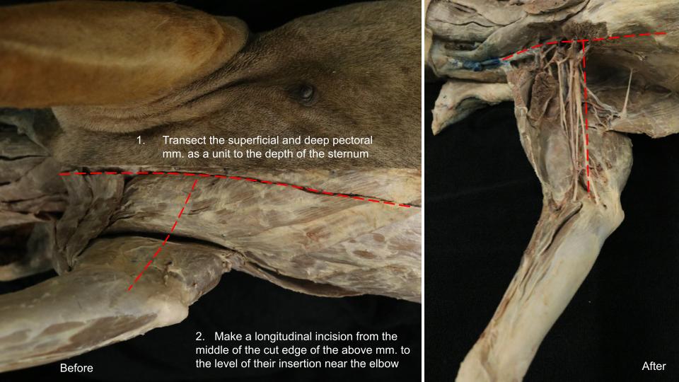
Dissect: If not already completed, transect the superficial and deep pectoral mm. together as a unit and gently reflect these muscles toward the limb. Doing so will allow some exposure to the vessels and nerves on the medial aspect of the arm. Take care to leave the nerves and arteries (red latex) located here intact. We will dissect and identify them next. While the muscles may be transected as needed on this limb, the goal is to keep the vessels and nerves intact this time around.
Video showing the initial dissection cuts. Only watch ~1 min, it quickly shifts to discuss vasculature.
Nerves of the Thoracic Limb
All of the following eleven nerves contain somatic efferent (i.e., motor) neurons to striated muscles and afferent (i.e., sensory) neurons from these muscles and surrounding structures. Cutaneous somatic afferents are found only in the musculocutaneous, axillary, radial, median, and ulnar nerves.
As you study these nerves, realize that groups of muscles with similar actions (muscle action groups) are typically innervated by the same nerve! Of course there are a few exceptions to be aware of. As you dissect, practice naming the muscles and muscle action groups each nerve innervates. Do the flip exercise as well; practice pointing to a muscle and name the nerve which innervates it. This is a valuable exercise that trains your brain to go both directions!
Cranial pectoral
Cranial pectoral nerves are derived from ventral branches of the sixth, seventh, and eighth cervical spinal nerves. They innervate the superficial pectoral muscle. The cranial pectoral nerves do not need to be dissected. Be aware of what muscle they innervate, however.
Dissect: Bluntly dissect the following nerves (see subsections for each nerve for descriptions). It may be beneficial to remove portions of the axillary vein (blue latex) in order to better visualize the arteries and nerves in this area.
- Suprascapular n.
- Subscapular n.
- Axillary n.
- Musculocutaneous n.
- Radial n.
- Ulnar n.
- Median n.
- Thoracodorsal n.
- Long thoracic n.
- Lateral thoracic n.
Suprascapular
The suprascapular nerve leaves the sixth and seventh cervical spinal nerves and courses between the supraspinatus and subscapularis muscles near the neck of the scapula. It passes across the scapular notch, where it is subject to injury from external compressive forces. It supplies the supraspinatus and infraspinatus muscles.
-
- Dog brachial plexus, right thoracic limb, medial aspect. 1
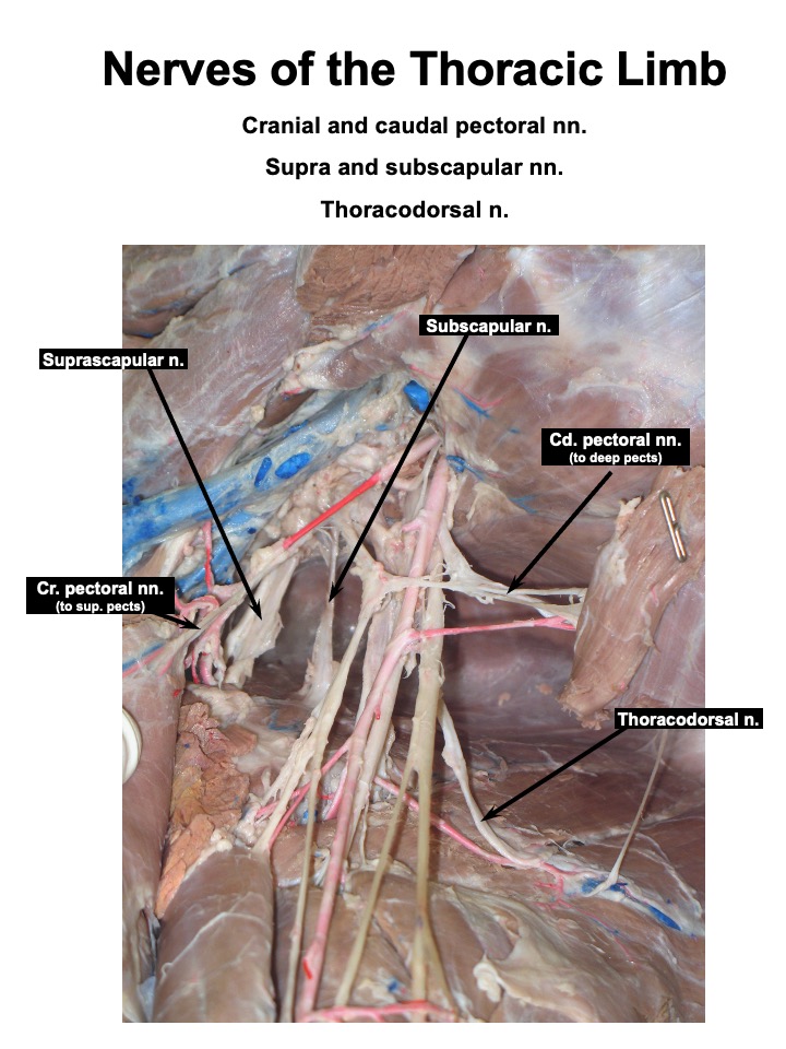
Subscapular
The subscapular nerve is a branch from the sixth and seventh cervical spinal nerves to the subscapularis muscle. Frequently, two subscapular nerves are seen entering the muscle belly of subscapularis m.
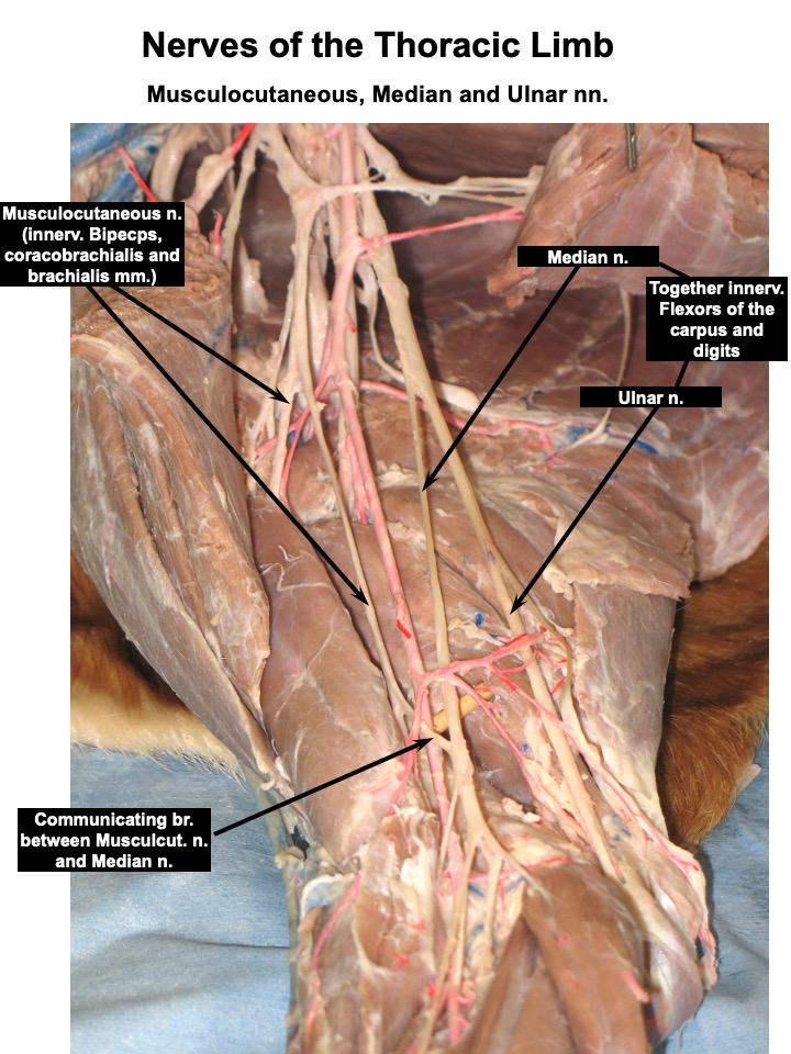
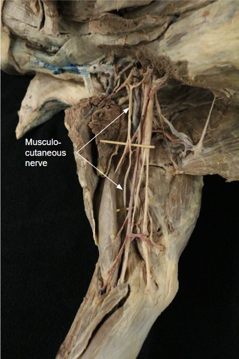
Musculocutaneous
The musculocutaneous nerve arises from the sixth, seventh, and eighth cervical spinal nerves. Throughout the brachium the musculocutaneous nerve lies between the biceps brachii cranially and the brachial vessels caudally. It supplies the coracobrachialis, the biceps brachii, and the brachialis mm. A branch communicates with the median nerve proximal to the flexor surface of the elbow. The musculocutaneous nerve courses deep to the insertion of the biceps. It supplies the distal end of the brachialis m. and gives off the medial cutaneous antebrachial nerve. This nerve is sensory to the skin on the medial aspect of the antebrachium. You may find it still hanging on with the subcutaneous tissue of the medial aspect of the antebrachium, though it is oftentimes removed during the skinning process. *Ultimately you are not responsible for identifying the medial cutaneous antebrachial nerve in the carnivore.*
-
- Dog brachial plexus, right thoracic limb, medial aspect. 1
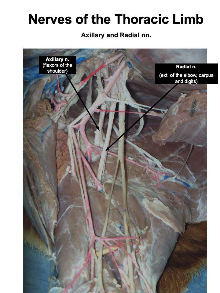
Axillary
The axillary nerve arises as a branch from the combined seventh and eighth cervical spinal nerves. It enters the space between the subscapularis and the teres major mm. on a level with the neck of the scapula. The following muscles are supplied by the axillary nerve: the teres major, teres minor, the deltoideus mm. and part of the subscapularis m.. There are cranial cutaneous antebrachial branches of this nerve that supply the skin on the cranial surface of the forearm. The latter overlap with cutaneous antebrachial branches of the superficial branch of the radial nerve laterally and the musculocutaneous nerve medially.
-
- Dog brachial plexus, right thoracic limb, medial aspect. 1

Thoracodorsal
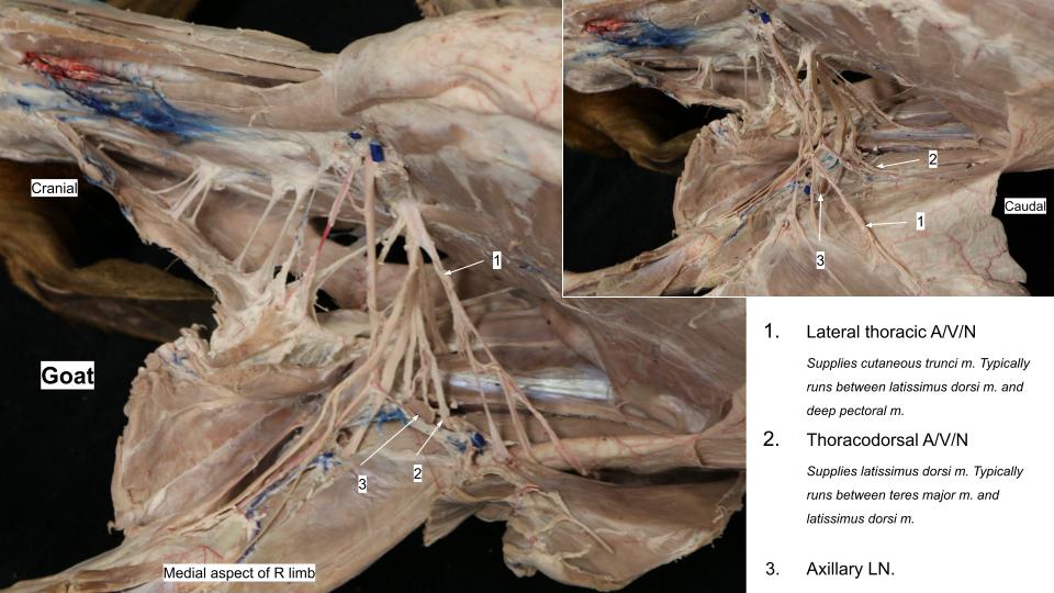
The thoracodorsal nerve arises primarily from the eighth cervical spinal nerve. It supplies the latissimus dorsi muscle. It courses with the thoracodorsal vessels on the medial surface of the muscle.
-
- Dog brachial plexus, right thoracic limb, medial aspect. 1

Radial
The radial nerve arises from the last two cervical and first two thoracic spinal nerves, runs a short distance distally with the trunk of the median and ulnar nerves, and enters the triceps distal to the teres major. The radial nerve supplies all of the extensor muscles of the elbow, carpal, and phalangeal joints.
Observe: Observe the branches of the radial n. to the triceps on the medial side of the brachium.
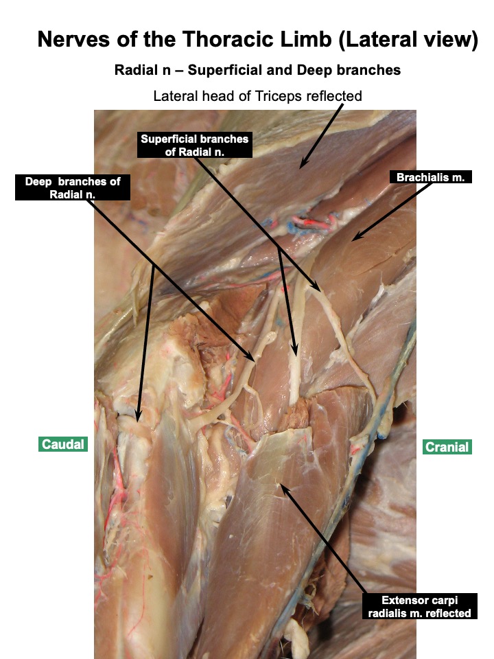
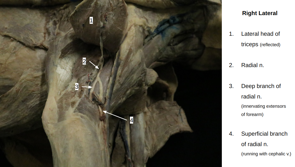
The radial nerve then spirals around the humerus on the caudal surface and then on the lateral surface of the brachialis muscle. On the lateral side at the distal third of the arm, the radial nerve terminates as a deep (motor) and a superficial (sensory) branch.
Dissect: Transect the lateral head of the triceps at its origin and reflect it to expose the following terminal branches:
a. The deep branch of the radial nerve crosses the medial surface of the extensor carpi radialis on its course into the forearm with the brachialis muscle. This deep branch is motor to the extensor carpi radialis, the common digital extensor, the supinator, the lateral digital extensor, the abductor digiti I longus, and the ulnaris lateralis mm.
b. The superficial branch divides into a lateral cutaneous antebrachial nerve and medial and lateral branches. The small medial branch follows the medial ramus of the cranial superficial antebrachial artery and continues distally in the forearm on the medial side of the cephalic vein. The lateral branch becomes associated with the lateral side of the cephalic vein and enters the forearm with the lateral branches of the cranial superficial antebrachial artery. These branches continue to the paw on either side of the accessory cephalic vein.
These medial and lateral branches are sensory to the skin on the cranial and lateral surface of the forearm and the dorsal surface of the carpus, metacarpus, and digits . They terminate in dorsal common digital nerves in the paw.
-
- Dog brachial plexus, right thoracic limb, medial aspect. 1
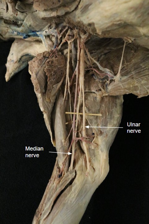
The median and ulnar nerves arise by a common trunk from the eighth cervical and the first and second thoracic spinal nerves. The common trunk lies on the medial head of the triceps between the brachial vein caudally and the brachial artery cranially.
Median
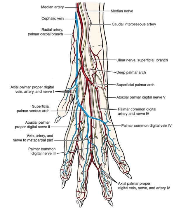
The median nerve, the cranial division of the common trunk, runs to the antebrachium in contact with the caudal surface of the brachial artery. It receives a branch from the musculocutaneous nerve at the level of the elbow. The brachial a. and v. and median n. pass just cranial to the medial epicondyle to enter the forearm. In the cat, the median n. and brachial a. (but not the vein) travel together through the supracondylar foramen of the humerus, a foramen not present in the canid. The median nerve innervates the pronator teres, the pronator quadratus, the flexor carpi radialis, the superficial digital flexor, and the radial head and parts of the humeral and ulnar heads of the deep digital flexor. The median nerve passes through the carpal canal with the median artery and branches to supply sensory innervation to the palmar surface of the forepaw.
Ulnar
The ulnar nerve, the caudal division of the common trunk, separates from the median nerve in the distal arm and crosses the elbow caudal to the medial epicondyle of the humerus. It picks up with the collateral ulnar a. The caudal cutaneous antebrachial nerve leaves the ulnar near the middle of the arm and runs caudo-distally across the medial surface of the triceps and olecranon. This cutaneous branch supplies the skin of the distal medial aspect of the brachium and the caudal aspect of the antebrachium. The ulnar nerve enters the antebrachial muscles caudal to the medial epicondyle of the humerus and is distributed to the flexor carpi ulnaris, and parts of the ulnar and humeral heads of the deep digital flexor mm.. The ulnar nerve lies on the caudal surface of the deep digital flexor deep to the humeral head of the flexor carpi ulnaris m.. In the middle of the antebrachium the sensory dorsal branch of the ulnar nerve arises and becomes subcutaneous on the lateral surface. It is distributed to the lateral surface of the metacarpus and the fifth digit. The palmar branch of the ulnar nerve continues into the forepaw after the dorsal branch arises. It divides into superficial and deep branches at the carpal level and then these further divide in the proximal metacarpus into branches supplying sensory innervation to the palmar surface of the forepaw and motor innervation to the intrinsic muscles of the forepaw.
-
- Dog brachial plexus, right thoracic limb, medial aspect. 1
Caudal pectoral
Caudal pectoral nerves are derived from the ventral branches of the eighth cervical and first and second thoracic spinal nerves. They innervate the deep pectoral muscle and are often combined with the lateral thoracic nerve at their origin. The caudal pectoral nerves do not need to be dissected. Be aware of what muscle they innervate, however.
Lateral thoracic
The lateral thoracic nerve is derived from the ventral branches of the eighth cervical and first thoracic spinal nerves. It leaves the caudal portion of the brachial plexus and courses caudally between the latissimus dorsi and deep pectoral and is the sole innervation of the cutaneous trunci m. The lateral thoracic nerve passes with the lateral thoracic a./v. across the lateral surface of the axillary lymph node (the large lymph node located just caudal to the brachial plexus in the axilla region).
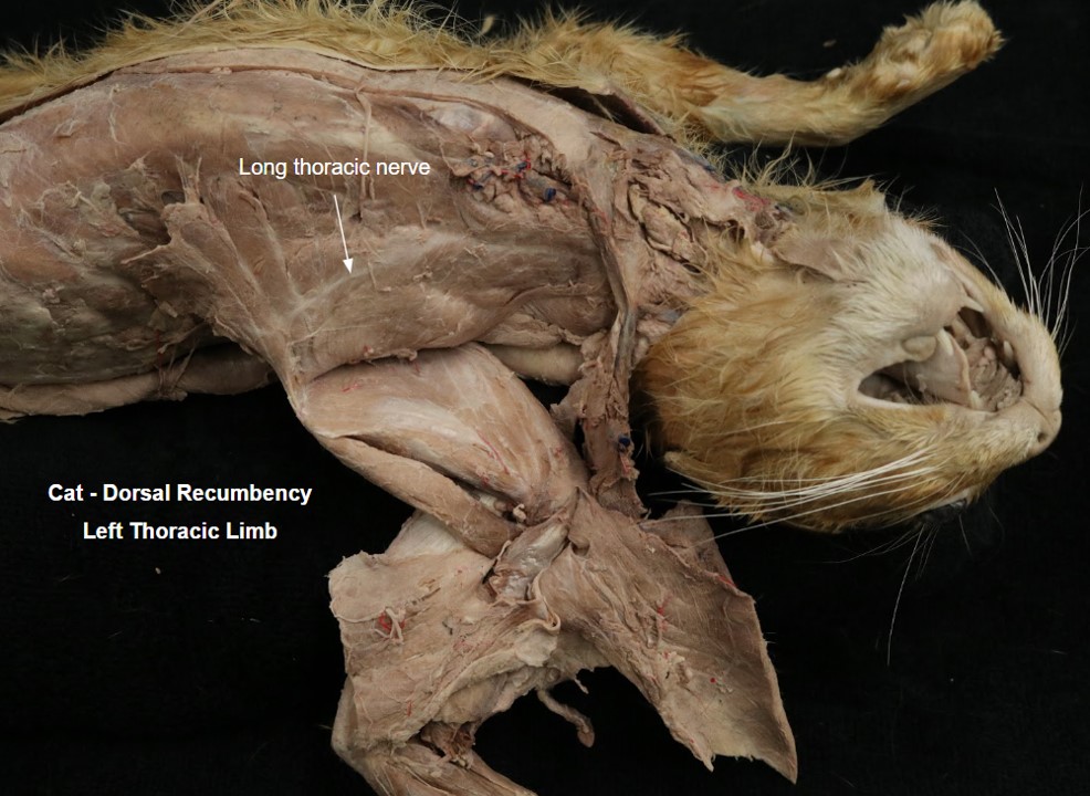
Long thoracic
The long thoracic nerve usually arises from the ventral branch of the seventh cervical nerve before it branches to aid in forming the brachial plexus. It runs largely horizontally on the superficial surface of the thoracic portion of the serratus ventralis m., which it innervates.
-
- Dog brachial plexus, right thoracic limb, medial aspect. 1
Sensory Nerves of the Antebrachium and Manus
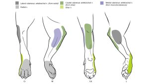
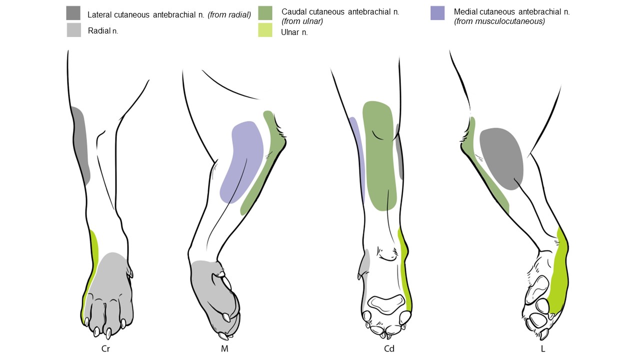
The skin of the antebrachium is innervated by sensory branches from four nerves: the cranial surface by the axillary and radial nerves; the lateral surface by the radial nerve; the caudal surface by the ulnar nerve; and the medial surface by the musculocutaneous nerve. There is considerable overlap in their cutaneous areas of distribution.
Although the terminal cutaneous distribution of nerves in the forelimb is difficult to dissect, it is important to know both the cutaneous areas and the autonomous zones of these nerves for local anesthetic procedures and for the diagnosis of nerve lesions. The cutaneous area is the entire area of skin innervated by a nerve. The autonomous zone is that area of skin innervated solely by a specific peripheral nerve with no overlap from adjacent nerves. We are particularly focused on autonomous zones in the paw, because at this location it is a common procedure of a neurology exam to perform toe pinches to assess pain sensation and hence the integrity of a specific nerve innervating the pinched digit.
In regards to the forepaw, the dorsal cutaneous surface of the first four digits is innervated by the radial nerve. The lateral surface of the fifth digit is innervated by the ulnar nerve. Much of the palmar cutaneous innervation is shared by the ulnar and median nerves, and thus cannot be considered an autonomous zone.
Observe: Practice identifying the autonomous zones based on pin locations on the forepaw.
-

- Autonomous zones of cutaneous innervation of thoracic limb of the dog. 1

-
- Thoracic limb autonomous zones L. Hudson
Observe: The distribution of nerves to the digits will not be dissected, but you should understand the pertinent terminology and how it is applied.
Specifically, use a bony model of a manus to practice the nomenclature and branching patterns of these nerves.
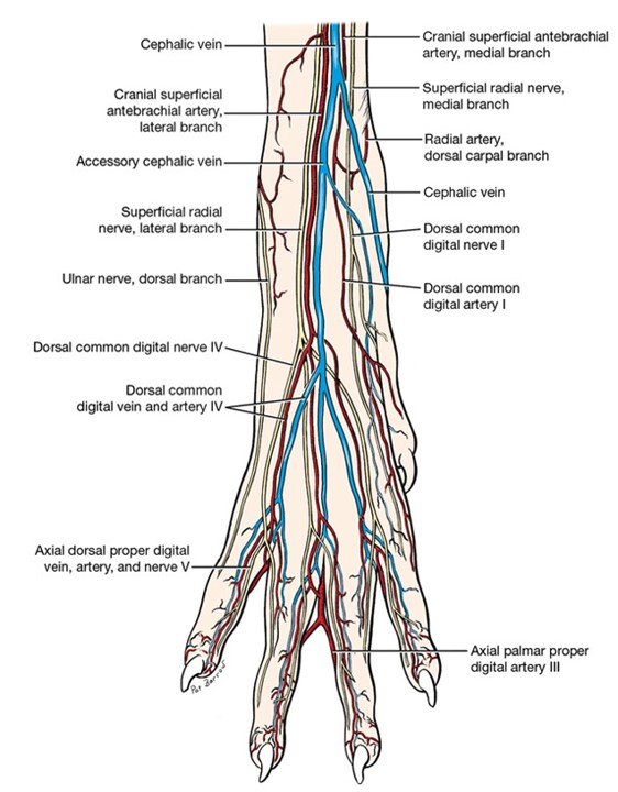
In the metacarpus there are superficial and deep branches of nerves on the palmar side, with only one set on the dorsal side. The superficial branches are called dorsal or palmar common digital nerves, and the deep branches are called dorsal or palmar metacarpal nerves (we won’t concern ourselves with these deep branches in the carnivore, but they’ll come back for the horse!). These nerves are numbered based on the digit on which they lie laterally. (For example, the dorsal common digital n. III lies on the lateral side of the third metacarpal bone).
At the metacarpophalangeal joint, the “common digital” and “metacarpal” branches unite to form a common trunk that maintains the name dorsal or palmar common digital n. on the dorsal and palmar surfaces, respectively. Each common digital nerve then divides into medial and lateral branches to the digits. These digital branches are called axial or abaxial dorsal or palmar proper digital nerves. Axial or abaxial refers to whether they are on the surface of the digit that faces toward the axis (axial) or away from the axis (abaxial) of the paw. The axis of the paw passes between digits III and IV.
Let’s review this with an example:
Passing along the dorso-lateral side of the fourth metacarpal bone would be the dorsal common digital n. IV. As it crosses the metacarpophalangeal joint, it then splits into an abaxial dorsal proper digital n. IV and an axial dorsal proper digital n. V. This would apply in the same way on the palmar side of the manus as well, just replace the word “dorsal” with “palmar”!
-

- Arteries, veins, and nerves of right forepaw of the dog, dorsal view. 1
-
- Arteries, veins, and nerves of right forepaw of the dog, palmar view. 1
Observe: Refer to lab models to help understand the location and naming of these structures.
Review Videos
Dog thoracic limb nerves – 10 min. Watch until 21 min.
Terms
| Terms | Species | Muscle(s) Innervated/Notes |
| Brachial plexus | All | Roots includes C6, C7, C8, T1, T2 |
| Cranial pectoral nn. | Know innervation, don’t identify |
|
| Caudal pectoral nn. | Know innervation, don’t identify |
|
| Thoracodorsal n. | Carnivore |
|
| Lateral thoracic n. | Carnivore |
|
| Long thoracic n. | All |
|
| Suprascapular n. | All |
|
| Subscapular n. | All |
|
| Axillary n. | All |
|
| Musculocutaneous n. | All |
|
| Medial cutaneous antebrachial n. | (Understand, don’t identify) | Cutaneous (sensory) innervation of medial arm |
| Radial n. | All |
|
| Deep branch | All | |
| Superficial branch | All | |
| Lateral cutaneous antebrachial n. | Carnivore (Understand, don’t identify) | Cutaneous (sensory) innervation of lateral arm |
| Median n. | All |
|
| Ulnar n. | All |
|
| Caudal cutaneous antebrachial n. | Carnivore (Understand, don’t identify) | Cutaneous (sensory) innervation of caudal elbow and antebrachium |
| Autonomous zones of the manus | Understand in the carnivore | Radial n. or ulnar n. |
| Dorsal/palmar common digital nn. (I-IV) | Understand on model or skeleton, do not identify in cadaver | |
| Dorsal/palmar axial/abaxial proper digital nn. (I-V) | Understand on model or skeleton, do not identify in cadaver |