Lab 8A: Spinal Cord & Meninges
Learning Objectives
- Identify the structures of the spinal cord, including: cross sectional anatomy, cervical/lumbosacral intumescences, dorsal and ventral roots, cauda equina, etc.
- Relate the cross sectional anatomy of the spinal cord to efferent and afferent nerve pathways.
- Define and identify the meninges and their associated spaces.
Lab Instructions:
In order to observe the spinal cord and its surrounding structures, we will need to access the vertebral canal (recall that the vertebral canal is the combined space created by the vertebral foramina when the vertebrae are in articulation). To do so, we are going to access the vertebral canal from the dorsal side by removing the vertebral arches (including the spinous processes) of a couple of vertebrae.
Dissect: If possible, turn your cadaver to a sternal recumbency to provide easier access to the back. This is more likely done with the feline cadavers; the teams with a canine cadaver may need to work as a team to flip and/or lift your cadaver at times to perform this dissection.
Begin by palpating the spinous processes of the vertebrae and locate the first five lumbar vertebrae (L1-L5). (Remember that the last thoracic vertebrae is articulating with the last rib!)
On both the right and left sides, make a longitudinal incision through the epaxial musculature directly lateral to the spinous process, making sure your scalpel blade makes contact with the underlying bone. The bone that you just made contact with is the lamina of the vertebra. Continue to reflect the epaxial muscles laterally to fully expose the spinous processes and lamina of L1-L5.
Using bone cutters, carefully cut through the lamina bilaterally. Transversely cut through the interspinous ligaments and ligamenta flava to remove the vertebral arches. Congratulations, you’ve just performed a laminectomy!
-

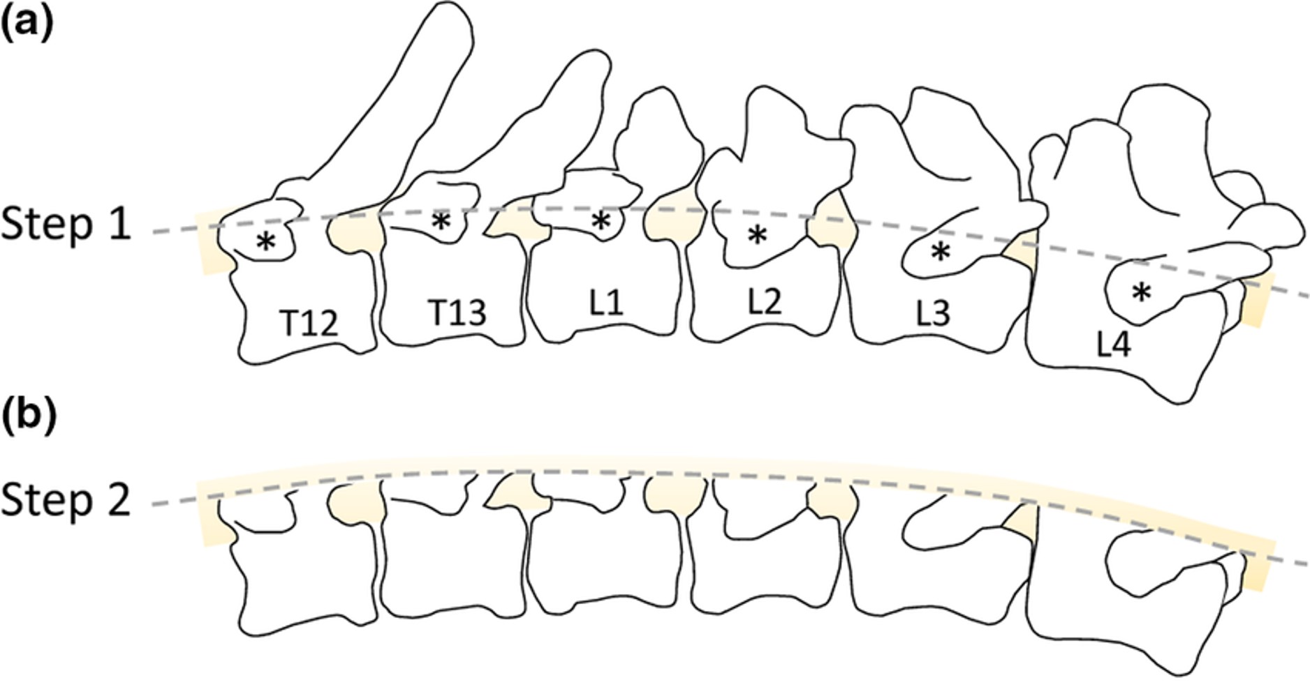
- Schematic diagram for dorsal laminectomy dissection of canine dorsal root ganglia. (a) Lateral view of canine thoracolumbar vertebral column and spinal cord position, (*) denotes the transverse processes as the dorsal landmarks for accessing the underlying pedicles to transect for (b) exposing the spinal cord. 24
-
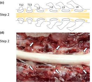
- (c) Dorsal view of canine thoracolumbar vertebral column with the cut surface of pedicle outlined. (d) Dorsal view following dissection, with the cut surface of pedicle outlined for landmark orientation and white arrows pointing at in situ DRG. Thoracic (T) and lumbar (L) vertebrae labeled for referencing. 24

Observe: Observe the spinal cord with its dural covering.
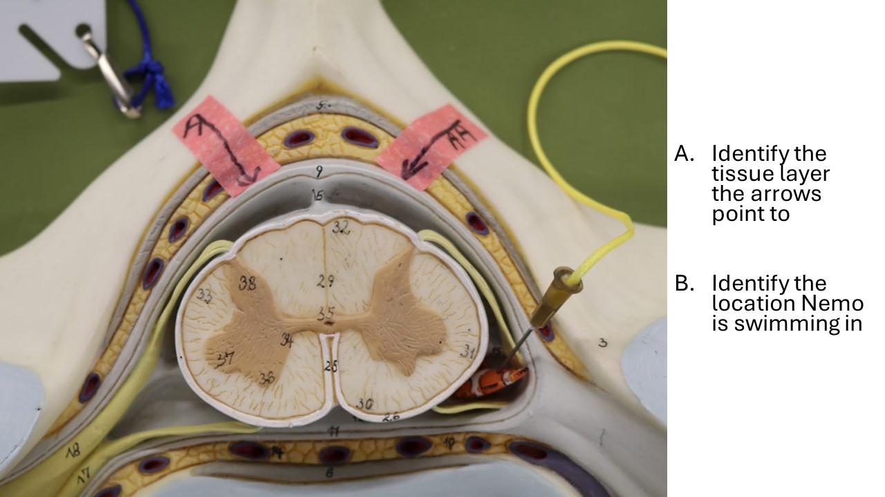
Between the dura of the spinal cord and the periosteum of the vertebrae is the epidural space, which contains loose connective tissue, fat, and blood vessels.
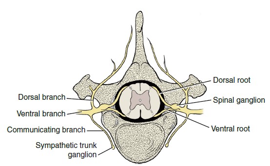
The spinal cord is divided into segments. A group of dorsal and ventral rootlets leave each spinal cord segment on each side and combine, respectively, to form the spinal nerve at the level of the intervertebral foramen. The dorsal rootlets converge into a spinal ganglion. Note the spinal ganglia in the intervertebral foramina.
-
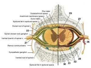
- Schematic transverse section of spinal cord and associated spinal nerves with meninges and vessels at the level of an intervertebral foramen. 4
-
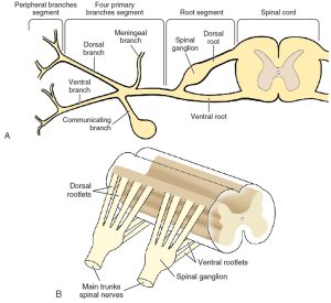
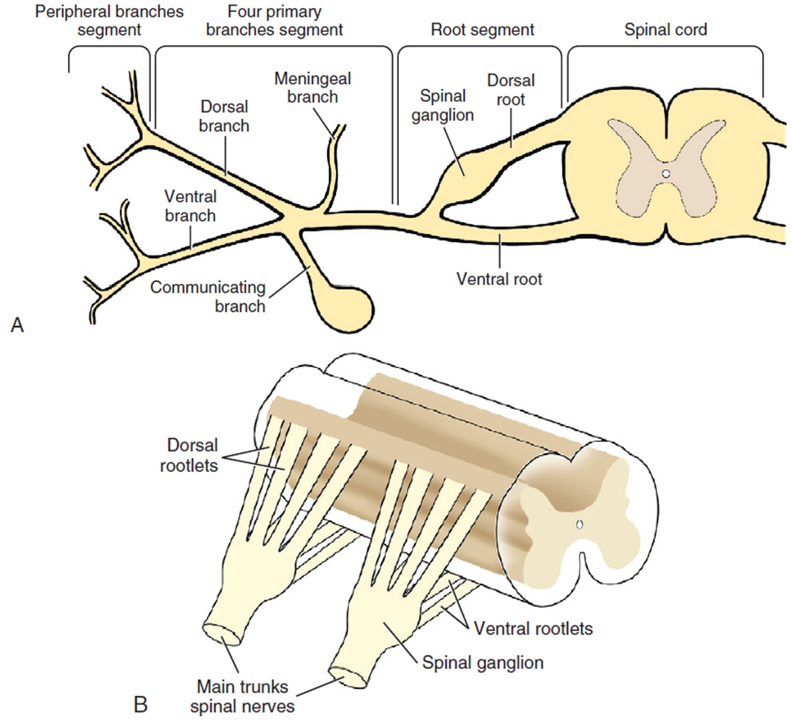
- A, Schematic drawing of the longitudinal segments of a typical spinal nerve. B, Schematic drawing of the dorsal and ventral rootlets of a typical spinal nerve. 3
-
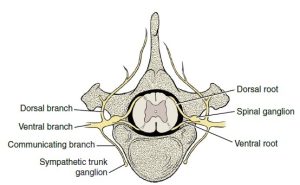
- Diagram of spinal nerve. 3
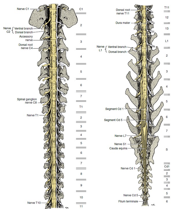
There are 8 cervical spinal cord segments, 13 thoracic, 7 lumbar, 3 sacral, and about 5 caudal. The first cervical spinal nerves leave the vertebral canal through the lateral vertebral foramen in the arch of the atlas. The second cervical spinal nerves leave caudal to the atlas. The cervical spinal nerves of segments 3 through 7 leave the vertebral canal through the intervertebral foramina cranial to the vertebra of the same number. The spinal nerves of the eighth cervical segment pass caudal to the seventh (last) cervical vertebra. The spinal nerves of all the remaining spinal cord segments pass through the intervertebral foramina caudal to the vertebra of the same number.
-
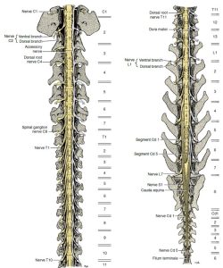
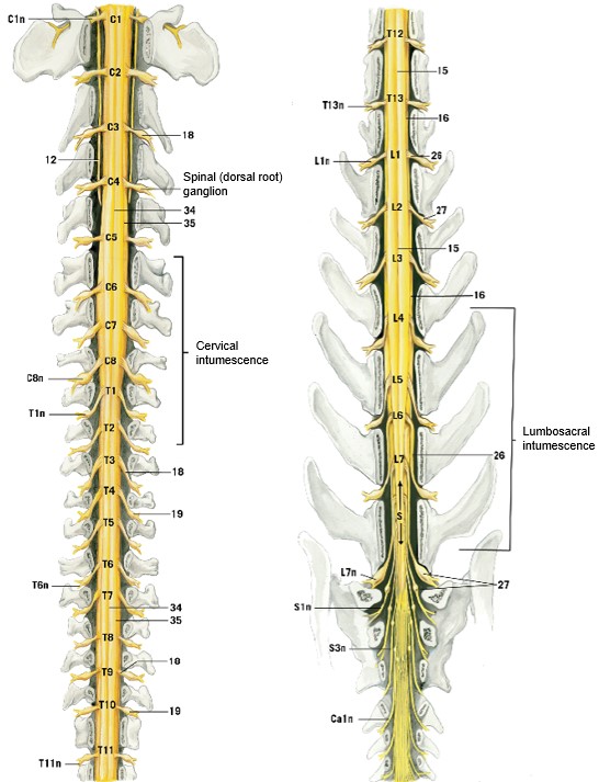
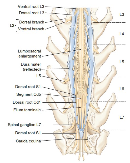
- Dorsal roots of spinal nerves and spinal cord segments. Dorsal view; vertebral arches removed. Dura removed on left side of both images. Notations on the right represent levels of vertebral bodies. 3
-
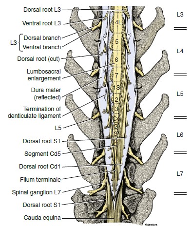
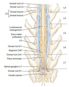
- Enlarged schematic view of terminal spinal cord with dura reflected. 3
-

- Cat spinal cord in situ, with dorsal aspect of vertebrae and meninges removed, dorsal view. 4
-

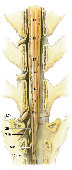
- Enlargement of caudal region of the cat spinal cord. Has dorsal roots removed on the right side. 4
The only spinal cord segments that are found entirely within their corresponding vertebrae are the last two thoracic and the first two (or occasionally three) lumbar segments. All other spinal cord segments reside in the vertebral canal cranial to the vertebra of the same number. This is most pronounced in the caudal lumbar and sacrocaudal segments of the spinal cord. In general, the three sacral segments lie within the fifth lumbar vertebra and the caudal segments lie within the sixth lumbar vertebra. There is breed variation in the length of the spinal cord. In small breeds it extends about one vertebra farther caudally, and in large breeds one vertebra farther cranially. The nerve roots of the first 10 thoracic segments and those caudal to the third lumbar segment are long because of the distance between their origin at the spinal cord and their passage through the intervertebral foramen.
Observe: Observe the relationship of the spinal cord segments to the corresponding vertebrae.
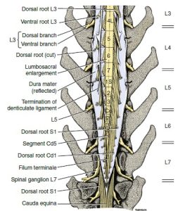
In the caudal cervical region over the fifth to seventh cervical vertebrae, there is an enlargement of the spinal cord that nearly fills the vertebral canal. This is the cervical intumescence. Its presence is due to an increase in white matter and cell bodies that are associated with the innervation of the thoracic limb. This intumescence occurs from the sixth cervical segment of the spinal cord through the first thoracic segment. Another enlargement occurs in the midlumbar vertebral region for the innervation of the pelvic limb. The lumbar intumescence that begins at about the fourth lumbar segment and gradually narrows caudally as the spinal cord comes to an end near the intervertebral space between the sixth and seventh lumbar vertebrae. The narrow caudal end of the parenchyma of the spinal cord is known as the conus medullaris. The spinal cord terminates in the filum terminale, which is a narrow cord of meninges that may include a long extension of the neural tube and central canal. This attaches the conus medullaris to the caudal vertebrae. The cauda equina ([L], “horse’s tail”) includes the conus medullaris together with the adjacent caudal lumbar, sacral, and caudal roots that extend caudally in the vertebral canal. It resembles a horse’s tail, hence “cauda equina”.
-
- Dorsal roots of spinal nerves and spinal cord segments. Dorsal view; vertebral arches removed. Dura removed on left side of both images. Notations on the right represent levels of vertebral bodies. 3
-
- Enlarged schematic view of terminal spinal cord with dura reflected. 3
-
- Cat spinal cord in situ, with dorsal aspect of vertebrae and meninges removed, dorsal view. 4
-
- Enlargement of caudal region of the cat spinal cord. Has dorsal roots removed on the right side. 4
-
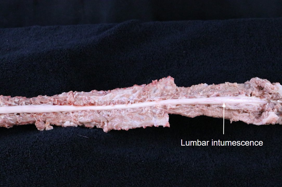
- Cauda equina of the dog. 9
Observe: Referring to the models and full length spinal cord prosections made available to you in the lab, observe the cervical and lumbar intumescences, conus medullaris, and cauda equina.
You should be able to observe the lumbar intumescence and conus medullaris and the cranial aspect of the cauda equina on your cadavers.

Meninges
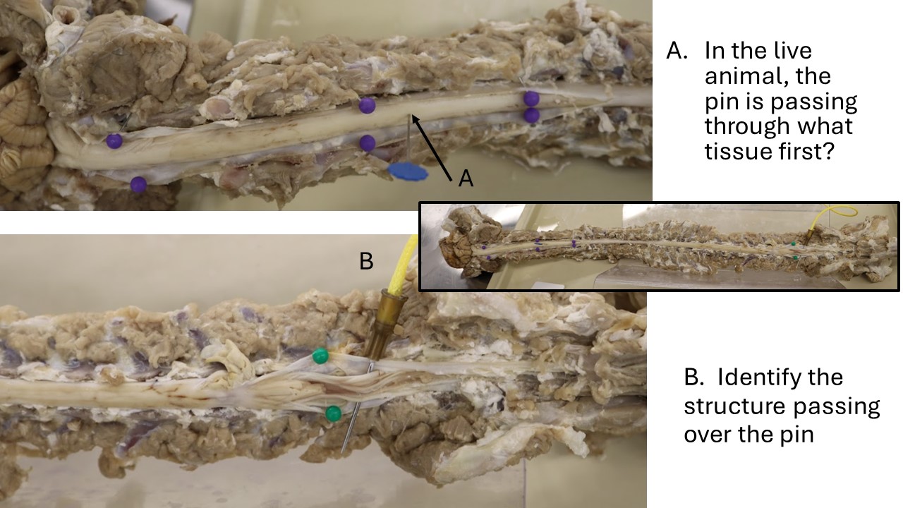
Returning to your cadaver, observe the thick, fibrous dura mater, the outermost layer of the meninges.
Dissect: Make a longitudinal incision along the dorsal aspect of the entire length of the exposed dura mater.
The thin arachnoid membrane and pia mater are opposed to each other in the embalmed specimen, with the arachnoid membrane adhering to the deep surface of the dura mater and the pia mater remaining on the surface of the spinal cord itself. For this reason, upon incising and opening the dura mater, we have now entered the subarachnoid space. Recall that through the subarachnoid space flows cerebrospinal fluid (CSF). On the lateral surface of the spinal cord, the pia thickens and forms a longitudinal cord of connective tissue called the denticulate ligament. This ligament segmentally attaches to the arachnoid and dura laterally, midway between the roots of adjacent spinal cord segments.
-
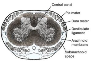
- Enlarged transverse section of seventh lumbar segment of the dog. 1
-
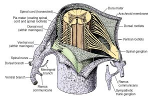
- Transected spinal cord with meninges incised to reveal a spinal cord segment, ventral view. Spinal cord roots attach to the spinal cord as a series of rootlets. Bilaterally, dorsal and ventral roots join to form a spinal nerve that immediately divides into four primary branches (dorsal, ventral, meningeal, and ramus communicans). A spinal ganglion is located on the dorsal root, and the ramus communicans joins a sympathetic trunk ganglion. 3
For the following material, refer to the calf cadavers made available to you in the lab.
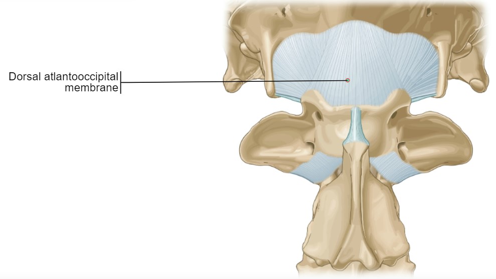
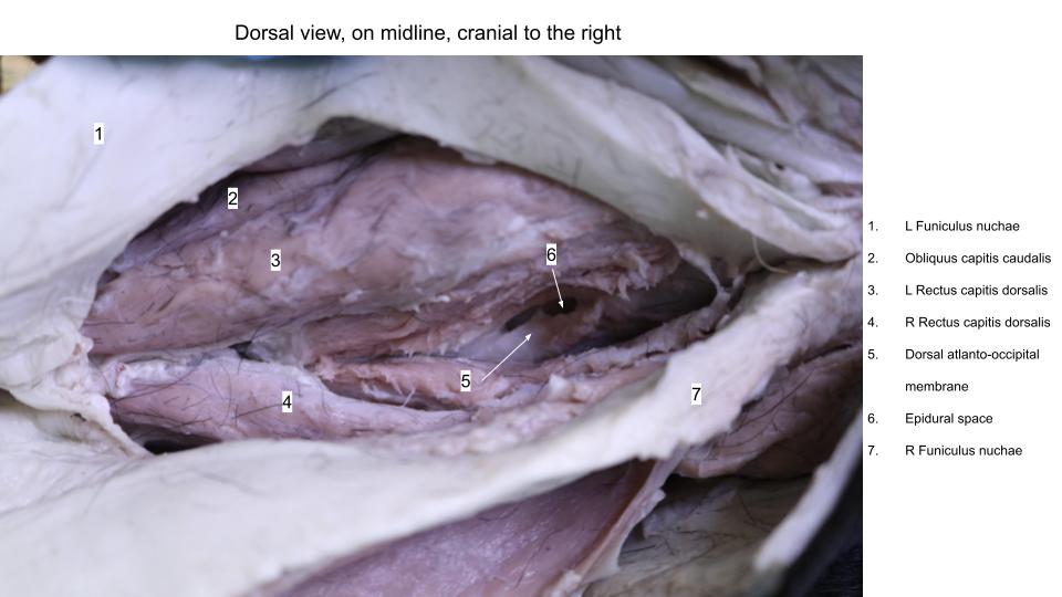
At the level of the atlanto-occipital membrane in the calf, reflecting the muscles of the poll region exposes a piece of tissue known as the dorsal atlanto-occipital membrane, which extends from the occipital bone to the dorsal arch of the atlas (C1). Retracting this membrane exposes the epidural space and dura mater.
Observe: Observe the dorsal atlanto-occipital membrane, epidural space, and dura mater in one of the instructors’ prosected calves.
-
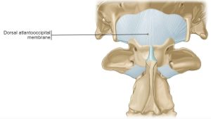
- Dorsal atlanto occipital membrane. 25
Clinical relevance:
Cerebrospinal fluid (CSF) samples can be obtained by puncture of the atlanto-occipital membrane, dura mater, and arachnoid to enter the subarachnoid space. In addition, a radiopaque contrast medium can be injected into the subarachnoid space to produce a myelogram for diagnostic purposes. Ultrasound-guided approaches to the subarachnoid space at the A-O or C1-C2 (AA) articulations in standing horses have been described. Cervical vertebral canal endoscopy has also been described in the horse using an A-O approach (FYI: Prange et al Endoscopic anatomy of the cervical vertebral canal in the horse: A cadaver study. EVJ 2011; 43(3):317-323.)
Transverse Sections of the Spinal Cord
The gray matter of the spinal cord in transverse section is in the shape of a butterfly or the letter H. It consists primarily of neuronal cell bodies. The dorsal extremity on each side is the dorsal horn, which receives the entering dorsal (sensory) rootlets. The ventral extremity is the ventral horn, which sends axons out via the ventral (motor) rootlets. In the thoracolumbar region, the lateral horn projects laterally from the gray matter midway between the dorsal and ventral gray horns. This lateral horn contains the cell bodies of the preganglionic sympathetic neurons. In the center of the gray matter of the spinal cord is the central canal. This remnant of the embryonic neural tube is continuous rostrally with the fourth ventricle. At the caudal end of the conus medularis, there is a small communication with the subarachnoid space.
The white matter of the spinal cord can be divided into three pairs of funiculi. Dorsally, a shallow, longitudinal groove extends the entire length of the spinal cord. This is the dorsal median sulcus. The longitudinal furrow along which the dorsal rootlets enter the spinal cord is the dorsolateral sulcus. Between these two sulci is the dorsal funiculus of the spinal cord.
Between the dorsolateral sulcus and the line of exit of the ventral rootlets, the ventrolateral sulcus, is located the lateral funiculus. The ventral funiculus is the white matter between the line of exit of the ventral rootlets and the longitudinal groove on the ventral side of the spinal cord, the ventral median fissure. In some species the funiculi have been subdivided topographically into specific cranially and caudally projecting tracts. Such anatomical information in domestic animals is still incomplete.
-
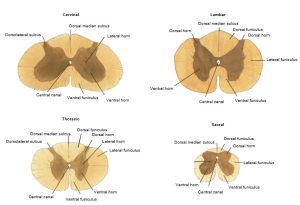
- Transverse sections of cat spinal cord segments. 1
Observe: Study lab models and provided transverse sections of the spinal cord at different locations. Compare the shape of the gray matter of these segments and relate it to their areas of innervation.
Review Videos & Practice
Terms
| Terms | Species |
| Vertebral foramen | All |
| Vertebral canal | All; combination of vertebral foramina in articulated vertebral column through which passes the spinal cord |
| Intervertebral foramen | All; foramen for spinal nerves to pass through to and from the spinal cord |
| Spinal cord segments | Understand, don’t identify |
| Cervical intumescence C6-T2 | Carnivore; enlargement of spinal cord related to brachial plexus |
| Lumbar intumescence L4-S3 | Carnivore; enlargement of spinal cord related to lumbosacral plexus |
| Conus medullaris | Carnivore; tapered caudal end of spinal cord |
| Cauda equina | Carnivore; bundle of nerve roots at the caudal end of the spinal cord resembling “horse’s tail” |
| Spinal nerves | Carnivore |
| Dorsal/ventral rootlets | Identify on models |
| Spinal ganglion (i.e., Dorsal root ganglion) | Identify on models |
| Dorsal atlanto-occipital membrane | Calf |
| Epidural space | Carnivore; calf |
| Dura mater | Carnivore; calf |
| Arachnoid membrane | Identify on models |
| Subarachnoid space | Carnivore |
| Cerebrospinal fluid (CSF) | Know that it flows through the subarachnoid space |
| Pia mater | Carnivore, also identify on models |
| White matter | Carnivore; Also be prepared to identify on models |
| Dorsal funiculus | Identify on models/slides |
| Lateral funiculus | Identify on models/slides |
| Ventral funiculus | Identify on models/slides |
| Grey matter | Carnivore; Also be prepared to identify on models |
| Dorsal horn | Identify on models/slides |
| Lateral horn | Identify on models/slides |
| Ventral horn | Identify on models/slides |
| Central canal | Identify on models/slides |
| Ascending tract | Understand, don’t identify |
| Descending tract | Understand, don’t identify |