Lab 5: Lymph nodes and Vasculature of the Thoracic Limb
Learning Objectives
- Identify lymph nodes of the thoracic limb and discuss the afferent and efferent flow of lymph to/from these nodes
- Identify the main channel blood supply of the thoracic limb
- Identify selected arterial branches of the main channel
- Describe blood flow in the manus
Dissect: For this lab, use the still-attached thoracic limb on your cadaver, i.e. the side opposite your thoracic cavity dissection.
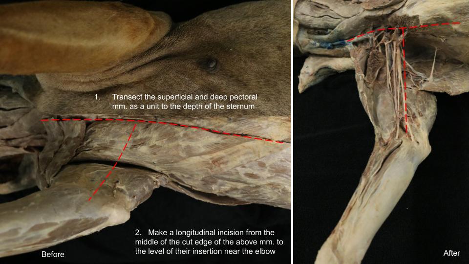
If needed, skin the limb to below the carpus (you can extend this as needed later). Transect the superficial and deep pectoral mm. together as a unit and gently reflect these muscles toward the limb. Doing so will allow some exposure to the vessels and nerves on the medial aspect of the arm. Take care to leave the nerves and arteries (red latex) located here intact. While the muscles may be transected as needed on this limb, the goal is to keep the vessels and nerves intact this time around.
Video showing the initial dissection cuts. Only watch ~1 min, it quickly shifts to discuss vasculature.
Lymph Nodes of the Carnivore Thoracic Limb
Observe: Identify the bolded lymph nodes below, and recall their afferent and efferent lymph nodes/structures.
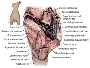
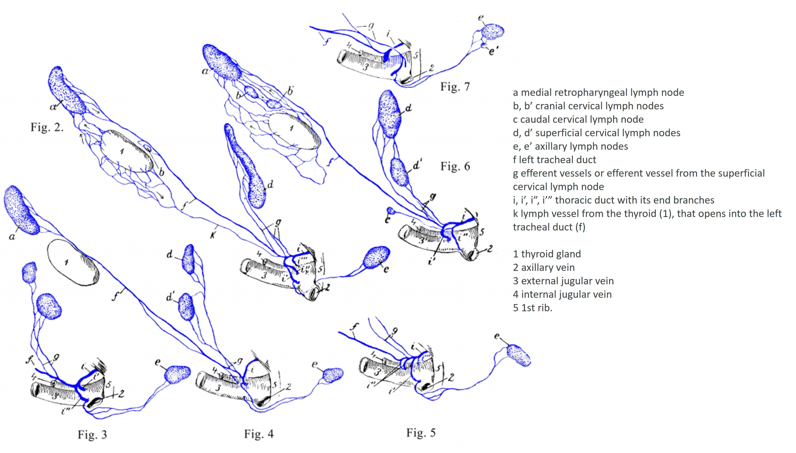
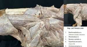
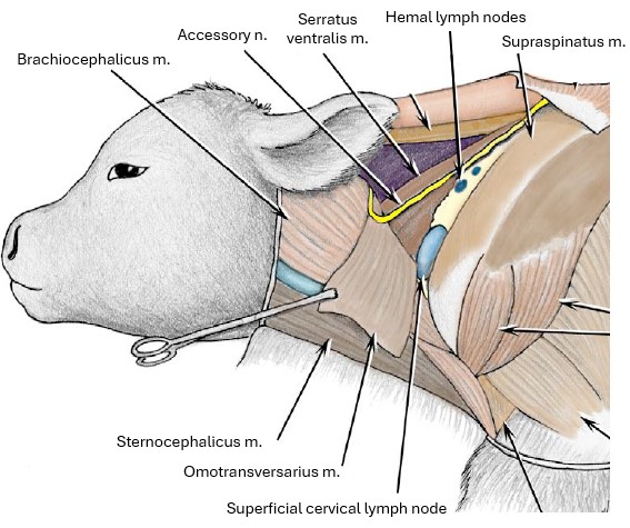
There are usually two superficial cervical lymph nodes, which lie on the serratus ventralis and scalenus cranial to the supraspinatus and covered by the omotransversarius and the cleidocephalicus. These nodes receive the afferent lymph vessels from the superficial part of the lateral surface of the neck, the caudal surface of the head including the ear and pharynx, and the thoracic limb. Efferent lymph from the superficial cervical lymph nodes may take several different routes to the tracheal trunk or thoracic duct or directly into the venous system.
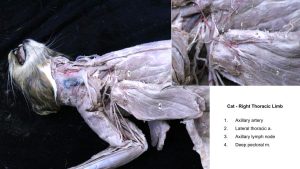
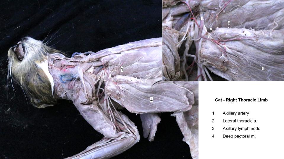
(Proper) Axillary lymph node – is located in the axilla closely related to nerves and vessels. The node lies dorsal to the deep pectoral m. and caudal to the axillary v. Thoracic wall and deep structures of the medial thoracic limb drain to this node. Like those from the superficial cervical lymph node, efferent lymph vessels from the axillary lymph node may take several different routes to the tracheal trunk or thoracic duct or directly into the venous system. The lateral thoracic a. typically crosses over the lateral surface of the axillary lymph node.
Accessory Axillary lymph nodes – are located deep to latissimus dorsi m. along the lateral thoracic v. between the 3rd and 6th intercostal space. These nodes drain the medial and cranial thoracic limb, skin of lumbar region, lateral thoracic wall, and cranial half of mammary glands. Efferents from these nodes travel to the axillary lymph nodes.
-
- Branches of the superficial cervical artery of the dog. 1

-
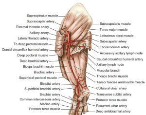
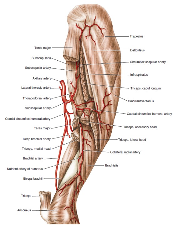
- Arteries of the right brachium of the dog, medial aspect. 1

-
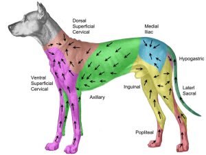
- Direction of lymph flow of the dog.H. Suami

-
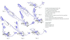
- Schemata of the variations of the left tracheal duct of the dog. Baum
-
- Dog superficial cervical lymph node

-
- Cat axillary lymph node
Clinical Application: Aspiration of an Efferent Lymph Node
Lymph nodes draining a site of infection will often enlarge as cells and fluid accumulate in response to antigenic stimulation. As the nodes enlarge, their capsules become stretched and the nodes become hot and painful. Focal sites of infection such as abscesses or upper respiratory infection often will lead to enlargement of the local nodes draining those sites. Neoplasia (cancer) can also cause enlargement due to a primary cancer of the node itself (lymphoma) or due to metastasis of neoplastic cells from a nearby structure(s). Therefore, it is very important from a clinical standpoint to know lymph node locations, normal sizes, and drainage sites.
Question: You are presented with a twelve year old, intact, female domestic shorthair cat. On physical examination, you noted an approximately 1 cm, firm, non-movable mass on the left, cranial-most mamma/teat. Afferent lymph from the affected region flows to which lymph node first? Where does efferent lymph from this node go next?
Arteries of the Carnivore Thoracic Limb
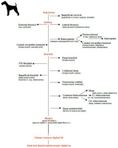
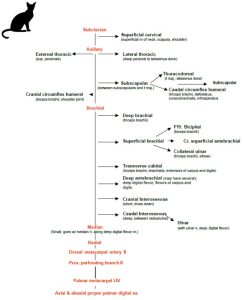
Primary vessels of the thoracic limb (canine map)
- Subclavian
- Vertebral
- Costocervical trunk
- Internal thoracic
- Superficial cervical
- Axillary
- External thoracic
- Lateral thoracic
- Subscapular
- Caudal circumflex humeral
- Thoracodorsal
- Cranial circumflex humeral
- Brachial
- Deep brachial
- Bicipital
- Collateral ulnar
- Superficial brachial
- Transverse cubital
- Common interosseous
- Ulnar
- Cranial interosseous
- Caudal interosseous
- Deep antebrachial (author’s experience)
- Median
- Radial
- Deep palmar arch
- Superficial palmar arch
Notes: As you trace the arteries distally (and the veins proximally-we generally follow vessels in the direction of blood flow), keep in mind that there can be significant individual and species variation in branching patterns. Use the described branching pattern as a starting point but realize that ultimately, vessels will be named for the structures they supply, not for where they branch off of the main channel artery.
As you know, nerves will only innervate the muscles and structures in which they insert. On the other hand, vessels will generally supply blood to all of the muscles and structures (via very small, unnamed branches and capillaries) close to them. You need not memorize every muscle which is supplied by a given artery, but you should be able to name a region/regions supplied by a given artery and vice versa.
Use the blood supply diagrams and diagrams below as a starting point as you follow the arteries distally.
-
- Dog arteries

-
- Cat arteries

-

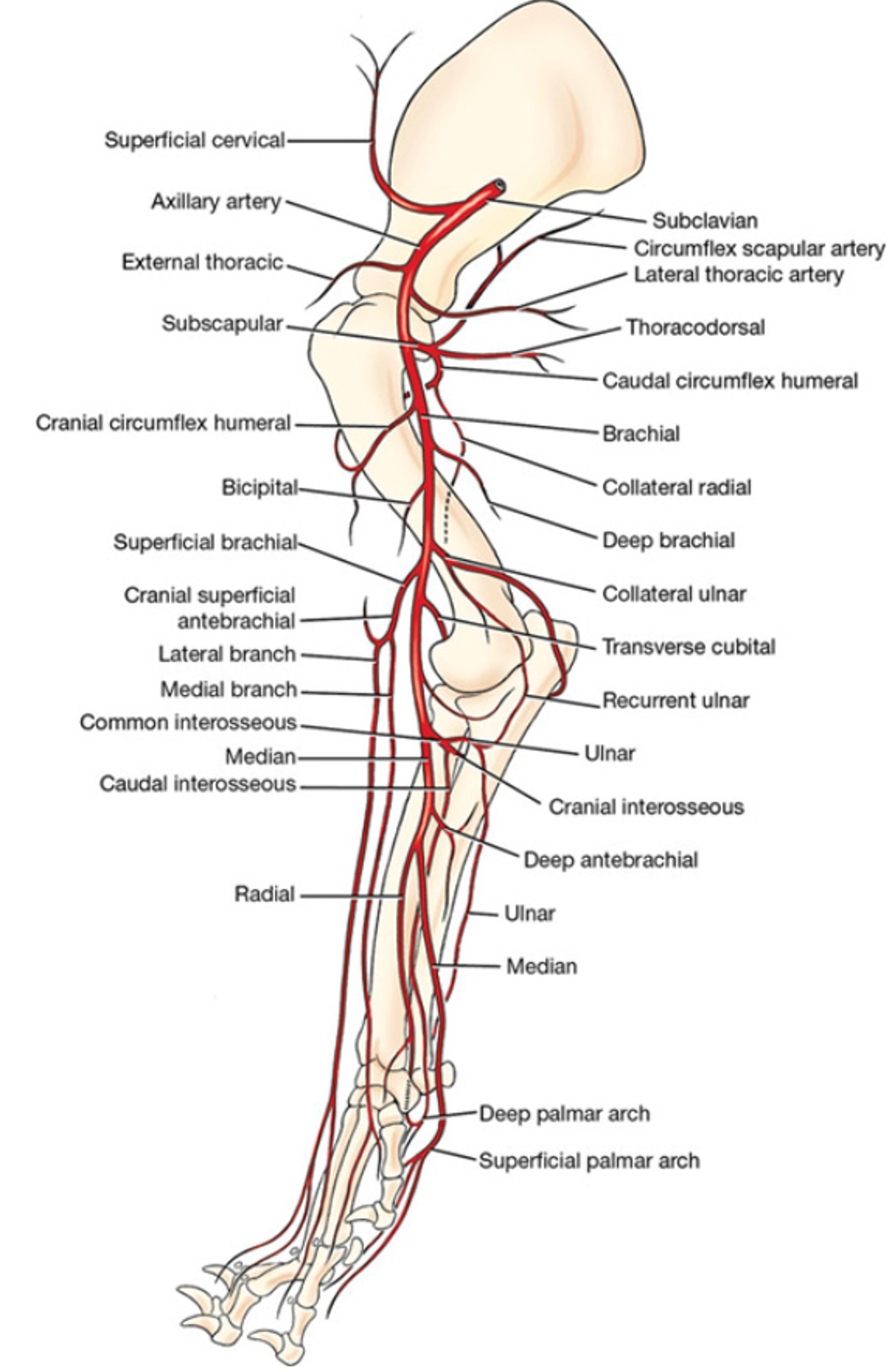
- Arteries of right forelimb of the dog, medial view. 1
Dissect: Using the attached thoracic limb on your cadaver, bluntly dissect, clean, and follow the following bolded arteries distally, as described below. Satellite veins (veins that accompany their same-named artery) may be removed if necessary to better view the arteries. It is particularly useful to transect the axillary vein to gain more abduction of the limb from the trunk.
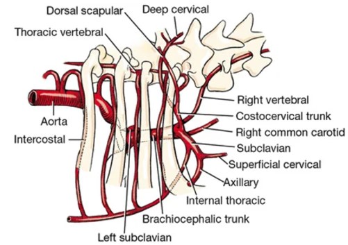
The main arterial blood supply to the thoracic limb arises within the thorax as the subclavian artery, which is a terminal branch of the brachiocephalic trunk on the right side and a direct branch of the aorta on the left side (in the carnivore). Recall that the subclavian artery has four branches: vertebral, superficial cervical, costocervical trunk, and internal thoracic. The superficial cervical artery arises from the subclavian just inside the thoracic inlet. It runs craniodorsal between the scapula and the neck. It supplies the superficial muscles of the base of the neck, the superficial cervical lymph nodes, the muscles of the scapula, and the shoulder.
-
- Branches of the superficial cervical artery of the dog. 1
-

- Branches of brachiocephalic trunk of the dog, right lateral view. 1
-
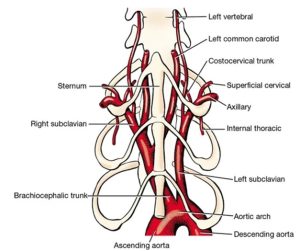
- Branches of aortic arch of the dog, ventral view. 1

Axillary artery and branches
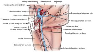
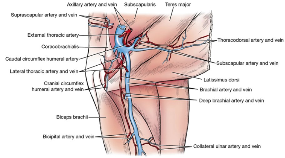
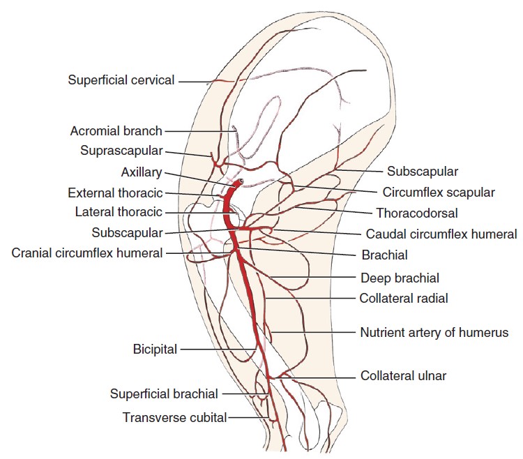
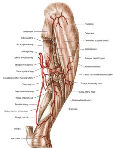
The axillary artery is the continuation of the subclavian and extends from the first rib to the conjoined tendons of the teres major and latissimus dorsi. It has four primary branches: the external thoracic, the lateral thoracic, the subscapular, and the cranial circumflex humeral.
Remember! In the following discussion some variability may be encountered in the origin of specific blood vessels. However, although the origin may vary, the area or structures supplied are usually consistent.
FYI: observe the following branches of the axillary artery as reference:
- The external thoracic artery leaves the axillary near its origin. The external thoracic artery curves around the craniomedial border of the deep pectoral with the nerve to the superficial pectorals and is distributed almost entirely to the superficial pectorals. It may arise from a common trunk with the lateral thoracic artery, or it may arise from the deltoid branch of the superficial cervical artery. Note: In the cat, the external thoracic artery often branches directly from the superficial cervical artery.
- The lateral thoracic artery runs caudally across the lateral surface of the axillary lymph node and along the dorsal border of the deep pectoral ventral to the latissimus dorsi. It usually arises from the axillary artery distal to the external thoracic. The vessel may arise distal to the subscapular artery. It supplies parts of the latissimus dorsi, deep pectoral, and cutaneous trunci muscles, and the thoracic mammae.
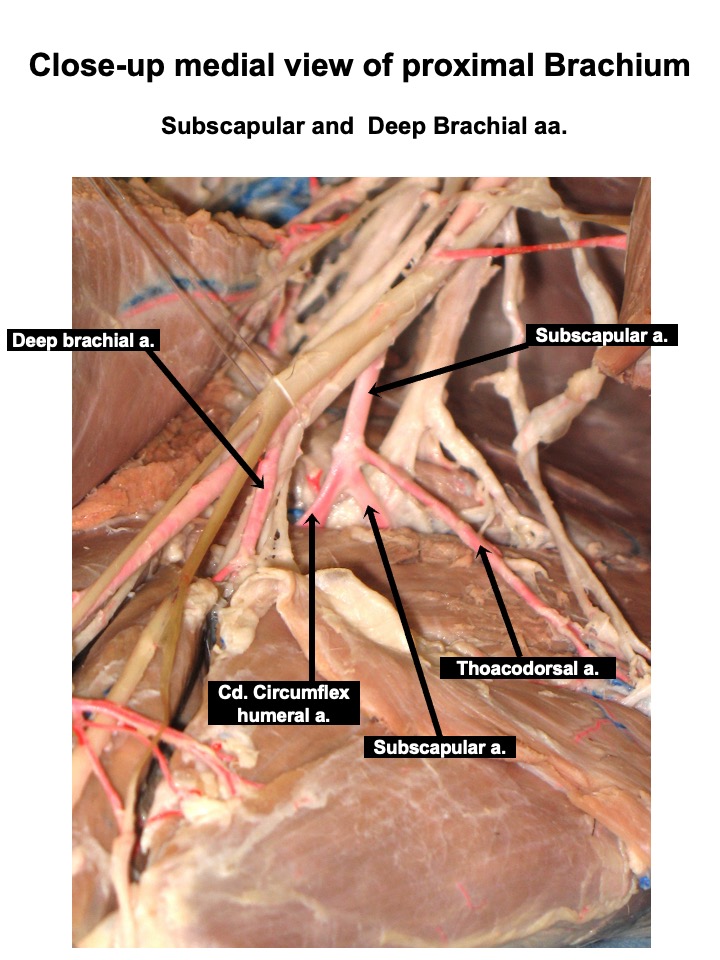
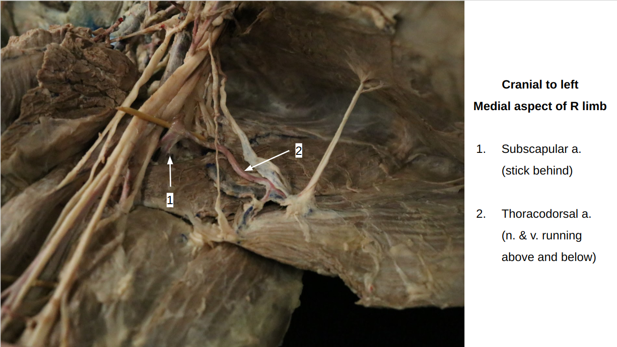
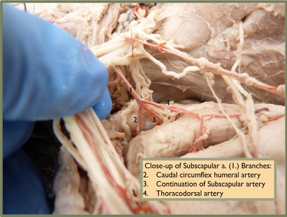
- The subscapular artery is larger than the continuation of the axillary in the arm. Only a short part of the subscapular is now visible. It passes caudo-dorsally between the subscapularis and the teres major and becomes subcutaneous near the caudal angle of the scapula. Each bone is supplied by at least one main artery, which enters through a nutrient foramen in the cortex of the bone. The nutrient artery is a branch of an adjacent artery, which for the scapula is the subscapular artery.
Isolate the subscapular artery and the following branches thereof:
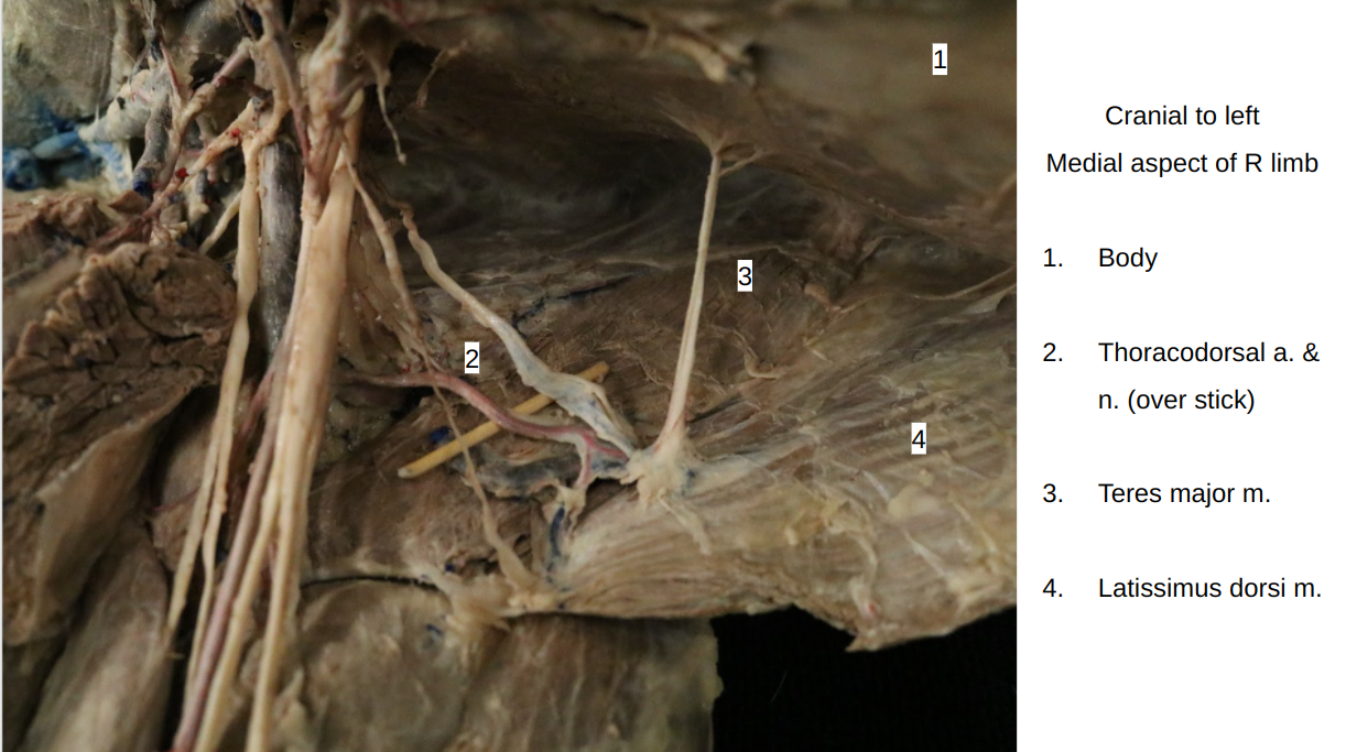
A. The thoracodorsal artery leaves the dorsal surface of the subscapular a. near its origin. It supplies a part of the teres major and latissimus dorsi and ends in the skin. It is readily seen on the deep surface of the latissimus dorsi.
B. The caudal circumflex humeral artery leaves the subscapular opposite the thoracodorsal and courses laterally between the head of the humerus and the teres major. The caudal circumflex humeral supplies the triceps, deltoideus, coracobrachialis and infraspinatus muscles and the shoulder joint capsule.
4. The cranial circumflex humeral artery arises from the axillary artery distal or proximal to the subscapular artery. It courses cranially to supply the biceps brachii and the joint capsule of the shoulder.
-
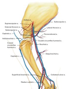
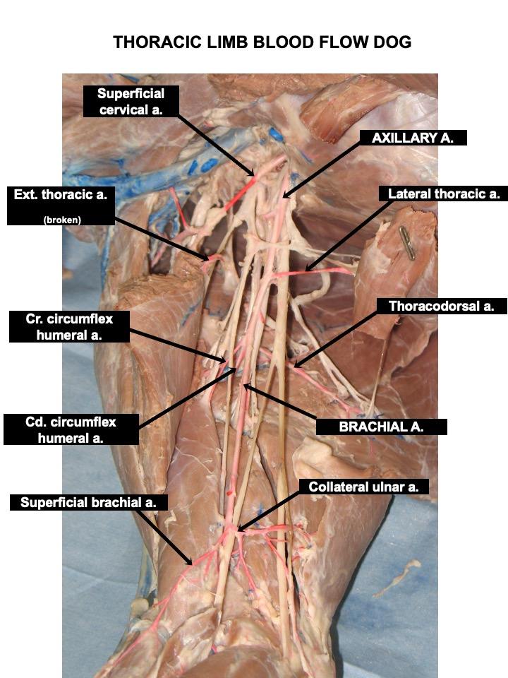
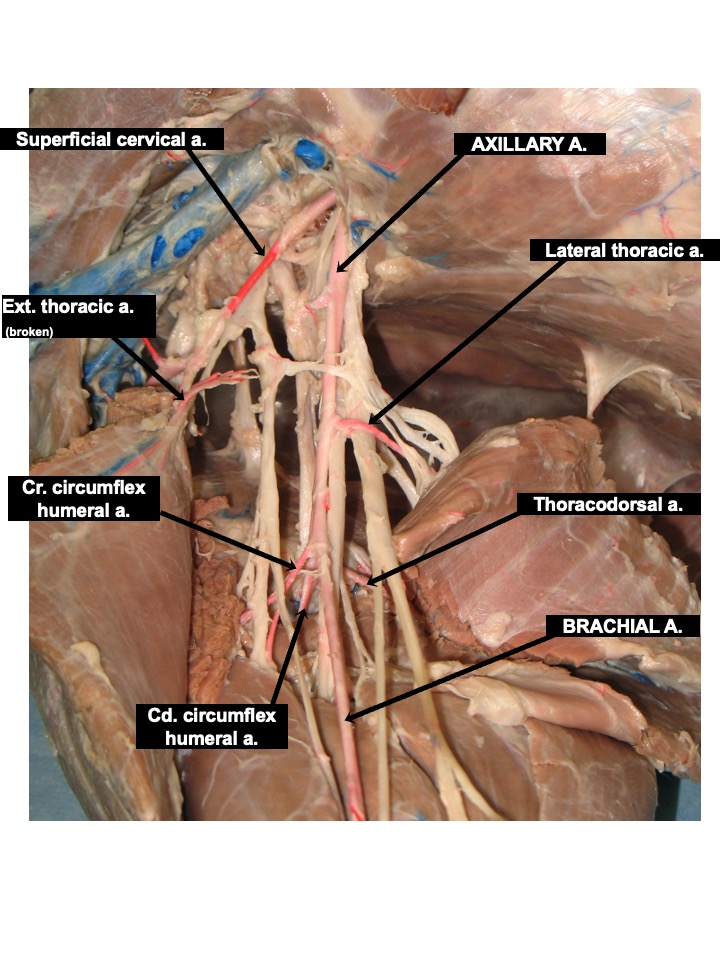
- Vessels of right axillary region of the dog, medial view. 1
-

- Diagram of the arteries of the right brachium of the dog, medial aspect. 1
-
- Arteries of the right brachium of the dog, medial aspect. 1
-
- Arteries of the right brachium of the dog, caudolateral aspect. 1
-
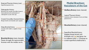
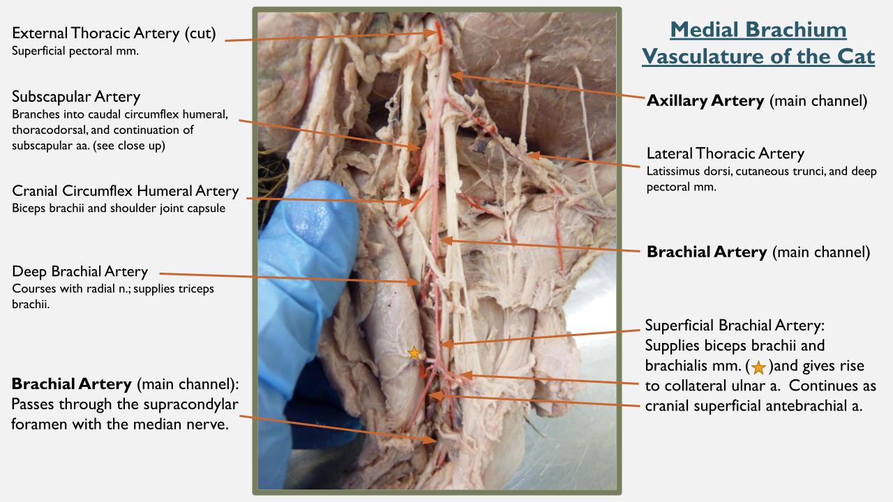
- Axillary and brachial artery branches of the cat. 4

-

- Dog proximal arteries
-
- Subscapular a.
-

- Thoracodorsal a.
-
- Cat brachial arteries
-
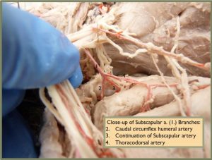
- Cat subscapular branches
Observe: Identify the axillary artery to its termination at the conjoined tendons of the teres major and latissimus dorsi muscles.
Brachial artery and branches
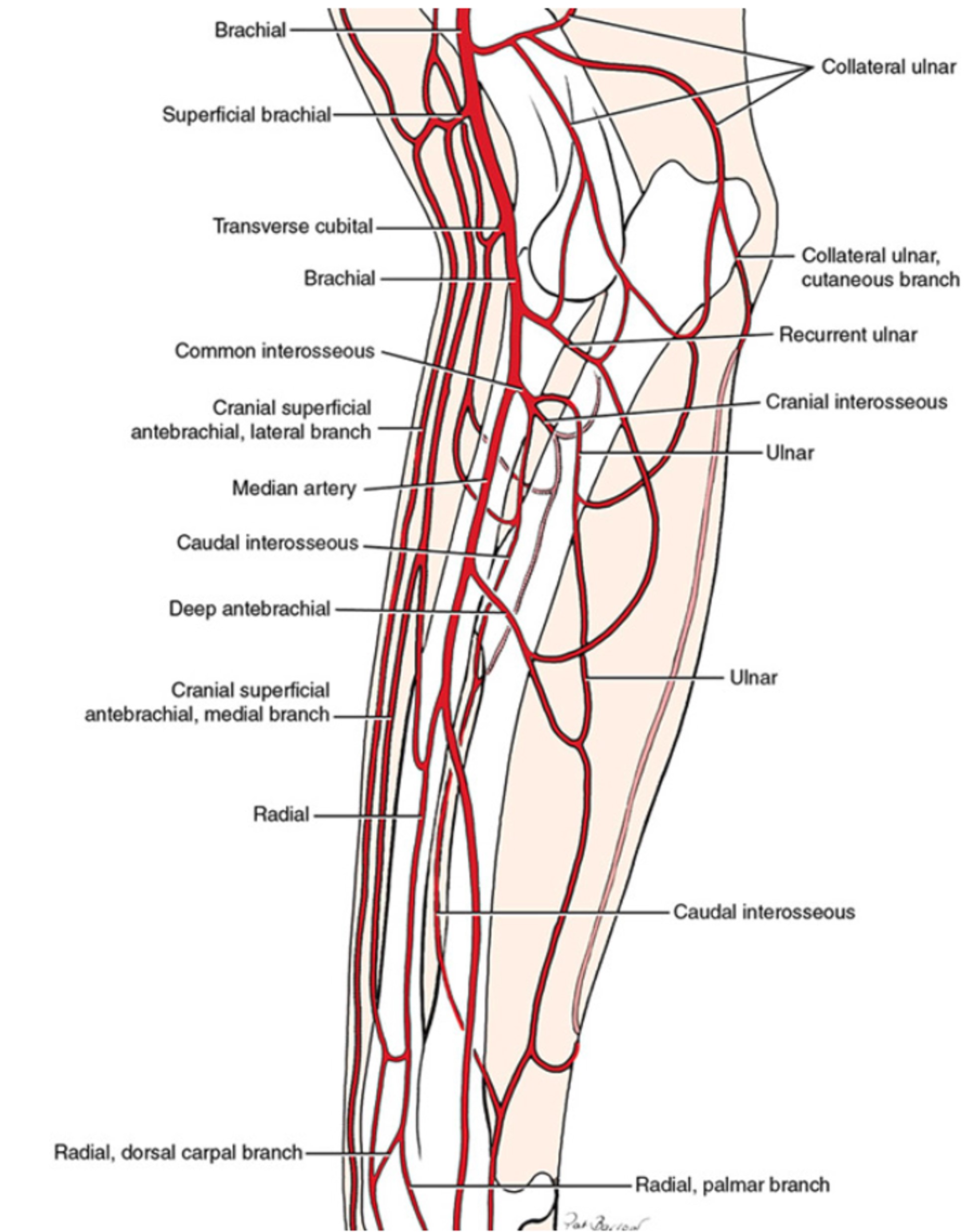
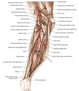
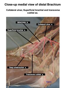
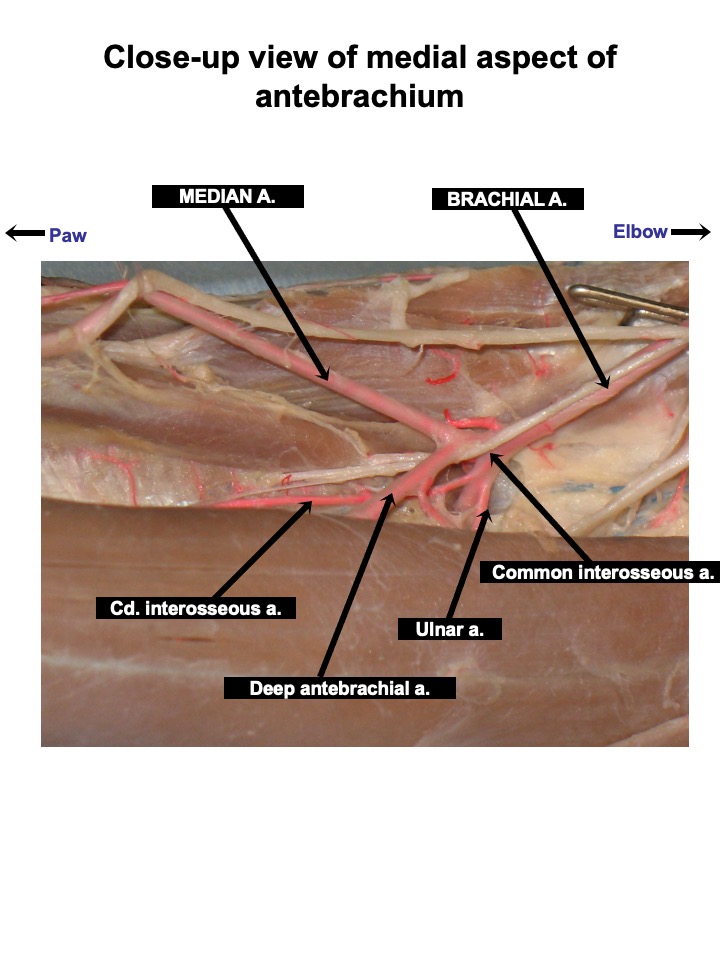
The brachial artery is a continuation of the axillary from the conjoined tendons of the teres major and latissimus dorsi. It courses distally across the body of the humerus to reach the craniomedial surface of the elbow, where it gives rise to several branches. The deep brachial and bicipital arteries are muscular branches of the brachial in the arm. Other branches are the collateral ulnar, superficial brachial, transverse cubital, and deep antebrachial arteries. The brachial artery continues into the proximal forearm and gives off its largest branch, the common interosseous, at which point the main channel vessel changes name to the median artery.
Identify the brachial artery. Note: In the cat, the median n. and brachial a. (but not the vein) travel together through the supracondylar foramen of the humerus, a foramen not present in the canid.
FYI: Trace the following branches of the brachial artery.
- Deep brachial is the caudal muscular branch passing into the triceps brachii m.
- The collateral ulnar artery is a caudal branch of the brachial in the distal third of the arm. It supplies the triceps, the ulnar nerve, and the elbow.
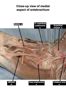
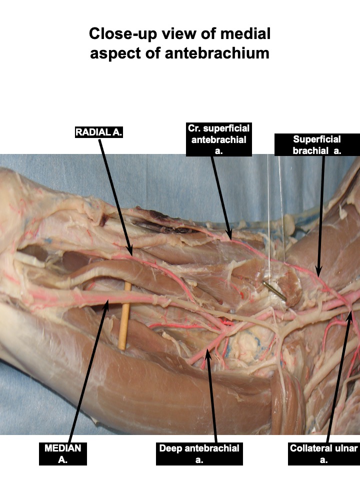
- The superficial brachial artery loops around the cranial surface of the distal end of the biceps brachii, deep to the cephalic vein. It continues in the forearm as the cranial superficial antebrachial artery. Medial and lateral rami arise from the latter, and both course distally on either side of the cephalic vein accompanied by the medial and lateral branches of the superficial radial nerve. These vessels supply blood to the dorsum of the forepaw via the dorsal common digital arteries.
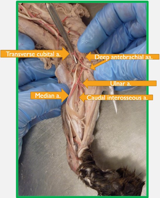
FYI: Retract the brachial artery at the level of the elbow so as to externalize the transverse cubital artery, which dives laterally, almost perpendicularly away from the brachial artery. NOTE: The recurrent ulnar artery (FYI) usually branches from the brachial artery in between the transverse cubital and common interosseous arteries. Take care not to confuse it for any of the need-to-know arteries.
4. Transverse cubital a. – located on the cranial aspect of the elbow (cubital joint). A sizable branch that passes lateral across the joint and supplies the elbow and adjacent muscles.
-
- Diagram of the arteries of the right brachium of the dog, medial aspect. 1
-
- Arteries of the right brachium of the dog, medial aspect. 1
-

- Diagram of the arteries of the right antebrachium of the dog, medial aspect. 1
-
- Arteries of the right antebrachium of the dog, medial aspect. 1

-
- Axillary and brachial artery branches of the cat. 4
-
- Dog brachial artery branches

-
- Cat brachial arteries
-

- Cat antebrachium
Proximal arteries of the antebrachium
Canine Only
The brachial artery in the forearm gives rise to the common interosseous and at this point, the main channel continues as the median artery. The median extends to the superficial palmar arch in the paw.
Observe: Identify the common interosseous and median arteries, as described below.
- The common interosseous artery is short and passes to the proximal part of the interosseous space between the radius and ulna before dividing into three branches. NOTE: recall the recurrent ulnar artery (FYI) usually branches from the brachial artery in between the transverse ulnar and common interosseous arteries. Take care not to confuse it for either of the need-to-know arteries. Once the common interosseous artery branches, the main channel changes from the brachial to the median artery.
FYI: Retract the brachial artery medially to see the branches of the common interosseous artery, which are the ulnar, caudal interosseous, and cranial interosseous arteries, described below. The caudal interosseous artery should be observed on instructor prosections. The pronator quadratus m. will have been excised to expose the caudal interosseous artery which runs parallel to it.
A. The ulnar artery courses caudally. The common interosseous and ulnar arteries may arise together from the brachial. A recurrent branch extends proximally between the humeral and ulnar heads of the deep digital flexor. The ulnar artery continues distally with the ulnar nerve between the humeral head of the deep digital flexor and the flexor carpi ulnaris. It supplies the ulnar and humeral heads of the deep digital flexor and the flexor carpi ulnaris.
B. The caudal interosseous artery lies between the opposed surfaces of the radius and ulna. It dives deep into the pronator quadratus m., portions of which are excised to expose the artery as it travels distally. In its course down the forearm, the artery supplies many small branches to adjacent structures. It passes through the lateral side of the carpal canal and in the carpometacarpal region it joins with branches of the radial and median arteries to form arches that supply the palmar surface of the forepaw.
C. The cranial interosseous artery passes through the proximal part of the interosseous space cranially to supply the muscles lying laterally and cranially in the forearm. It almost immediately runs directly laterally away from its origin on the common interosseus artery. You need not dissect it beyond this point.
-
- Diagram of the arteries of the right antebrachium of the dog, medial aspect. 1
-
- Arteries of the right antebrachium of the dog, medial aspect. 1
-

- Dog antebrachium
-
- Dog caudal antebrachium
-
- Cat antebrachium
Feline Only
- In the cat, the common interosseous artery is usually absent. Instead, the cranial interosseous artery branches directly from the brachial artery and passes through the proximal part of the interosseous space cranially to supply the muscles lying laterally and cranially in the forearm.
FYI: Observe the cranial interosseous artery as it branches from the brachial artery. It almost immediately runs directly laterally away from its origin and it need not be dissected beyond this point.
2. The caudal interosseous artery branches directly from the brachial artery in the cat and demarcates the name change from brachial to median artery in this species. It lies between the opposed surfaces of the radius and ulna. It dives deep into the pronator quadratus m., portions of which are excised to expose the artery as it travels distally. In its course down the forearm, the artery supplies many small branches to adjacent structures. It passes through the lateral side of the carpal canal and in the carpometacarpal region it joins with branches of the radial and median arteries to form arches that supply the palmar surface of the forepaw.
Dissect: Identify the caudal interosseous artery as it branches from the brachial artery and follow it distally. The pronator quadratus m. will need to be excised to expose the caudal interosseous artery which runs parallel to it. Distal to the caudal interosseous artery, the brachial artery changes name to the median artery.
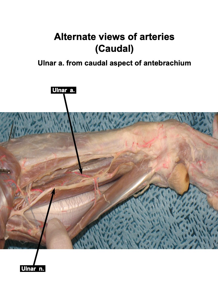
3. In the cat, the ulnar artery (FYI) is usually a branch of the caudal interosseous artery. The ulnar artery continues distally with the ulnar nerve between the humeral head of the deep digital flexor and the flexor carpi ulnaris. It supplies the ulnar and humeral heads of the deep digital flexor and the flexor carpi ulnaris. It courses over the ulna and is readily identifiable. You may find it first and then follow it back to the caudal interosseous artery from which it arose.
FYI: Observe the ulnar artery as it courses over the ulna.
-
- Antebrachial vasculature of the cat. 4

-
- Cat antebrachium
Distal Arteries of the Antebrachium
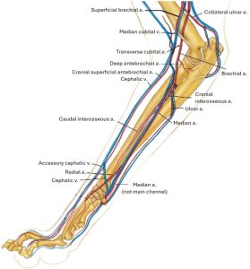
1. The median artery is the continuation of the brachial artery beyond the origin of the common interosseous (in the dog) or the caudal interosseous (in the cat). It gives off the deep antebrachial (there may be a few of these) and the radial artery in the forearm and continues into the paw deep to the flexor carpi radialis. The median a. is the principal source of blood to the paw in the canine (but not in the feline). It is accompanied by the median nerve along the humeral head of the deep digital flexor. The median artery and nerve pass through the carpal canal between the superficial and deep digital flexor tendons.
The median a. forms the superficial palmar arch in the proximal palmar metacarpus with a branch of the caudal interosseous a. This arch gives rise to the palmar common digital arteries, which course to the palmar surface of the forepaw.
2. The radial artery arises from the medial side of the median artery. in the middle of the forearm it follows the medial border of the radius. At the carpus it divides into palmar and dorsal carpal branches that supply the deep vessels of the forepaw.
NOTE: In the cat, the radial artery is the main channel artery supplying the carpus and manus whereas in the canine, the median artery remains the main channel arterial supply to the forepaw. Note that in the cat, the median artery drastically diminishes as it courses distally. Likewise, the radial artery tapers off drastically in the canine.
3. FYI: The deep antebrachial artery(ies) supply(ies) the flexor carpi radialis, the deep digital flexor, the flexor carpi ulnaris, and the superficial digital flexor mm. It is usually a caudal branch of the median artery in both the dog and cat. However, other arteries supplying these muscles may be observed branching from the brachial artery, proximal to the cranial interosseous artery, either instead of, or in addition to, those located distal to the median artery.
-
- Diagram of the arteries of the right antebrachium of the dog, medial aspect. 1
-
- Arteries of the right antebrachium of the dog, medial aspect. 1
-
- Antebrachial vasculature of the cat. 4
-
- Dog antebrachium
Clinical Application: Assessing Blood Supply in the Trauma Patient
In treating limb trauma patients, it is important to assess blood supply to the limb in order to determine viability of tissue going forward. At times, blood supply is compromised by the initial injury, but it is also important to consider how blood supply will be affected by treatment. There is a considerable amount of collateral circulation to the limb. Usually a back-up exists if one supply channel is disrupted. However, if a main channel artery is compromised in particular, all of the structures distal to the injury may not be viable. Likewise, when performing treatments such as skin flaps and grafting, one must assure that blood supply is maintained.
Vasculature of the Carnivore Manus
Observe: Use diagrams and lab models to describe the vasculature of the manus, as explained below, to include how many arteries are present in between each metacarpal bone, at the metacarpophalangeal joint, and in the digits.
The distribution of vessels to the digits will not be dissected, nor are you responsible for naming them all. However, you should be able to describe where they are located and how many are present in each region of the manus.
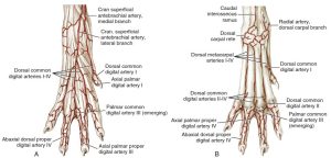
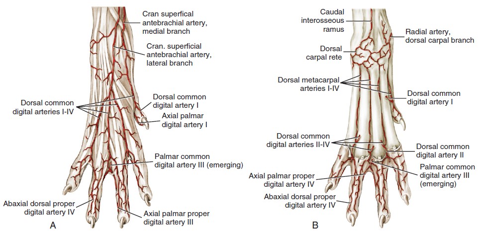
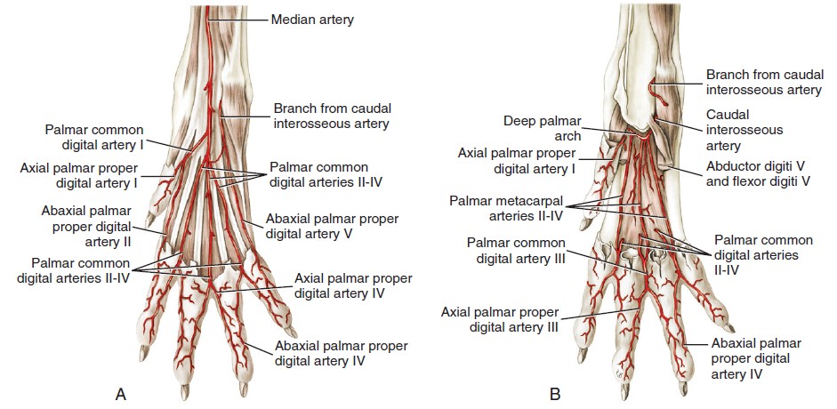
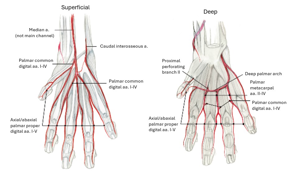
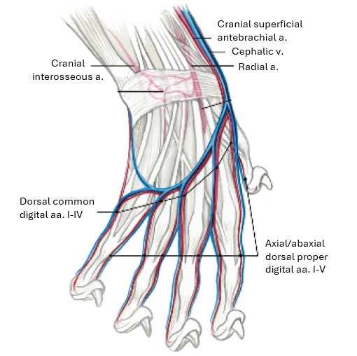
On the dorsal side of the metacarpus, in between each metacarpal bone, there are two vessels, one more superficial than the other. This pattern is repeated on the palmar side of the metacarpus. Therefore, in the metacarpus, there are four sets of four vessels, each set located between the metacarpal bones.
At the metacarpophalangeal joint, in between digits 2 and 3, 3 and 4, and 4 and 5, the two branches on the dorsal side of the manus combine, as do the two branches located on the palmar side. So now there exists one common trunk on the dorsal side and one common trunk on the palmar side, in between each metacarpal bone 2-5.
Each common trunk then divides into medial and lateral branches to the digits. Instead of medial or lateral, vessels on the digits are called axial or abaxial, which refers to whether they are on the surface of the digit that faces toward the axis (axial) or away from the axis (abaxial) of the paw. The axis of the paw passes between digits III and IV.
-
- Superficial and deep arteries of the right forepaw of the dog, dorsal aspect. 1
-
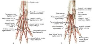
- Superficial and deep arteries of the right forepaw of the dog, palmar aspect.
-

- Superficial and deep vasculature of the forepaw of the cat, palmar view. 4
-
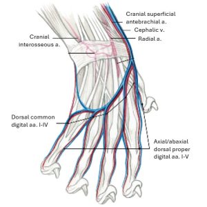
- Superficial and deep vasculature of the forepaw of the cat, dorsal view. 4
Clinical Application: Digit Amputation and Lymph Node Aspiration
Toe amputation is a common surgical procedure often performed to resect a mass that is affecting function of the foot and/or is neoplastic (cancerous). Consideration of the large blood supply to the digits should be made when performing a digit amputation, as their transection is cause for a significant amount of hemorrhage . Some clinicians opt to place a tourniquet proximally to maintain hemostasis (decrease blood loss) during the procedure. Cauterization of the vessels may also be utilized. Regardless, isolation of each individual artery is usually not possible or warranted.
It is also important to note that digits three and four are both weight-bearing in the carnivore. Therefore, while it likely will improve with healing, lameness in the affected limb may not fully resolve if one of these digits is amputated. In patients with both of these removed, they may not weight-bear at all.
Questions: You are scheduled to perform a digit amputation on your patient’s right third digit due to a progressive, non-painful mass located at the level of the nail bed.
How many arteries provide blood supply to this digit at the level of the third phalange?
You are concerned that the mass you are removing may be neoplastic and, if so, may have already metastasized. You have obtained approval from your patient’s owners to submit the mass for histopathological analysis, which should confirm whether or not the mass is cancerous.
On physical examination, you had noted that the ipsilateral superficial cervical lymph node was enlarged. What is another important diagnostic step you should take in this case?
What information are you obligated to give about the patient given you will be removing the third phalanx in particular?
Venous Return of the Carnivore Thoracic Limb
The cephalic vein begins on the palmar side of the paw from the superficial palmar venous arch. The accessory cephalic vein arises from small veins on the dorsum of the paw and joins the cephalic on the cranial surface of the distal third of the forearm.
At the flexor surface of the elbow, the median cubital vein forms a connection between the cephalic and brachial veins. From this connection the cephalic continues proximally on the craniolateral surface of the arm in a furrow between the brachiocephalicus cranially and the origin of the lateral head of the triceps caudally.
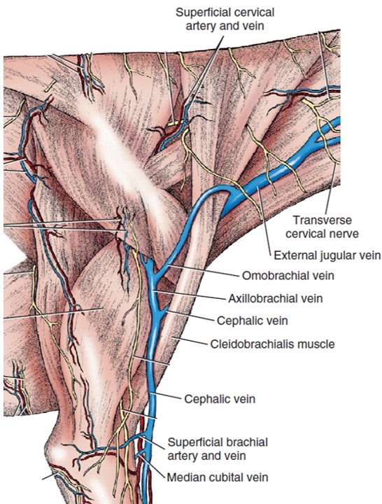
FYI: In the middle of the arm, the axillobrachial vein leaves the caudal aspect of the cephalic vein. The cephalic vein runs deep to the brachiocephalicus and enters the external jugular near the thoracic inlet. The axillobrachial vein continues proximally and passes deep to the caudal border of the deltoideus to join the axillary vein at this site. The omobrachial vein (not usually present in cats) arises from the axillobrachial vein and continues subcutaneously across the cranial surface of the arm and shoulder and brachiocephalicus muscle before entering the external jugular vein cranial to the cephalic vein.
-
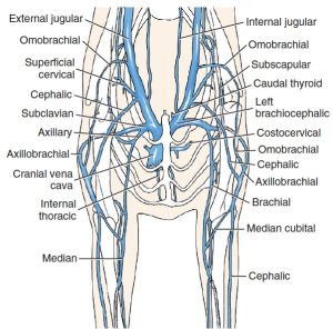
- Veins of the neck and shoulders of the dog, cranial aspect. 1

-
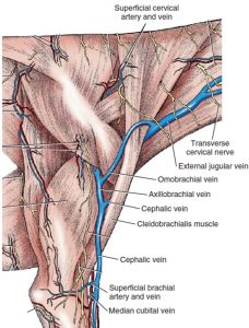
- Superficial structures of the scapula and arm of the dog, lateral view. 1
-
- Veins of the right antebrachium of the dog, cranial aspect. 1
Clinical Application: Venipuncture of the Cephalic Vein
The cephalic vein is a common site for venipuncture in the canine patient. These veins are generally too small in the domestic feline to allow for venipuncture; the hindlimb or external jugular vein are usually used in felines instead. Watch the video linked below and be able to answer questions about it.
Lymph Nodes of the Ungulate Thoracic Limb
Observe: In the horse, goat, and pig observe the superficial cervical lymph nodes (in a similar location as in the carnivore) and the axillary lymphocenters, as described below.
The axillary lymphocenters, as can be anticipated given their name, are generally observed to be a cluster of nodes located in the axillary region of the ungulate forelimb. Afferent lymph to these nodes arrives from the deep structures of the TL; recall the superficial drainage of the TL mostly goes to the superficial cervical lnn.
Lymph nodes of the axillary lymphocenter are located:
1) close to the axillary a. continuation as the brachial a. (axillary lnn., not present in pig)
2) near the 1st rib (axillary ln. of 1st rib, found in ruminants, pig, cat)
3) near 3rd/4th rib (accessory axillary lnn., found in carnivores, some ruminants).
-

- Superficial cervical lymph node of the calf. 2
-
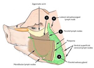
- Lymph nodes of the pig. 2

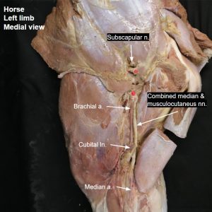
Observe: In the equine only, isolate the cluster of lymph nodes medial to the brachial vessels in the distal part of the brachium near the elbow. These are the cubital lymph nodes.
-
- Horse cubital lymph node
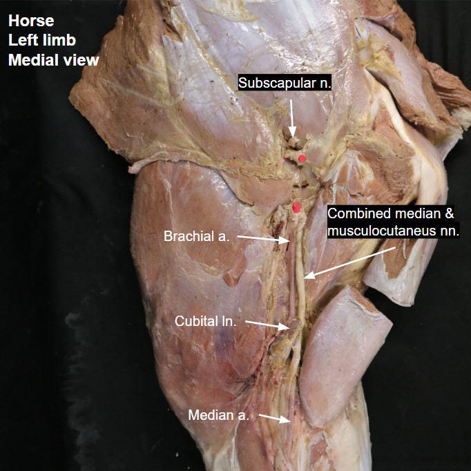
Vasculature of the Ungulate Thoracic Limb
Observe: Observe the following main channel arteries on the horse, goat, and pig instructor pro-sections.
Axillary artery – usually the artery which has been transected in order to remove the limb (if observing on a detached limb).
(Identify the subscapular artery. It is not a main channel artery. Rather, it is a main branch of the axillary artery, diving directly into the subscapularis muscle.)
The name change from the axillary artery to brachial artery occurs distal to the cranial circumflex humoral artery. You need not identify the cranial circumflex humoral artery in the ungulate, but know that the main channel artery in the brachium is, conveniently…the brachial artery.
Brachial artery
Name Change: Common interosseous a. – large branch distal to the cubital joint.
Median artery
-
- Branches of the axillary artery of the horse, medial view. 2

-

- Branches of the axillary artery of the ox, medial view. 2
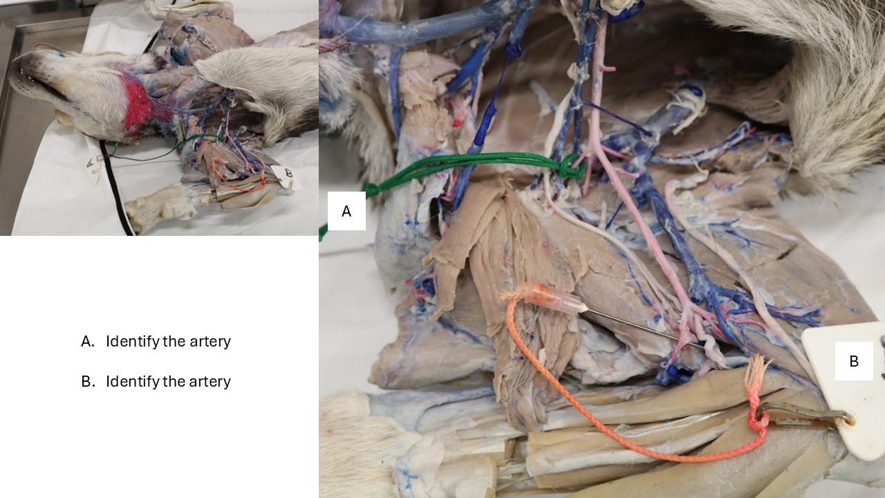
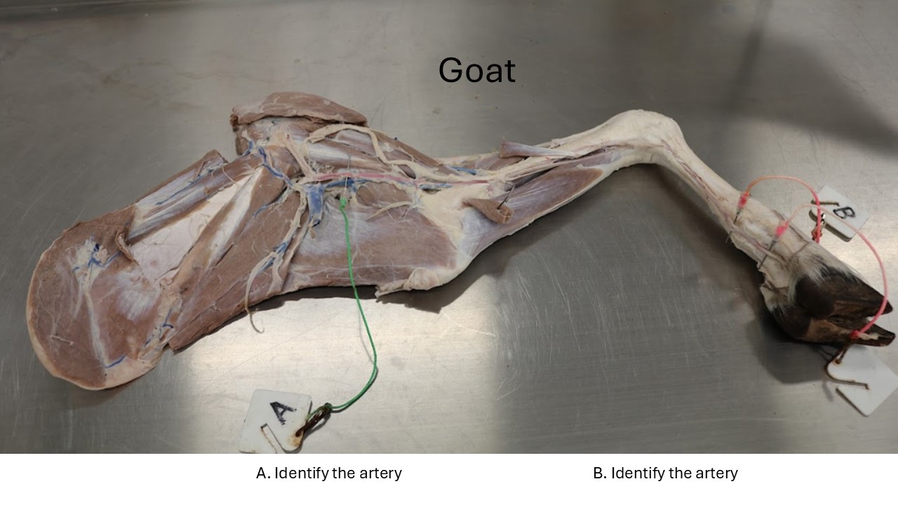
Observe: Observe the arterial supply to the goat distal limb as follows:
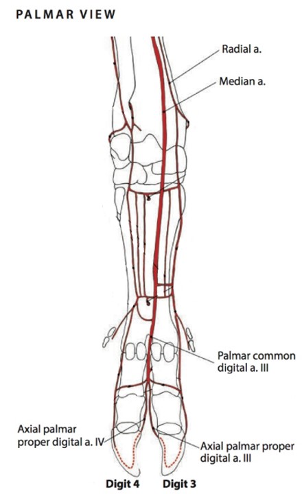
In artiodactyls, the main channel continuation of the median a. (name change just proximal to the metacarpophalangeal joints) is the palmar common digital artery 3, entering the interdigital space. The palmar common digital artery 3 bifurcates to form the axial palmar proper digital arteries of digits 3 and 4, which are main channel to the digits.
-
- Branches of the axillary artery of the ox, medial view. 2
-
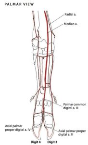
- Palmar arteries of the left distal limb of the ox. 5
Clinical Application: Digital pulses in the ruminant
As palmar common digital artery 3 crosses the SDF tendon, the artery is easily palpable as a large, elongated bulge. You can take the pulse of the ruminant at this site.
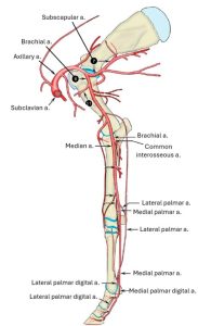
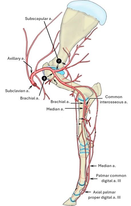
Observe: Observe the arterial supply to the equine distal limb as follows:
Continue to follow the median artery distally as it runs adjacent to the median n.
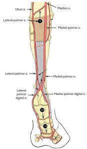
Proximal to the carpal canal, the median a. of the horse gives off a lateral palmar a., and the main channel continues as the medial palmar a. through the carpal canal and into the metacarpus.
The lateral palmar a. receives the collateral ulnar a. and then accompanies the lateral palmar n. into the metacarpus. Note that this lateral set travels outside of the carpal canal, passing through the flexor retinaculum of the carpus.
Continue to follow the medial and lateral palmar aa. distally through the metacarpus to the level of the fetlock, traveling closely associated with the deep digital flexor tendon (DDFT) most of the way. Recall the medial palmar a. is the main channel blood supply and is substantially larger than the lateral palmar a.
At the distal metacarpus the medial palmar a. inclines axially into the space between the DDFT and the suspensory ligament. Just proximal to the fetlock, the artery bifurcates into the medial and lateral palmar digital arteries, which diverge and continue over the abaxial sides of the proximal sesamoid bones. Here they join the palmar digital veins and palmar digital nerves.
-
- Branches of the axillary artery of the horse, medial view. 2
-
- Arteries of the distal limb of the horse, caudomedial view. 2

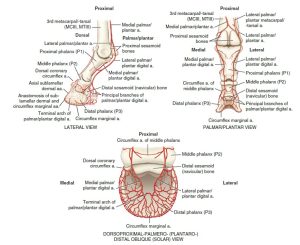
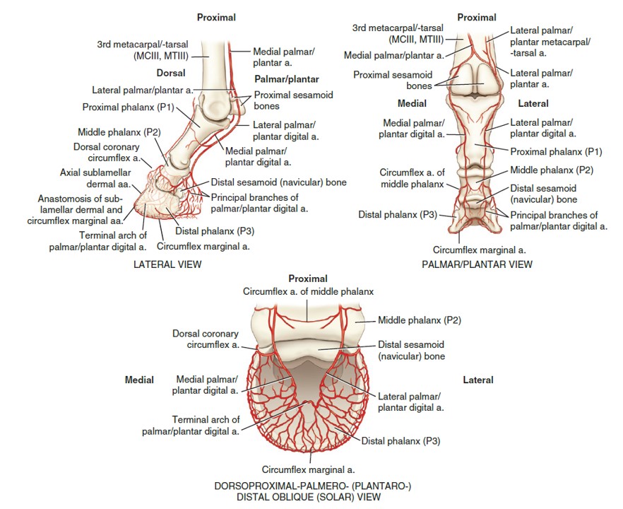
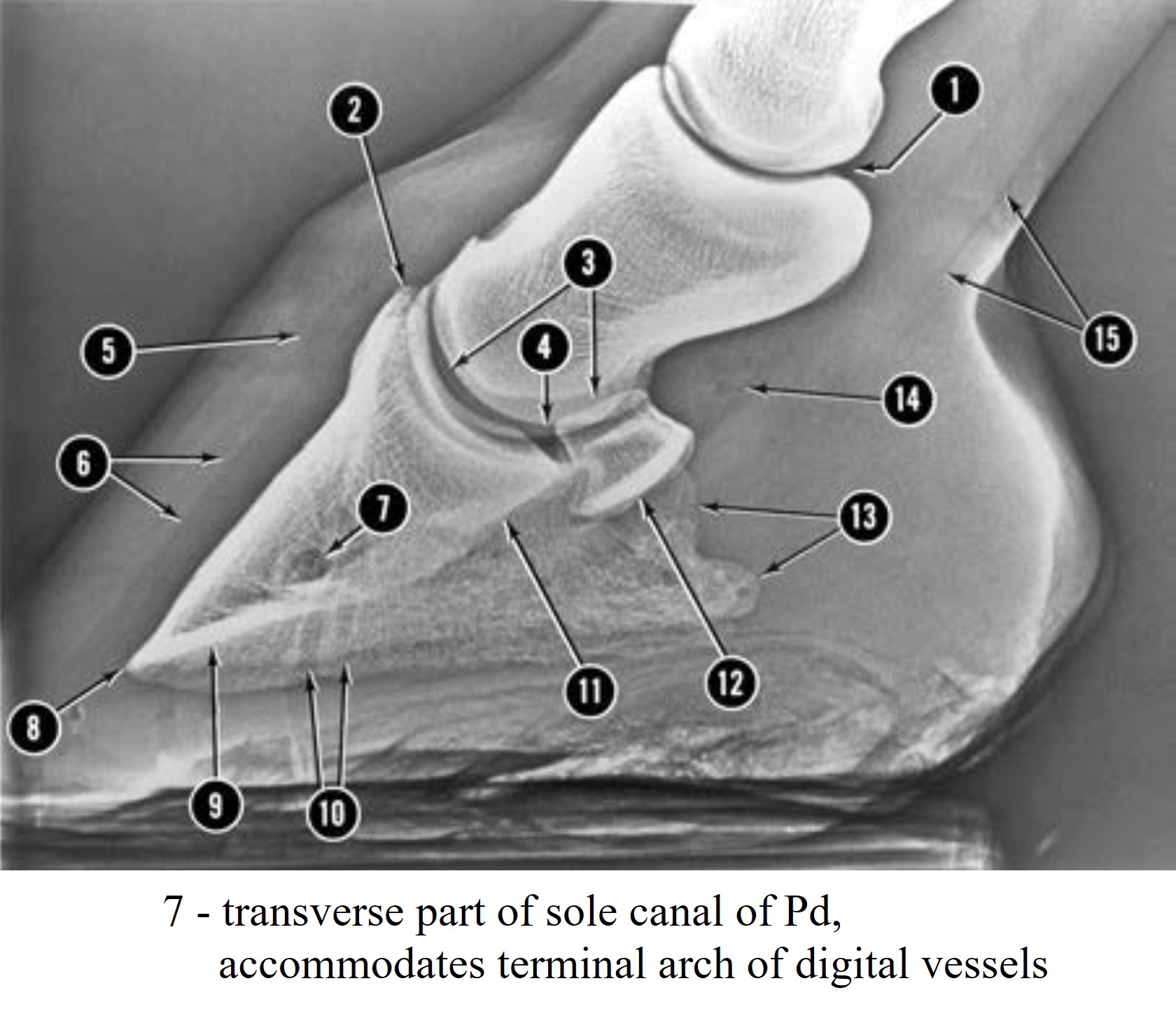
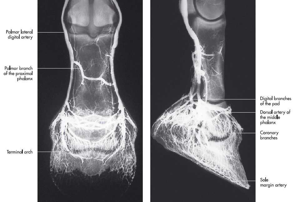
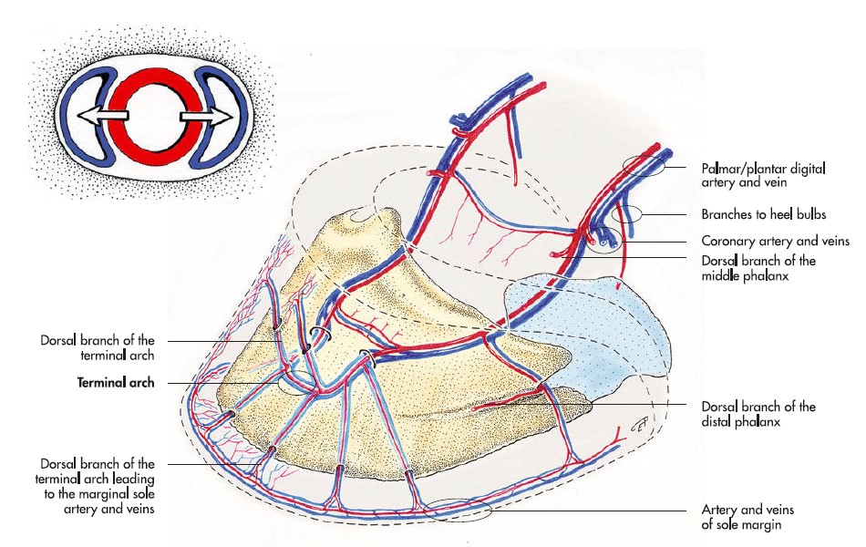
Observe: You need not dissect the M/L (palmar) digital arteries distal to the fetlock. However, you are responsible for knowing that they anastomose in the sole canal of the distal phalanx to form the terminal arch, which is functionally important if one of the arteries becomes occluded. Radiating from the sole canal are many vascular channels, which transmit vessels from the terminal arch to the parietal surface of the distal phalanx, especially to the sole border. The medial and lateral openings into the sole canal, located on the distal phalanx of the horse, are called the sole foramina. You are also responsible for identifying the sole canal and foramina on a distal phalanx.
-
- Arteries of the equine digit and hoof region. 9
-
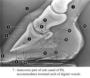
- Lateral radiograph of left horse forefoot. 2
-
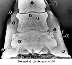
- Dorsopalmar radiograph of left horse forefoot. 2

-
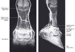
- Arteriogram of the foot of a horse, dorsopalmar (left) and lateromedial (right) projection. 7
-

- Blood vessels of the distal phalanx of the horse. 7
Clinical Application: Digital Pulses in the Horse and Digit Amputation in the Artiodactyls
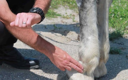
The digital vessels and nerves are palpable in the live horse as they cross the proximal sesamoid bones on their abaxial surface. Here, the digital pulse can be evaluated, and this is also the site of the abaxial sesamoid nerve block. The trio of structures lie closely related in sequence dorsally to palmarly as the vein, artery and nerve i.e. VAN.
-

- Taking a horse’s digital pulse Missy’s Bucket
-
- Cow digit amputation

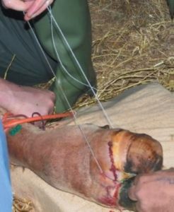
Question: Review the image on the right and ponder the following questions.
1) What common cutting device is being used to amputate the digit (claw)?
2) What ligament of the ruminant foot must be transected to allow removal of one digit, if amputation is through the mid shaft of the proximal phalanx?
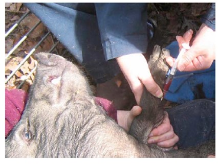
3) Note the green butterfly catheter and orange torniquet – what vessel has likely been catheterized? What drug may have been injected?
Venous Return of the Ungulate Thoracic Limb
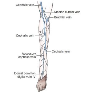
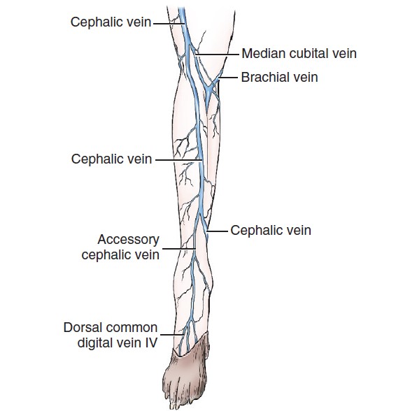
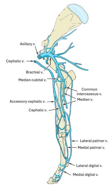
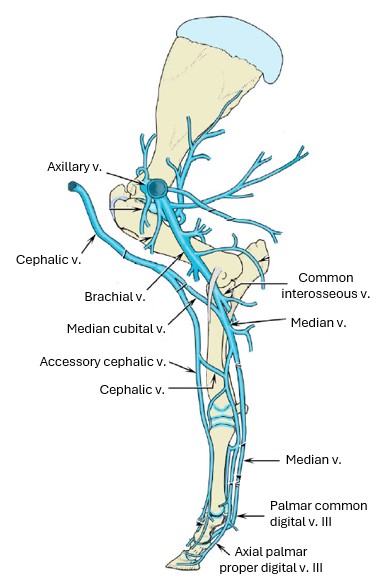
Observe: Using the diagrams below to identify them, observe the cephalic v. and accessory cephalic v. in the horse and goat instructor cadavers.
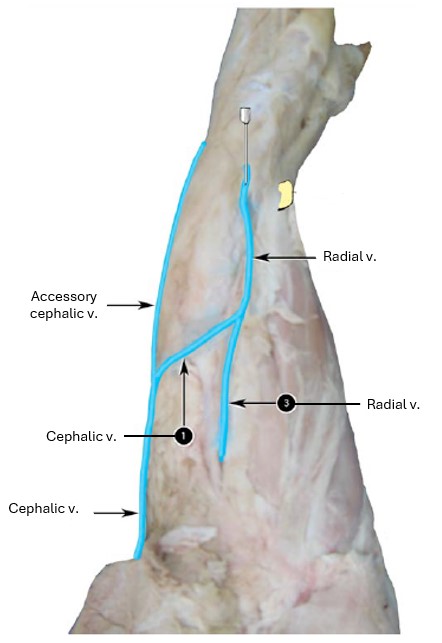
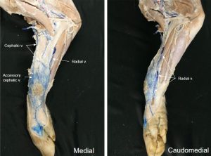
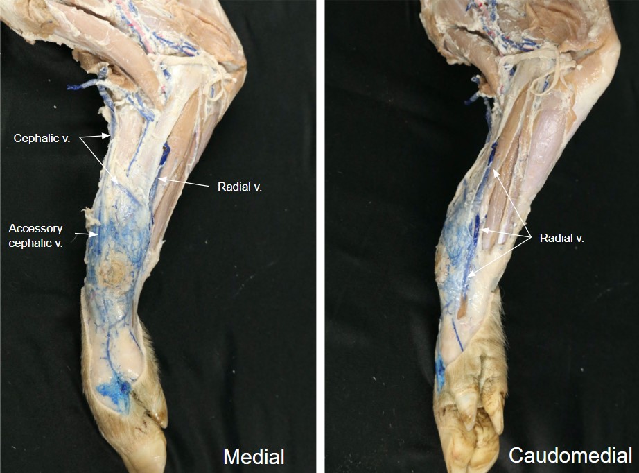
Differentiate the cephalic and accessory cephalic veins from the radial vein in the pig.
If it is still intact, note the passage of the cephalic v. in the horse in the lateral pectoral groove (between brachiocephalicus and superficial pectoral mm- descending part.)
-

- Branches of the cephalic and axillary veins of the horse. 2
-

- Branches of the cephalic and axillary veins of the ox. 2
-

- Veins of the forearm of the pig. 2
Clinical Application: Venipuncture in the Porcine Patient
A blood sample may be obtained from Vietnamese Pot-Bellied Pigs from the radial vein. Note the vein is not unique to VPB.
-
- Pig radial vein
Review videos
Dog thoracic limb vasculature – watch until 11:15 min
Horse thoracic limb vasculature – 2 min
Terms
| Thoracic Limb |
|
| Features | Species |
| Superficial cervical ln | All. Efferent and afferent flow. |
| Axillary ln | Focus on Carnivore. Efferent and afferent flow. |
| Accessory axillary ln | Cat. Efferent and afferent flow. |
| Subclavian artery | Observe in carnivore |
| Superficial cervical a | Observe in carnivore |
| Axillary a/v | All |
| Brachial a/v | All |
| Supracondylar foramen | Cat. ID on bone and know what artery and nerve pass through it. |
| Common interosseous a | Dog and ungulate. Cats generally lack a common interosseous a. |
| Caudal interosseous a | ID in cat, as name changer to median a. |
| Median a | All |
| Radial a | Carnivore (main channel in cat) |
| Palmar common digital a. III | Artiodactyls |
| Axial proper digital aa. III and IV | Artiodactyls |
| M/L palmar aa. | Horse |
| M/L palmar digital aa. | Horse |
| Terminal Arch | Horse (conceptual) |
| Sole canal | Horse distal phalanx, where terminal arch is located |
| M/L sole foramen | Horse distal phalanx. Openings of sole canal. |
| Cephalic v | All |
| Accessory cephalic v | All |
| Median cubital v | All |
| Radial vein | Pig |