Labs 8 and 9: Head and Brain
Learning Objectives
- Identify the main arteries and veins of the head and brain.
- Identify the thyroid glands; describe the parathyroid glands.
- Identify the main lymph nodes of the head, and know from where afferent flow arises and to where efferent flow runs from each node.
Carnivore
Thyroid gland and Parathyroid glands
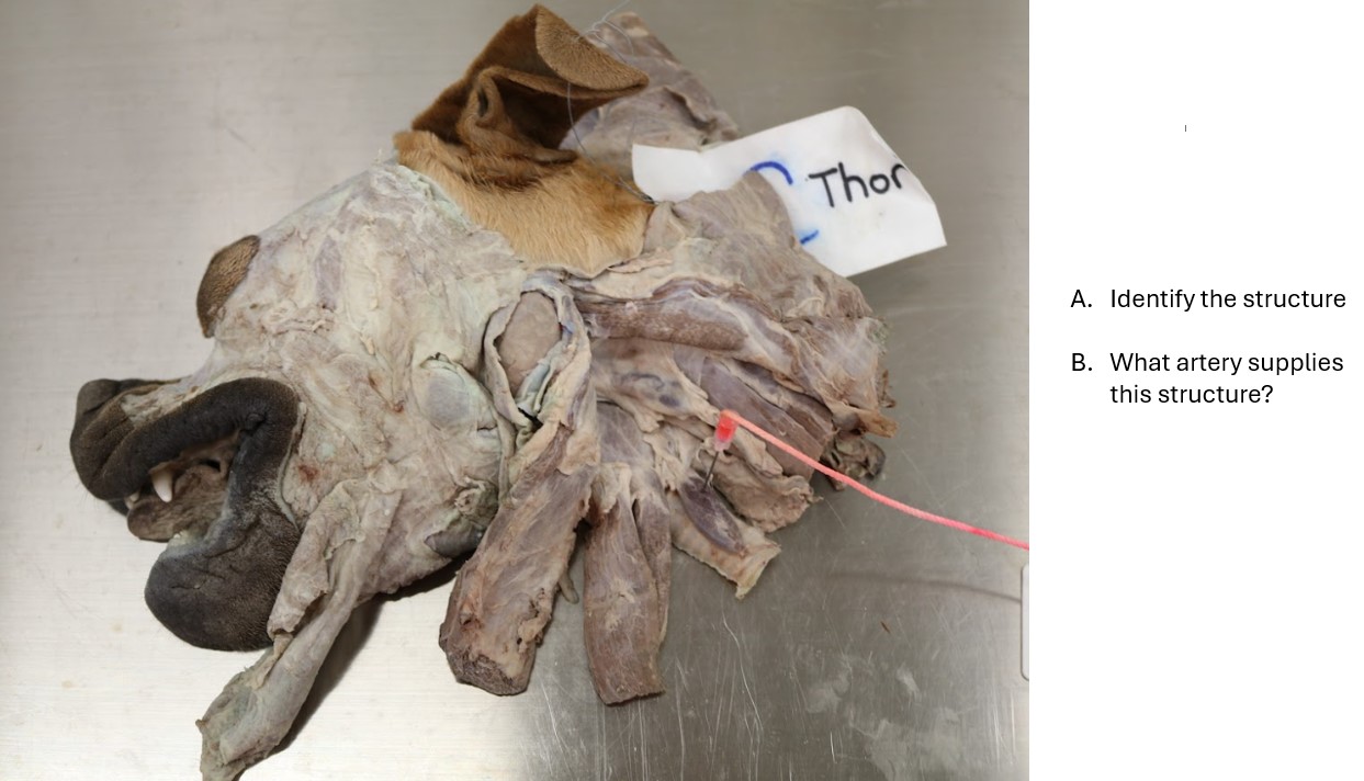
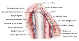
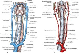
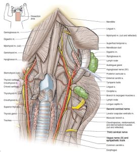
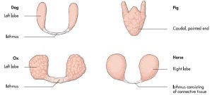
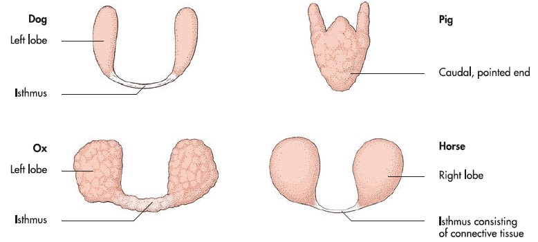
The thyroid gland is dark-colored and usually consists of two separate lobes lying lateral to the first five tracheal rings. Occasionally, a connecting isthmus is present.
There are two parathyroid glands associated with each thyroid lobe. They are small, light-colored, spherical bodies. The external parathyroid most commonly lies in the fascia at the cranial pole of the thyroid lobe. It may be entirely separate from the thyroid tissue or embedded in the cranial pole of the thyroid external to its capsule. The internal parathyroid lies deep to the thyroid capsule on the medial aspect of the lobe. Occasionally, it is embedded in the parenchyma of the thyroid and is difficult to locate. The location of these glands is subject to variation.
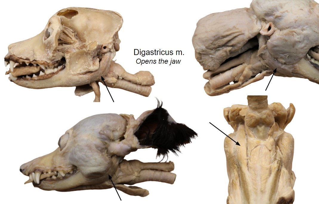
Dissect: Skin both halves of your cadaver’s head. Leave a narrow rim of skin around the margin of the eyelids and at the edge of the lips. Preserve the nose and the philtrum, which is the median groove separating the right and left parts of the nose and superior lips. You need only skin to the base of the ear.
We will use one half of the head to observe the superficial lymph nodes, vasculature, and salivary glands, and the other to perform a deep dissection of the arteries. You may begin the same dissection on both halves of the head if you wish, before choosing one to continue the deep dissection on.
Find the two lobes of the thyroid gland adjacent to the trachea, just caudal to the larynx. Do not spend much time (2-5 minutes maximum) looking for the external parathyroid glands. Do not spend any time looking for the internal parathyroids, but do know that they exist.
Clinical Application: Thyroid and Parathyroid Glands
- Both the thyroid and parathyroid glands are very important clinically. It is not uncommon for cats to suffer from hyperthyroidism and dogs to suffer from hypothyroidism, both of which are generally treated medically. Hyperparathyroidism results in hypercalcemia and is treated by surgically excising the affected, enlarged gland.
-
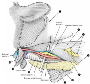
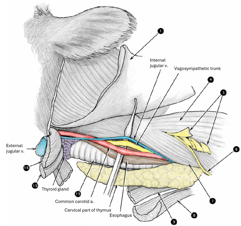
- Retropharyngeal lymph node and thyroid gland, lateral aspect of the neck. 1

-
- Neck of the dog, ventral view. 9
-
- Veins (A) and arteries (B) of the neck of the dog, ventral view. 1

-

- Thyroid gland of the cat, lateral view. 4
-
- Neck of the cat, ventral view. 5
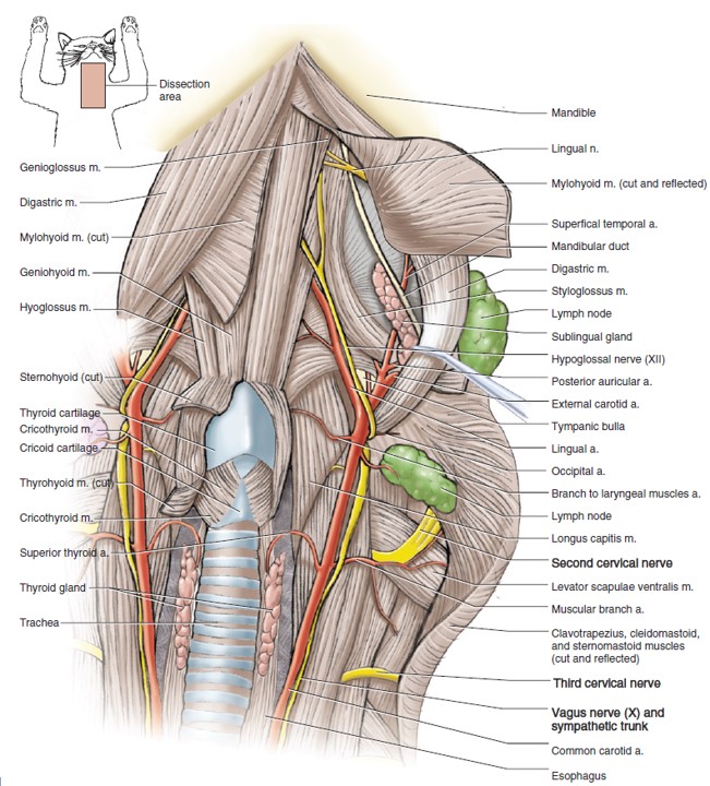
Common carotid artery and its branches; Medial Retropharyngeal Lymph Node
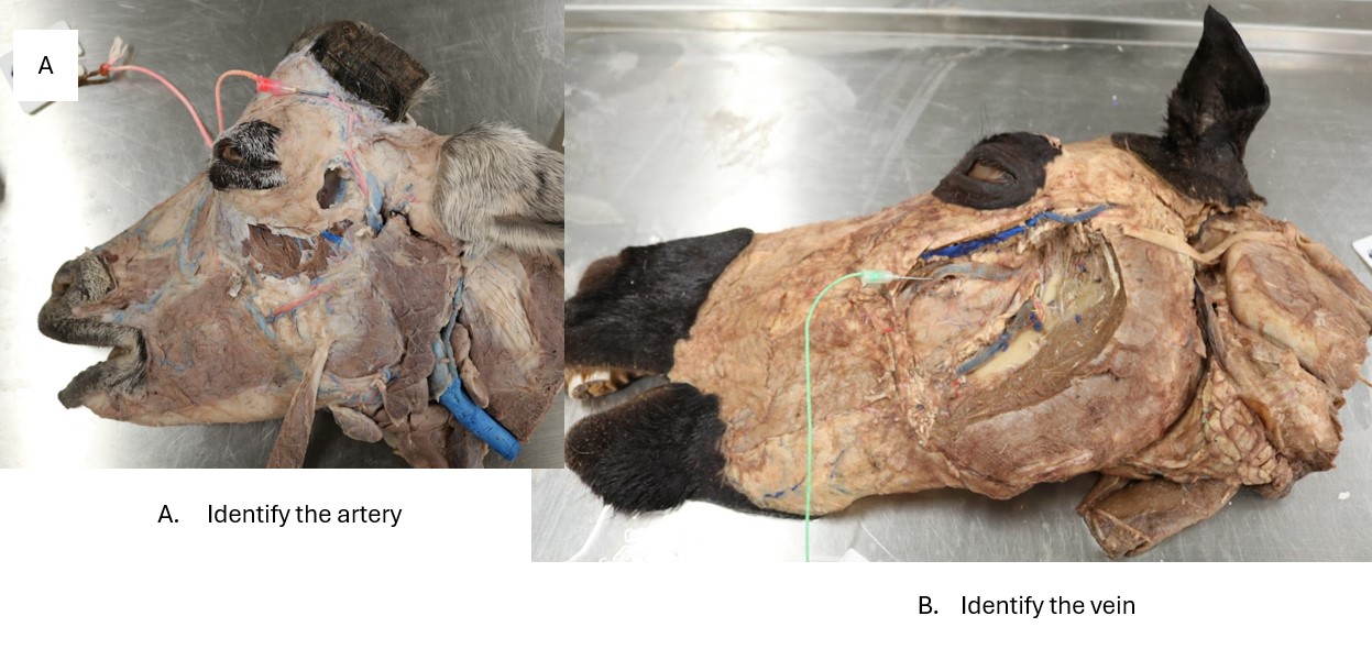
Dissect: Start this dissection on the medial aspect of the head. On the half of the head being used for deep dissection, expose the common carotid artery in the carotid sheath and observe the cranial thyroid a. branching from it. Look for it branching from the common carotid artery and running to the cranial aspect of the thyroid gland (note that at times the thyroid gland will have been removed during decapitation or both thyroid glands may be on one side of the head, if the split was off midline). The cranial thyroid artery is usually still identifiable.
Follow the common carotid artery rostrally by cleaning any fascia surrounding it and adjacent nervous and lymphatic structures.
WHILE FOLLOWING THE COMMON CAROTID ARTERY AND ITS BRANCHES, RECALL THAT CRANIAL NERVES IX-XII and THEIR BRANCHES AND ASSOCIATED STRUCTURES, ARE PROMINENT STRUCTURES IN THIS REGION. THEY WERE DESCRIBED IN DETAIL IN THE CRANIAL NERVE SECTION OF THE NERVOUS SYSTEM UNIT. TAKE THIS OPPORTUNITY TO REVIEW THESE NERVES AS YOU NOW LEARN THE ARTERIES LOCATED IN THIS REGION.
- FYI: The caudal thyroid artery has a variable origin from the major arterial branches in the thoracic inlet. It passes cranially on the trachea supplying the trachea, esophagus, and thyroid gland. It need not be dissected.
- The cranial thyroid artery arises from the ventral surface of the common carotid at the level of the larynx. It supplies the thyroid and parathyroid glands, the pharyngeal muscles, the laryngeal muscles and mucosa, the cervical parts of the trachea and esophagus, and the adjacent portions of the sternocephalicus and the mastoid part of the cleidocephalicus.
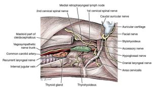
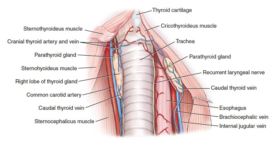
The large medial retropharyngeal lymph node is dorsal to the common carotid artery at the larynx and ventral to the wing of the atlas. Afferent vessels arise from the tongue, the nasal cavity, the pharynx, the salivary glands, the external ear, the larynx, and the esophagus. The tracheal trunk of each side arises from this lymph node.
Dissect: Clean and expose the medial retropharyngeal lymph node.
-
- Retropharyngeal lymph node and thyroid gland, lateral aspect of the neck. 1
-
- Neck of the dog, ventral view. 9
-
- Veins (A) and arteries (B) of the neck of the dog, ventral view. 1
-
- Thyroid gland of the cat, lateral view. 4
Dissect: Incise the rectus capitis ventralis muscle and remove it from the cadaver.
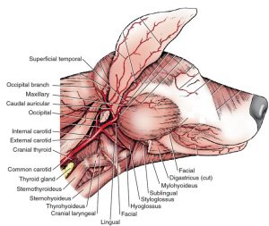
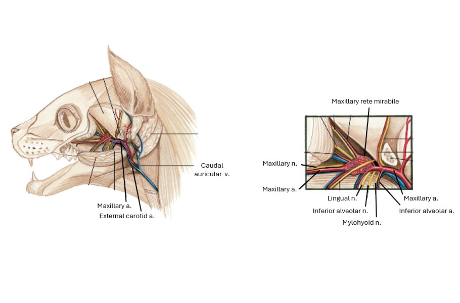
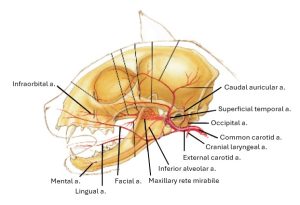
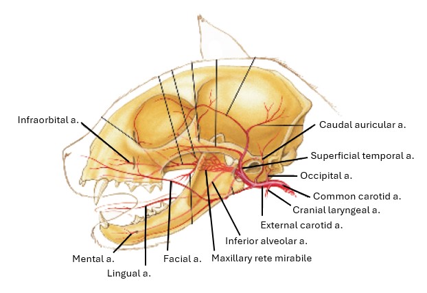
Identify the internal and external carotid arteries, which are the terminal branches of the common carotid artery. Note that in the cat, the internal carotid a. degenerates postnatally. Typically, all that remains of this part of the artery in the adult cat is a thin fibrous cord. The function of this artery to supply the brain is taken over by the arteries of the maxillary rete mirabile, to be identified later in the lab.
Insert photo of medial side of split head to illustrate rectus capitis ventralis.
- The internal carotid artery is closely associated with the occipital artery, the first branch of the external carotid. FYI: A bulbous enlargement at the origin of the internal carotid artery is the carotid sinus, a baroreceptor. (The carotid body, a chemoreceptor, lies at the bifurcation of the carotid arteries.) Beyond this the internal carotid artery ascends across the lateral surface of the pharynx medial to the occipital artery. No branches leave the internal carotid in its extracranial course. It enters the carotid canal deep in the tympano-occipital fissure. Ultimately, it branches to form the cerebral arterial circle that supplies the brain. Its branches to the brain will be observed later in the lab.
-
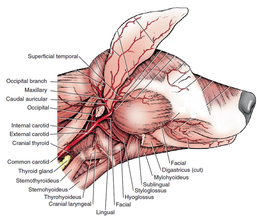
- Branches of the common carotid artery of the dog, lateral aspect. 1
-

- Branches of the common carotid artery in relation to the ventral aspect of the skull of the dog. 1
-

- Deep vasculature of the head of the cat with zygomatic arch removed, lateral view. 4
-
- Schematic illustration of head vasculature of the cat, lateral view. 4
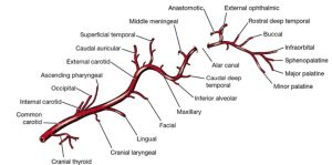
Dissect: The common carotid artery splits into the aforementioned internal carotid artery and the external carotid artery. Bluntly dissect connective tissue in order to follow the external carotid artery cranially . Dissect the following bolded branches thereof.
4. The occipital artery leaves the external carotid adjacent to the internal carotid and passes dorsally to supply the muscles on the caudal aspect of the skull and the meninges.
5. FYI: The cranial laryngeal artery is a ventral branch that supplies the adjacent sternomastoideus and pharyngeal muscles. It enters the larynx between the thyrohyoid bone and the thyroid cartilage to supply the mucosa and laryngeal muscles.
6. The lingual artery leaves the ventral surface of the external carotid and passes rostrally to supply the tonsil and tongue. On its course to the tongue it is accompanied by the hypoglossal nerve.
7. The facial artery leaves the external carotid beyond the lingual, medial to the digastricus. FYI: A branch, the sublingual artery, continues medial to the digastric muscle and is accompanied by the mylohyoid nerve. It runs rostrally into the tongue. The facial artery courses rostrolaterally between the digastric and masseter muscles to reach the cheek lateral to the mandible, where it supplies the lips and nose. Be sure to identify it on the superficially dissected head.
Dissect: Now move to the lateral side of the head. Reflect the caudal limb of the parotid gland to identify the caudal auricular artery. Continue to follow the external carotid artery rostrally. Transect the digastricus muscle and remove the caudal portion as needed to visualize the bolded vessels described below.
8. The caudal auricular artery usually arises from the external carotid at the base of the ear and courses dorsally under the caudal auricular muscles. Lateral, intermediate, and medial auricular branches course distally on the convex caudal surface of the ear. Occasionally, this artery arises closer to the origin of the external carotid.
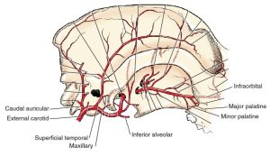
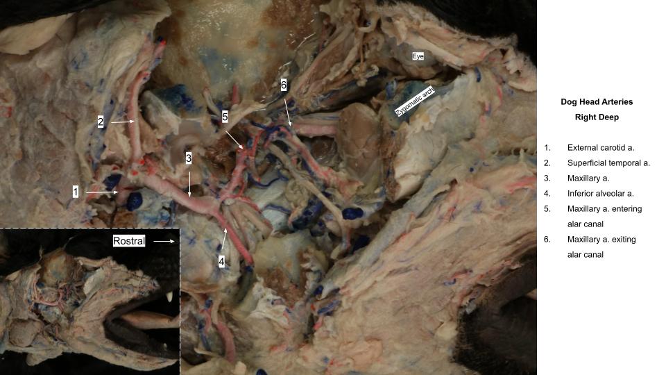
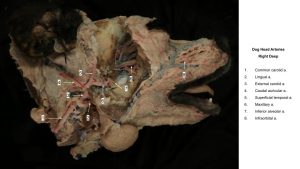
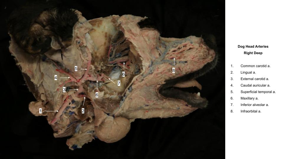
At the caudal border of the mandible, rostroventral to the annular cartilage of the ear, the external carotid artery terminates by dividing into the superficial temporal and maxillary arteries.
9. The superficial temporal artery arises rostral to the base of the auricular cartilage at the caudodorsal border of the mandible and courses dorsally. It supplies the parotid gland, the masseter and temporal muscles, the rostral auricular muscles, and the eyelids.
-
- Branches of the common carotid artery of the dog, lateral aspect. 1
-
- Branches of right common carotid artery of the dog, deep view. 1

-
- Arteries of the head in relation to lateral aspect of the skull of the dog. 1

-
- Branches of the common carotid artery in relation to the ventral aspect of the skull of the dog. 1
-
- Deep vasculature of the head of the cat with zygomatic arch removed, lateral view. 4
-
- Schematic illustration of head vasculature of the cat, lateral view. 4
Maxillary Artery and Branches
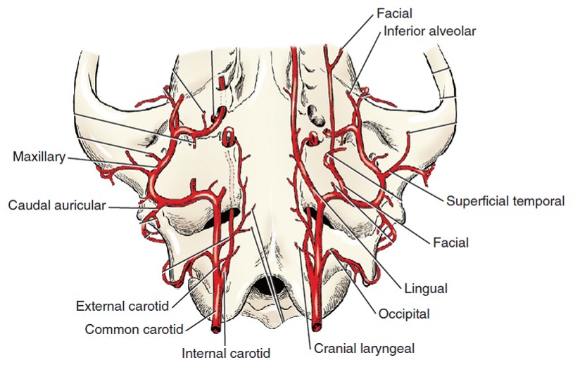
The maxillary artery is the larger terminal branch of the external carotid artery. It is deeply placed and is closely associated with a number of cranial nerves. From its origin with the superficial temporal, it passes rostromedially ventral to the temporomandibular joint medial to the retroarticular process in its course to the alar canal. It enters the the alar canal via the caudal alar foramen and exits via the cranial alar foramen along with the maxillary nerve (branch of CNV), which exits the skull into the alar canal via the round foramen.
NOTE that the cat does not possess an alar canal!
Dissect: Like the dissection of the nerves, there is a large volume of muscle to be resected in this dissection. Review prosections to observe how deeply the dissection occurs before beginning.
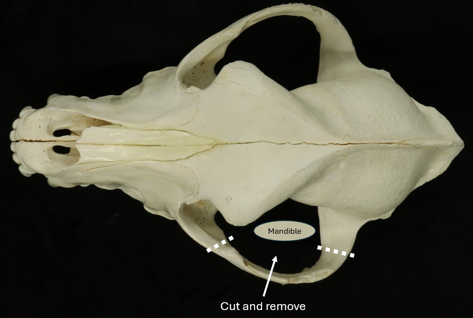
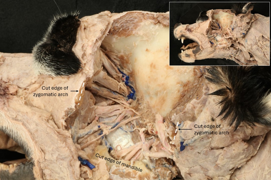
On the lateral aspect of this head half, cut all of the attachments of the temporal and masseter muscles to the zygomatic arch. Cut through the bony arch rostrally and caudally and remove all of the arch rostral to the temporomandibular joint.
Include diagram and/or cadaver illustrating where to incise muscles and to apply bone cutters.
Remove the temporalis m. from its wide origin by sharply excising the bulk of it with a scalpel. Once closer to the surface of the skull, muscle can be scraped off the bone with the blunt end of thumb forceps or a scalpel handle (with no blade loaded!). Be vigorous; there is a large amount of muscle to remove.
Outline temporalis in a diagram or photograph and outline what needs to be resected.
Transect the masseter m. as close to its attachment on the zygomatic arch as possible and use this incision as the starting point to incise completely around the muscle as depicted below. Remove the entire masseter m. all the way medially to the coronoid process.
Diagram of where to cut around masseter.
With bone cutters, remove most of the zygomatic arch.
Diagram or photograph showing where to cut and respect zygomatic arch.
Now, transect any muscle attachments still remaining on the dorsal aspect of the dorsolateral aspect of the coronoid process. Starting on the exposed dorsal aspect of the coronoid process, use bone cutters to remove approximately half of the coronoid process.
Diagram as to how far ventrally to cut process.
Deep to the coronoid process there will be more muscle to remove. As you do so, expose the underlying pterygoid muscles. Note that there are a number of important arteries and nerves (the maxillary artery/nerve are the most obvious of these structures) located just lateral (superficial) to the pterygoid muscles that will need to be identified, so proceed with caution. Take a moment to have a look at a prosected deep dissection to guide you through this process.
Complete the disarticulation of the temporomandibular joint and reflect the ramus of the mandible laterally, ultimately removing the entire ramus. You may find that there is still some temporalis muscle tissue that needs to be removed in order to readily identify the deeper structures.
See image below to get a sense of the view that you should be able to see in your cadaver. Arteries and nerves passing over the shiny white fascia of the pterygoid muscles indicates that you are deep enough!
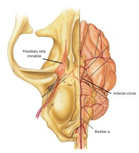
In the cat, you will notice what looks to be a red splotch of small arteries. This is the maxillary rete mirabile, and replaces the maxillary artery in this location in the cat! Behind the eye orbit you should find a dull, white, fibrous tissue. This is the periorbita and we’ll leave it intact for now.
Visualize the rostral continuation of the maxillary artery. Reflect the ramus of the mandible laterally and identify the following branches of the maxillary artery.
The first three arise before the maxillary artery enters the alar canal.
-
- The inferior alveolar artery enters the mandibular foramen with the inferior alveolar nerve and courses through the mandibular canal. It supplies branches to the roots of the teeth in the lower jaw. Mental branches supply the skin.
2. FYI: The caudal deep temporal artery arises near the inferior alveolar artery and enters the temporal muscle. Only the origin of this artery may be seen.
3. FYI: The middle meningeal artery passes through the oval foramen and courses dorsally in a groove on the inside of the calvaria.
FYI: The external ophthalmic artery arises from the maxillary on its emergence from the alar canal and penetrates the apex of the periorbita adjacent to the orbital fissure. The external ophthalmic artery gives rise to the vessels that supply the structures within the periorbita. You also may observe the buccal artery as it passes cranioventrally adjacent to the buccal nerve.
-
- Arteries of the head in relation to lateral aspect of the skull of the dog. 1
-
- Branches of right common carotid artery of the dog, deep view. 1
-
- Superficial temporal a.
-
- Inferior alveolar a.

Dissect: In the dog, identify the maxillary artery adjacent to the maxillary nerve as they exit the alar canal. Note that the cat possesses neither an internal carotid artery nor an alar canal. In the cat, interposed between the maxillary a and the cerebral arterial circle is a network of small, interconnected aa known as the maxillary rete mirabile. Efferent branches from this rete pass through the orbital fissure to anastomose with the basilar a. and form the arterial circle that supplies much of the brain. In the cat, observe the maxillary rete mirabile and the continuation of the maxillary artery distal to it.
-
- Deep vasculature of the head of the cat with zygomatic arch removed, lateral view. 4
-
- Schematic illustration of head vasculature of the cat, lateral view. 4
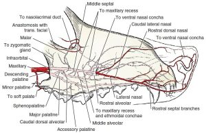
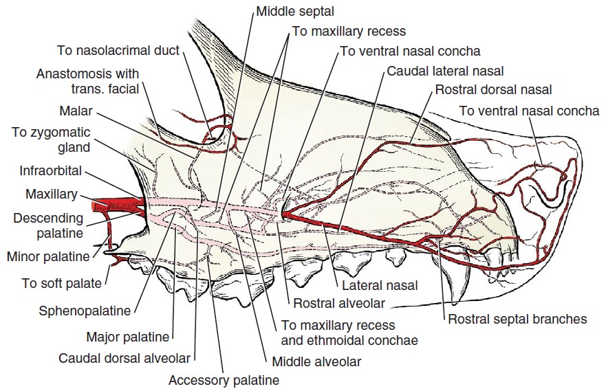
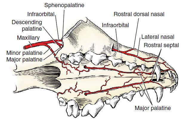
You need not follow the maxillary artery any farther rostrally from this location. However, you are responsible for knowing the main channel arterial supply to the maxillary aspect of the mouth, which is Maxillary; descending palatine; major palatine arteries. The infraorbital and major palatine arteries will be visualized from other locations.
FYI: In both the dog and cat, you may observe the minor palatine artery as it branches from the maxillary. The minor palatine artery passes ventrally, caudal to the hard palate, and is distributed to the adjacent soft and hard palates. Just caudal to the maxillary foramen, the maxillary artery bifurcates into the descending palatine and infraorbital arteries, the former of which becomes the main channel artery to the rostral head.
The infraorbital artery supplies the malar artery to the eyelids and dental branches to the caudal cheek teeth. It then enters the maxillary foramen and passes through the infraorbital canal. Within the canal, dental branches arise. These supply the premolars, the canine teeth, and the incisor teeth. The infraorbital artery emerges from the infraorbital foramen and terminates as the lateral and rostral dorsal nasal arteries, which supply the nose and the superior lip.
-
- Arteries of the head in relation to lateral aspect of the skull of the dog. 1
-
- Branches of right common carotid artery of the dog, deep view. 1
-
- Schematic illustration of head vasculature of the cat, lateral view. 4
-
- Infraorbital a.
Optional Dissection. Concepts are need to know: Reflect the zygomatic salivary gland to find where the maxillary artery splits into the infraorbital and the descending palatine aa. Note again that the descending palatine artery is the main channel artery. Observe the infraorbital artery as it enters the maxillary foramen and courses through the infraorbital canal.
Now, move back to where the maxillary artery split into the descending palatine and the infraorbital artery.
FYI: The sphenopalatine artery branches from the descending palatine and passes through the sphenopalatine foramen to the interior of the nasal cavity. It need not be dissected.
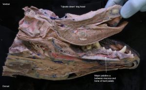
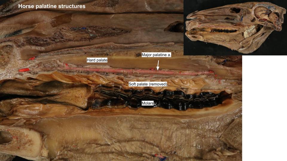
The major palatine artery, the continuation of the descending palatine beyond the sphenopalatine, represents the main channel artery to the hard palate. It enters the caudal palatine foramen and passes through the major palatine canal to supply the hard palate.
Dissect (not optional): Incise the hard palate parallel to the left maxillary arcade and reflect it to visualize the major palatine artery.
Palpate the infraorbital foramen through the gingival surface dorsal to the maxillary teeth and incise the superficial muscles of the face to observe the infraorbital artery and its branches as it exits the infraorbital canal through the infraorbital foramen.
Show photograph of where to palpate and of infraorbital a. On rostrally aspect of the face.
-
- Scheme of the terminal branches of the maxillary artery of the dog, lateral aspect. 1
-

- Terminal branches of the maxillary artery of the dog. 1
-
- Major palatine a.
Clinical Application: Major Palatine Artery
- It is very important to use care when using dental elevators during maxillary dental extractions. If the instrument slips, it is easy to incise the major palatine artery, leading to hemorrhage which can be difficult to control.
Vasculature of the Brain
Observe: Examine the arteries of the brain on a latex-injected wet brain specimen.
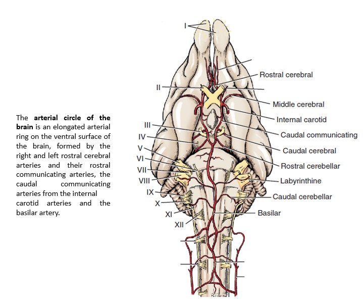
The arteries to the cerebrum and cerebellum are branches from the vessels on the ventral surface of the brain. These major arteries are not accompanied by veins. The basilar artery is formed by the terminal branches of the vertebral arteries, which enter the floor of the vertebral canal through the lateral vertebral foramina of the atlas. It is continuous caudally with the ventral spinal artery of the spinal cord. The basilar artery courses along the midline of the ventral surface of the medulla oblongata and pons and then divides into two branches that form the caudal portion of the arterial circle of the brain (eponym: “circle of Willis”).
The internal carotid arteries are the other main source of blood to the arterial circle of the brain (not in the cat or the ruminant). Remember that in the cat, the other main source of blood to the arterial circle is the maxillary rete mirabile. Each internal carotid artery divides into a middle cerebral, rostral cerebral, and caudal communicating artery. The small caudal communicating arteries from each internal carotid course caudally and join the terminal branches of the basilar artery. Rostrally, the two rostral cerebral arteries anastomose, completing the arterial circle on the ventral surface of the brain.
The cerebral arterial circle surrounds the pituitary gland, which receives small branches from the circle as well as directly from the internal carotid artery.
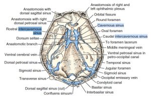
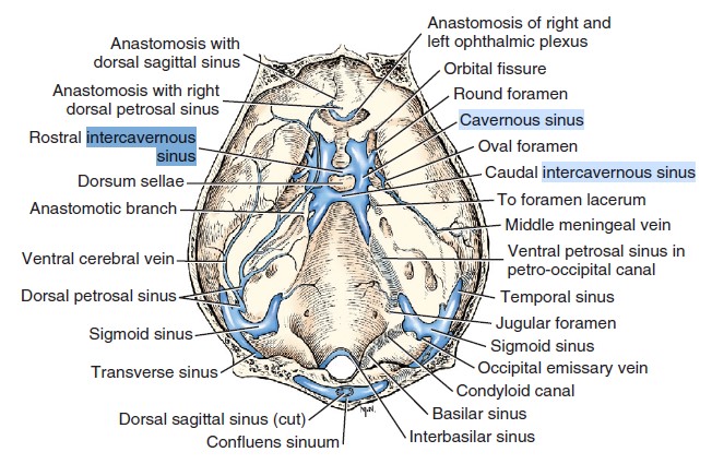
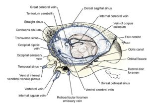
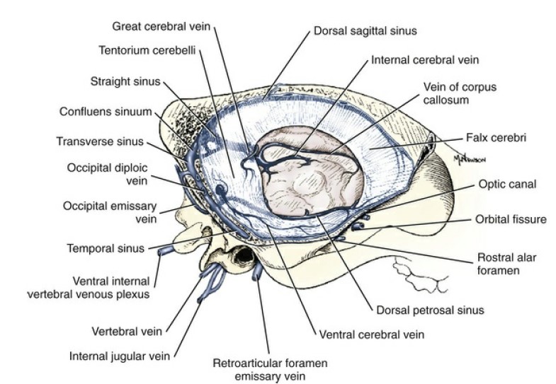
Venous sinuses are large veins in the brain that drain blood from the brain to the heart. They are located between the layers of the dura mater, the brain’s outermost covering. On one half of the head, a fold of dura will be found extending ventrally from the midline in the longitudinal cerebral fissure between the two cerebral hemispheres. This is the falx cerebri, which contains the dorsal sagittal sinus dorsally. The paired cavernous sinus lie on the respective sides of the floor of the middle cranial fossa. The two cavernous sinuses are connected medially by means of the large but short rostral and caudal intercavernous sinuses.
Observe the dorsal sagittal sinus on the medial aspect of the cadaver heads. Observe the model of the cranial venous sinuses, as photographed below.
Include cut cadaver head pointing to dorsal sagittal sinus. Include photo of cavernous sinus model.
-

- Arterial circle of brain of the dog, ventral view. 1
-
- Arterial circle of brain of the cat, ventral view. 4

-
- Cranial venous sinuses of the dog, dorsal aspect; calvaria removed. 1
-
- Cranial venous sinuses, lateral aspect. 1
Superficial Veins of the Carnivore Head and Mandibular Lymph Nodes
If students are using new heads again, consider moving these sections up to the beginning of the lab as description for the “superficial head dissection.”
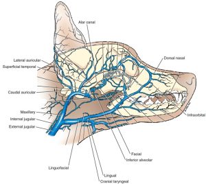
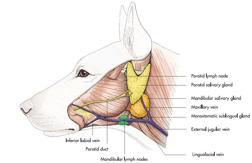
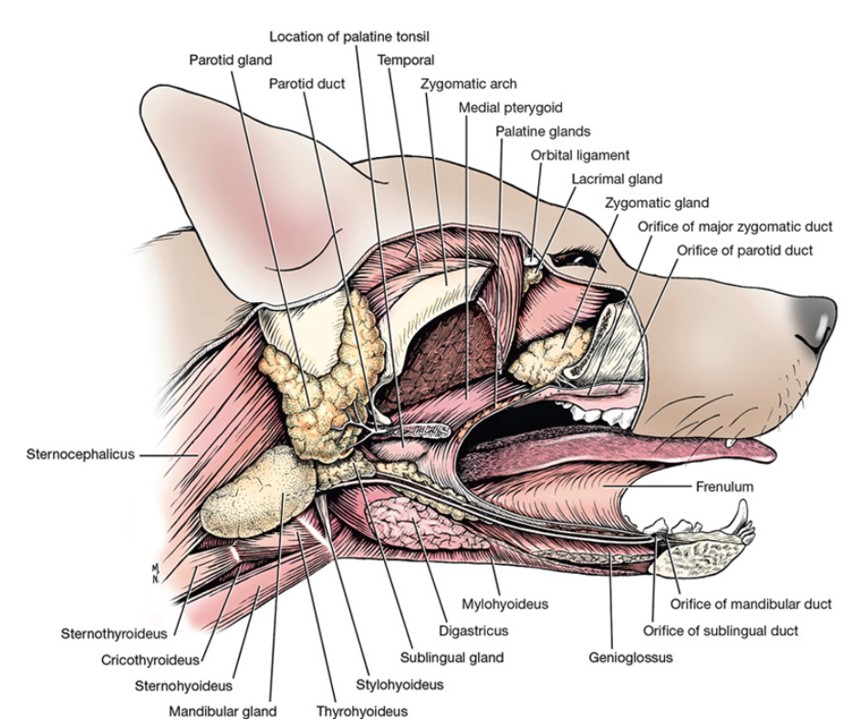
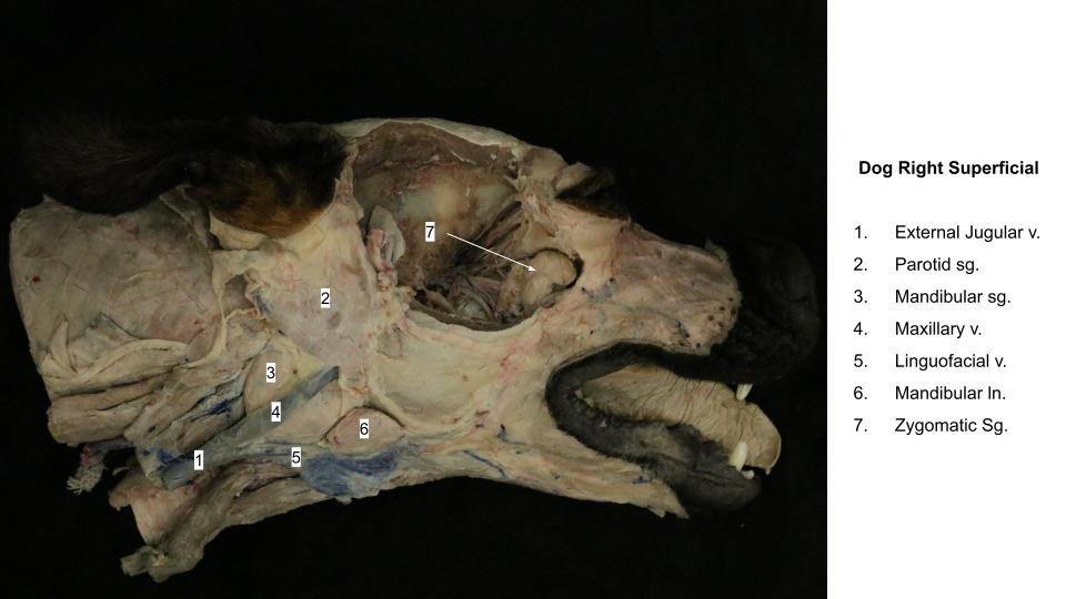
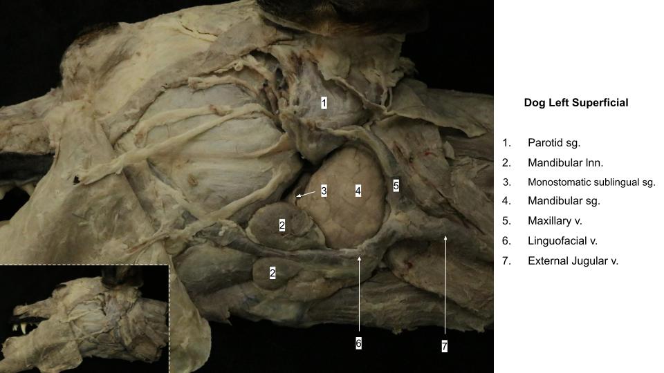
The external jugular vein is formed by the confluence of the linguofacial and maxillary veins caudal to the mandibular salivary gland, which lies between these two veins.
The maxillary vein drains the ear, orbit, palate, nasal cavity, cheek, and mandible as well as the cranial cavity.
The lingual vein is the first large tributary that enters the linguofacial vein ventrally. Its branches drain blood from the tongue, the larynx, and part of the pharynx. These branches will not be dissected.
The facial vein is the other tributary of the linguofacial. The branches that form the facial vein lie on the dorsal surface of the muzzle. FYI: One of these, the dorsal nasal, runs caudally from the nares, whereas another, the angularis oculi, passes rostrally from the medial aspect of the orbit, where it is continuous with the ophthalmic plexus within the periorbita. Blood may drain from the face in either direction through the angularis oculi. There is also a large communication between the ophthalmic plexus and the facial vein, via the deep facial vein, which is ventral to the zygomatic bone.
Dissect: Use the superficial head dissection to isolate and observe the aforementioned bolded veins: external jugular, linguofacial, lingual, facial, maxillary vv.
-
- Veins of the head of the dog. 1

-

- Cat and dog veins

Lymph Nodes of the Carnivore Head
Recall that the large medial retropharyngeal lymph node is dorsal to the common carotid artery at the larynx and ventral to the wing of the atlas. It should have been located during the study of the common carotid artery and its branches. Afferent vessels arise from the tongue, the nasal cavity, the pharynx, the salivary glands, the external ear, the larynx, and the esophagus. The tracheal trunk of each side arises from this lymph node.
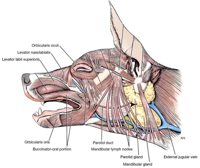
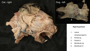
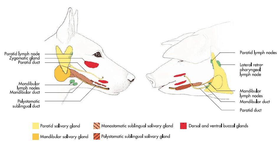
The two or three small mandibular lymph nodes are located along the linguofacial vein just rostroventral to the mandibular salivary gland. The salivary gland will be studied in the alimentary unit. The afferent lymph vessels to the mandibular lymph nodes come from all parts of the head not drained by the afferent lymph vessels of the parotid lymph node. The efferent lymph vessels of the mandibular lymph nodes go primarily to the ipsilateral medial retropharyngeal lymph node.
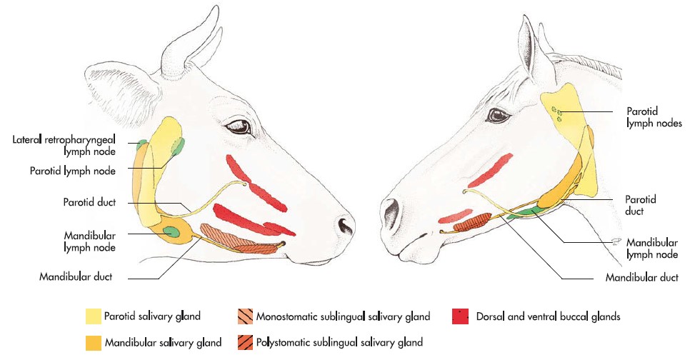
The parotid salivary gland lies between the mandibular gland and the ear and will be studied with the alimentary system. It is closely applied to the base of the auricular cartilage of the ear. A small parotid lymph node may be found along the rostral border of this gland.
The afferent lymph vessels to the parotid lymph node come from the cutaneous area of the caudal half of the dorsum of the muzzle and the side of the cranium, including the eyelids and associated glands, the external ear, the temporomandibular joint, and the parotid salivary gland. The parotid lymph center also drains the temporal, masseter, and zygomatic muscles; the muscles of the ear; the lacrimal apparatus; the nasal, frontal, parietal, zygomatic, and temporal bones; and the mandible. Its two or three efferent lymph vessels run between the digastric muscle and the parotid salivary gland to the large medial retropharyngeal lymph node. This flow can also drain into the lateral retropharyngeal lymph node, if the latter is present.
The lateral retropharyngeal lymph nodes are rarely missing in the cat. They are located just caudal to the parotid salivary gland, at the ventral border of the wing of the atlas, against the dorsal border of the mandibular gland, under the base of the ear.
FYI: Lateral retropharyngeal lymph nodes are present in approximately 30% of dogs. This lymph node is less than 10 mm in diameter and lies at the dorsal border of the horizontal part of the cartilaginous external acoustic meatus. It is completely or partially covered by the caudal part of the parotid salivary gland. The afferent lymph vessels of the lateral retropharyngeal nodes come from the structures lying adjacent to them. Their efferent vessels drain into the medial retropharyngeal lymph node.
Observe: Review the location of the medial retropharyngeal lymph nodes and identify the mandibular and parotid lymph nodes. In the cat, identify the lateral retropharyngeal lymph node. You may also observe a lateral retropharyngeal lymph node in the canine if the specimen you are dissecting happens to be one of the approximately 30% of individuals possessing them.
-

- Salivary glands and lymph nodes of the dog. 7
-

- Superficial muscles of the head of the dog, lateral aspect. 1
-

- Salivary glands of the dog: parotid, mandibular, sublingual, and zygomatic. Right mandible removed. 1
-

- Dog salivary glands
-

- Dog salivary glands
-
- Cat salivary glands

Clinical Application: Palpation of the mandibular lymph nodes
Palpation of the mandibular lymph nodes should be part of every physical examination. Lymphadenopathy (enlargement) of these nodes can be indicative of active viral or bacterial infection, inflammation, and/or neoplasia. The mandibular salivary glands, which are located just caudal to the lymph nodes of the same name are easily confused with the lymph nodes, which are relatively small compared to the salivary glands. To distinguish between the structures, find the easily palpated salivary glands first, and then slide your fingers cranially to locate the lymph nodes.
Vasculature and Lymph Nodes of the Ungulate Head
Superficial Vasculature of the Equine and Ruminant Head
NOTE: For this section, we will be focusing on the equine and the small ruminant as the specimens which have latex in their vessels. You need not identify the vasculature in the bovine or porcine. However, you are responsible for the lymphatic, thyroid and parathyroid anatomy on the all of the ungulate species.
When available, observe this section with one equine specimen and one goat specimen on which a superficial dissection has been performed.
Equine
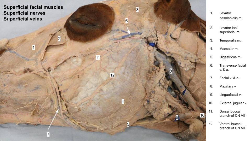
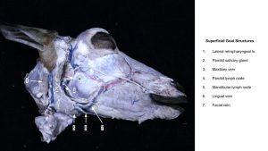
Observe: On the provided equine specimens on which a superficial dissection has been performed, identify the transverse facial a. and v. lying immediately ventral to the facial crest and zygomatic arch.
If possible, follow the transverse facial a. caudally, deep to the parotid ln. and sg. to its origin off the superficial temporal a. If the specimen you are using does not have the origin of the transverse facial artery readily accessible, perform this dissection now and/or make sure to observe this branching on a head on which the deep dissection has been performed!
At the ventral mandible on its medial aspect, identify the facial a. and v., passing with the parotid sg. duct and then crossing over the ventral cortex of the mandible and traveling dorsolaterally along the rostral margin of the masseter m. (note deep venous sinuses in horse face will be dissected on the deep dissection side).
-

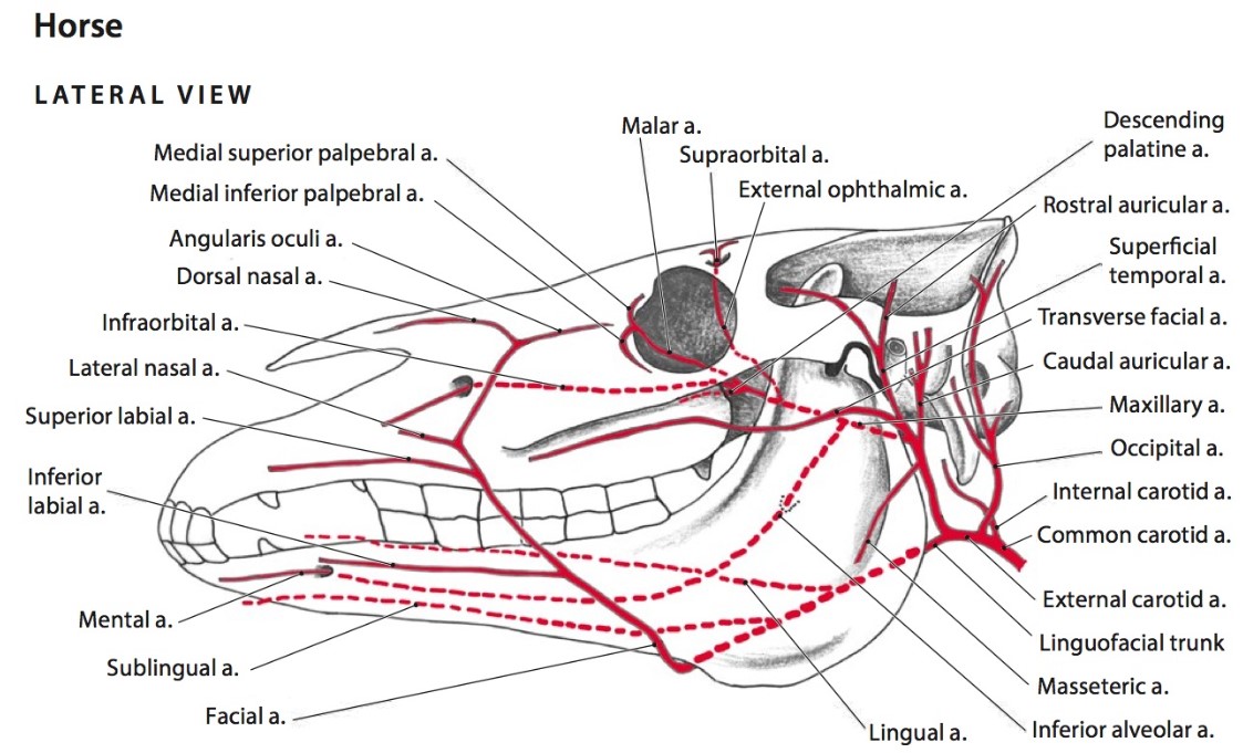
- Arteries of the head of the horse, lateral view. Dashed lines indicate vessels that are either deep to bone or inside bone. 6
-

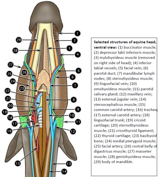
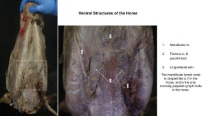
- Structures of the equine head, ventral view. 2
-

- Horse vasculature
-
- Horse superficial arteries
Clinical Application: Transverse facial and facial arteries
Arterial pulses can be palpated in the transverse facial a. and the facial a. Both of these vessels are also commonly catheterized for arterial blood pressure monitoring and collection of arterial blood for blood gas measurements during general anesthesia of the horse. The transverse facial v. is commonly used to obtain a small venous sample for blood gas analysis in the standing equine patient.
Ruminant
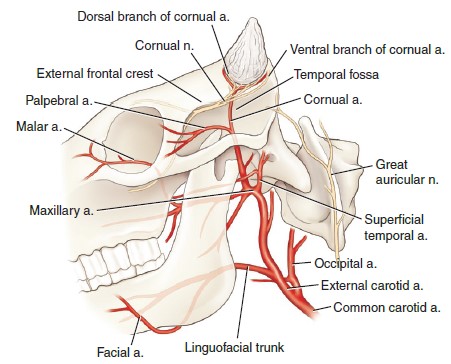
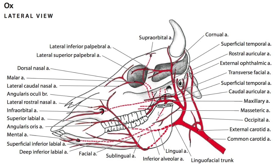
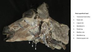
On a goat specimen on which a superficial dissection has been performed, trace the transverse facial vessels caudally to the parotid ln. Continue tracing the artery deep to the lymph node to identify the superficial temporal a. Follow the superficial temporal a. dorsally across the zygomatic arch and identify the cornual a. or arteries (often several vessels are present going to the horn base), which is/are given off toward the horn as the superficial temporal a. turns rostrally toward the eye. Follow the cornual artery(ies) for a few millimeters, tracing these vessels to the base of the horn if practical.
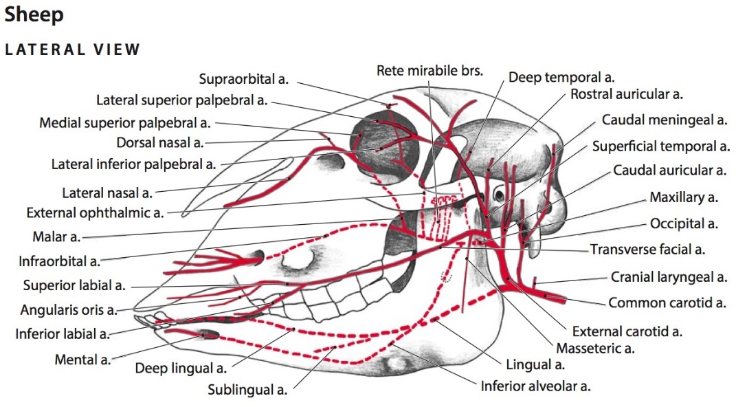
Note that the small ruminant, but not the bovine, is lacking a facial artery! Therefore, the transverse facial a. in these species is a significant blood supply to the rostral face. Later in the study of the vasculature of the ungulate head, note that the equine and bovine both possess a linguofacial trunk. The analogous branch from the external carotid artery is simply called the lingual artery in the small ruminant.
Confusingly, the small ruminant DOES possess a facial vein, which will be observed later in the unit with the other veins.
Clinical Applications: Superficial Arteries of the Ruminant Head
- The cornual a. branches can bleed extensively during dehorning of a mature animal. Do you recall the innervation to the horn?
- The pulse may be palpated on the facial (in bovine) or transverse facial a.
-
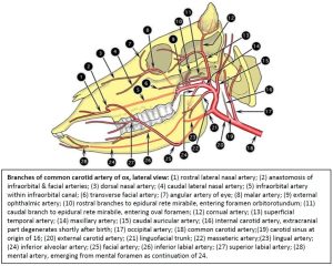
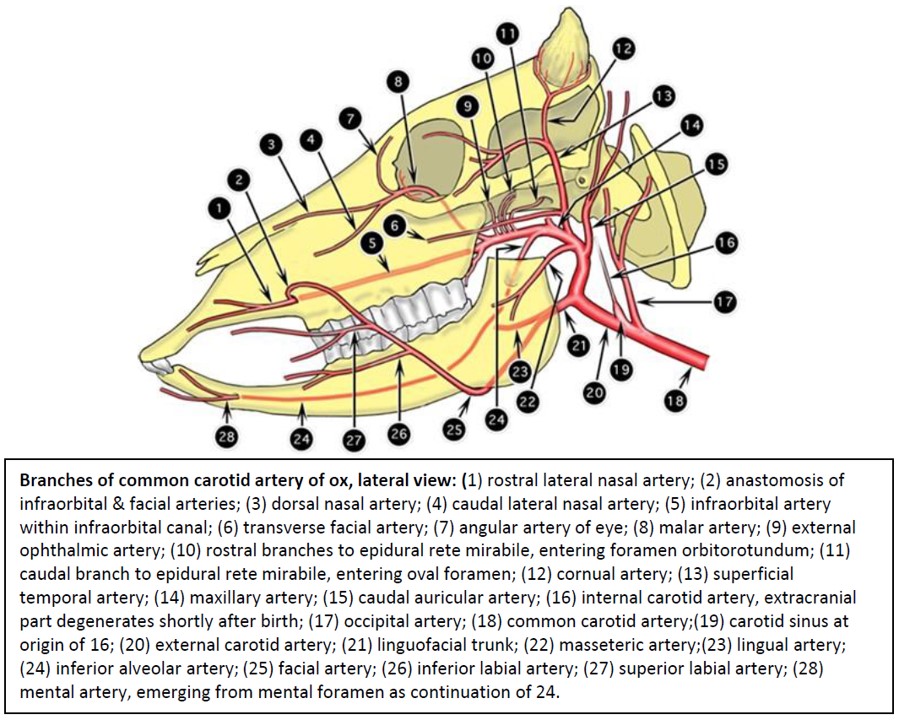
- Branches of common carotid artery of ox, lateral view. 2
-

- Arteries of the head of the ox, lateral view. Dashed lines indicate vessels that are either deep to bone or inside bone. 6
-
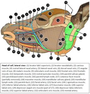
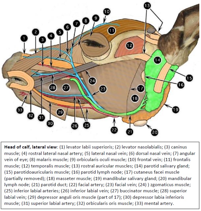
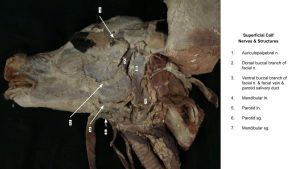
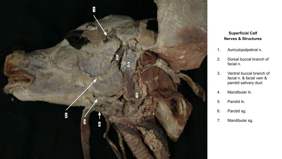
- Head of the calf. 2
-
- Arteries of the head of the sheep (small ruminant), lateral view. Dashed lines indicate vessels that are either deep to bone or inside bone. 6
-
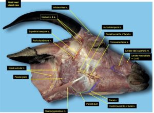
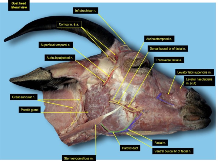
- Goat head, lateral view. 12
Superficial Veins of the Ungulate Species
Observe: Identify the facial v., linguofacial v., maxillary v., and external jugular v. on the superficial surface of both the horse and goat. Note that although the small ruminant lacks a facial artery, their venous anatomy is the same as in the other species.
-
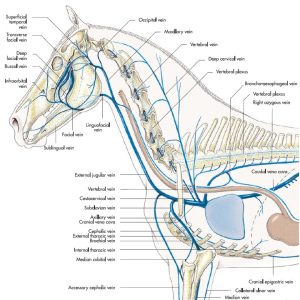
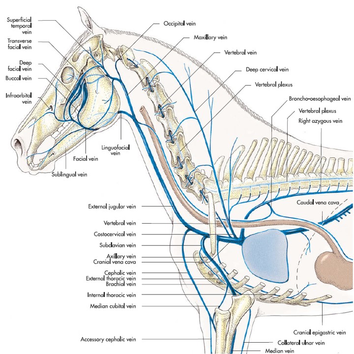
- Larger veins of the head, neck and the thoracic limbs and the tributaries to the cranial vena cava of the horse. 7
-
- Head of the calf. 2
-

- Horse superficial structures
-
- Goat veins

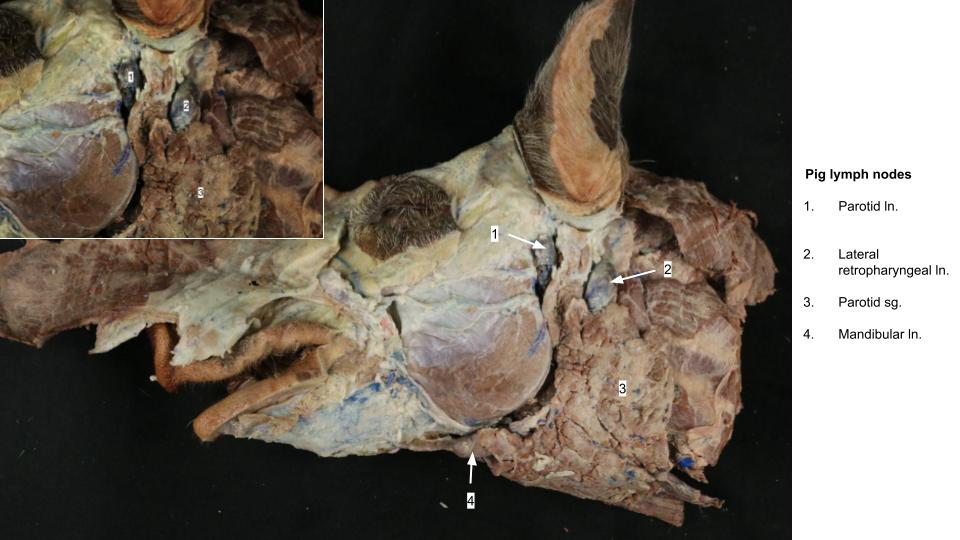
Superficial Lymph Nodes of the Equine, Ruminant, and Porcine Head
Observe: Confirm you can identify the parotid ln on the deep cranial margin of the parotid salivary gland. The parotid ln. in the horse is a cluster of 6-10 small nodes embedded deep in the cranial aspect of the parotid sg. – they can be quite hard to identify.
Isolate the mandibular lnn., which are found ventrally, related to the mandibular sg. and linguofacial v. in the ox, and facial v. in small ruminants. As is typical for equidae, these nodes are pea-sized and form a cluster medial to the body of the mandible. The right and left clusters converge rostrally to form a V-shaped mass in the intermandibular space. The mandibular lnn. of the horse are routinely palpated in the live animal.
In the pig and goat, identify the lateral retropharyngeal lymph node, closely related to the caudal border of the parotid sg. In the goat, this lymph node is sometimes difficult to find. Observe it here if you are able to do so. If not, it may be more accessible on another specimen or on the deep dissection. [The lateral retropharyngeal lnn. in the horse are best identified with the medial retropharyngeal lnn. when dissecting the guttural pouches (see deep dissection).]
-

- Salivary glands and lymph nodes of the ox and horse. 7
-

- Salivary glands and lymph nodes of the dog and pig. 7
-
- Head of the calf. 2
-
- Goat head, lateral view. 12
-
- Structures of the equine head, ventral view. 2
-
- Horse mandibular lymph node

-
- Goat lymph nodes

-
- Calf lymph nodes
-
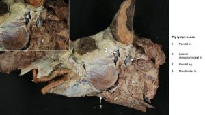
- Pig lymph nodes
Clinical Application: Lymph nodes as an index of health in food animals
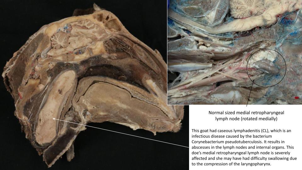
Knowledge of the lymph nodes of the head has special significance in food animals. Lymph nodes provide an index to the health of the animal and are therefore examined on the carcass and offal (viscera). Lymph nodes of the head receive considerable attention during meat inspection.
-
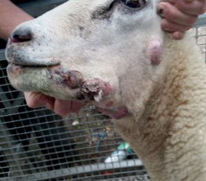
- Sheep with multiple abscessed lymph nodes from Caseous Lymphadenitis

-
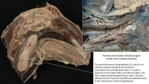
- Goat with abscessed medial retropharyngeal ln.
Deep Arteries of the Ungulate Head
Equine
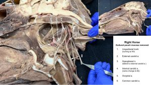
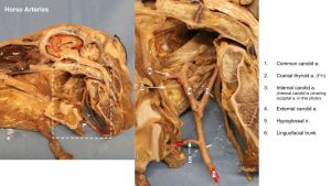
Observe: On the medial aspect of an equine specimen on which a deep dissection has been performed, locate the transected common carotid a.
Identify the cranial thyroid a. (look for it heading straight toward the thyroid gland).
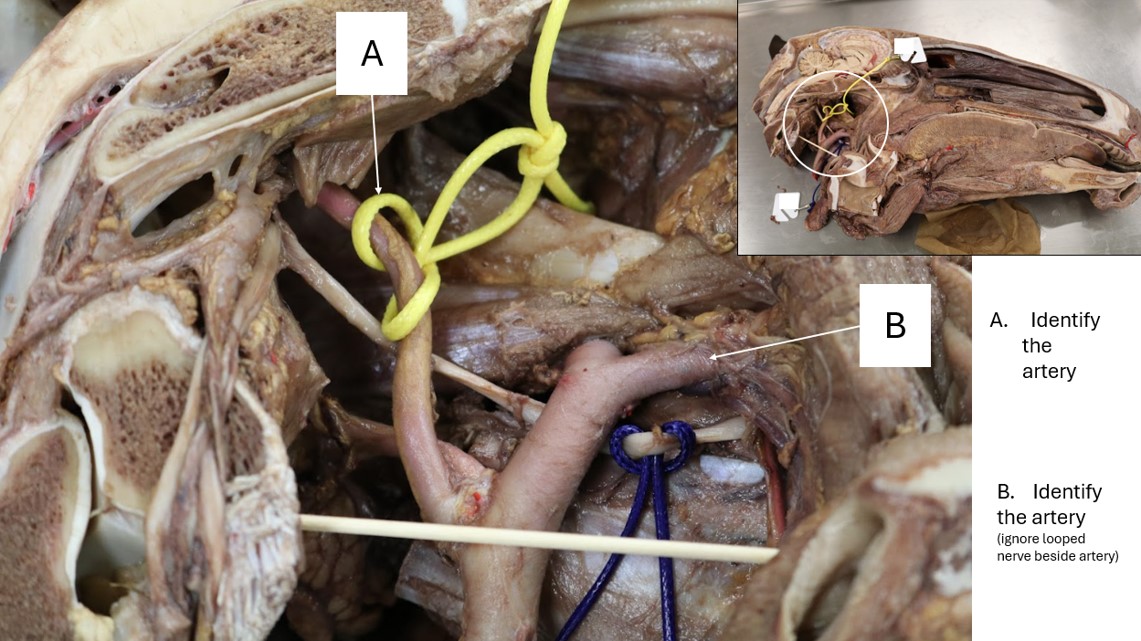
Continue cranially along the common carotid artery until the internal carotid a. branches from it. Once the internal carotid has branched from the common carotid, the latter changes name to the external carotid artery, which is the main channel artery.
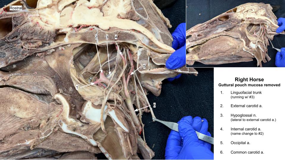
The first branch of the external carotid artery is the occipital a. Note that the occipital artery is aptly named and will head toward the occipital region of the head. Although it branches from the main channel cranial to the internal carotid artery, it runs more caudally.
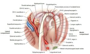
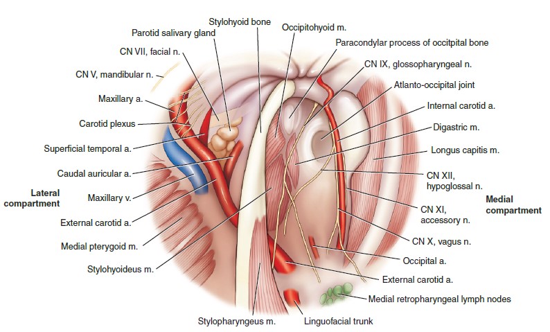
To visualize the following structures, disruption of the wall of the guttural pouch is necessary as viewed from the medial side. Remember the guttural pouches, which were also reflected to observe cranial nerves, are large, auditory-tube diverticula. They will be covered in depth during the respiratory unit. Removal of the longus capitis m. insertion should have been completed during the cranial nerve dissection. From here, you can opt to flip to the lateral side to keep following the external carotid a. , or stay on the medial side until you can no longer move cranially along the external carotid.
You are responsible for identifying the following branches of the external carotid artery in the equine:
Occipital artery (heading toward the occipital region)
Linguofacial Trunk
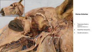
Superficial Temporal Artery (will have been transected so the temporalis muscle could be removed). Find the transverse facial artery, which is the first branch of the superficial temporal artery.
The continuation of the external carotid artery beyond the superficial temporal artery is the maxillary artery, which becomes the main channel artery at this location.
Branching from the maxillary artery, observe the inferior alveolar artery, running with the nerve of the same name, on its way to the mandibular foramen, through which it enters the mandibular canal.
The descending palatine artery branches from the maxillary artery more distally and becomes the main channel artery. Its continuation beyond the sphenopalatine artery is the major palatine artery. You need not observe these arteries as they branch from the maxillary artery, but you should observe the major palatine artery in the mouth, where the hard palate has been elevated to reveal it at that location.
-
- Arteries of the head of the horse, lateral view. Dashed lines indicate vessels that are either deep to bone or inside bone. 6
-
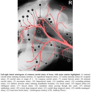
- Left-right lateral arteriogram of common carotid artery of horse, with major arteries highlighted. 2
-
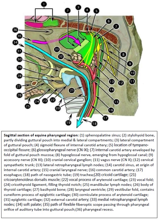
- Sagittal section of equine pharyngeal region. 2
-
- Anatomy of the internal wall of the equine guttural pouch and anatomical relationships with adjacent structures. 9
-
- Horse guttural pouch arteries
-
- Horse vasculature
-
- Horse superficial arteries
-
- Major palatine a.
-
- Horse arteries

Ruminant
While you are not required to identify all of the deep arteries in the ruminant, there are some significant species differences for which you will be responsible.
In the ruminant, as in the cat, the internal carotid a. degenerates to a fibrous cord in the neonate and its remnant may not be apparent at all. The occipital a. should be readily identifiable. The main blood supply to the brain is via the epidural rete mirabile.
Because the small ruminant does not possess a facial artery, the analogous artery branching from the external carotid artery is called the lingual artery. Note that the bovine species do possess a facial artery, and thus also possess a linguofacial trunk.
Observe: Identify the epidural rete mirabile in the goat on an available deep dissection of the goat. Be able to explain that the internal carotid artery degenerates in the neonatal ruminant and that the epidural rete mirabile is present instead. Be able to state that the artery analogous to the linguofacial trunk in the bovine and equine is called the lingual artery in the small ruminant.
-
- Arteries of the head of the ox, lateral view. Dashed lines indicate vessels that are either deep to bone or inside bone. 6
-
- Arteries of the head of the sheep (small ruminant), lateral view. Dashed lines indicate vessels that are either deep to bone or inside bone. 6
-
- Goat epidural rete mirabile

Clinical Application: Dental Extractions and the Major Palatine Artery in the Ungulate
As in the carnivore, avoidance of the major palatine a. during dental extractions of the maxillary arcades is of utmost importance.
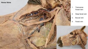
Deep veins of the Equine Head
Observe: Observe the following bolded veins on an equine head on which a deep dissection has been performed.
At the ventral mandible on its medial aspect dissect and identify the facial a. and v., passing with the parotid sg. duct and then crossing over the ventral cortex of the mandible and traveling dorsolaterally along the rostral margin of the masseter m. Zero in on the facial v. path along the rostral masseter.
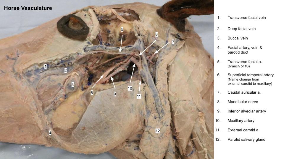
The masseter muscle will have been resected in order to visualize the large veins and their sinuses that emerge from under this muscle to join the facial v. From dorsal to ventral they are 1. the transverse facial vein (look just ventral and adjacent to the facial crest), 2. the deep facial vein, and 3. the buccal vein.
-
- Horse vasculature
-
- Horse deep veins

-

- Selected veins of equine head, lateral view. 2

Clinical Application: Transverse Facial Vein and Venous Sinuses of the Equine Patient
Venous blood samples may be collected from transverse facial v. Be aware of these large venous sinuses when contemplating surgery in the region e.g. to access a tooth via a buccotomy, or to repair a mandible fracture.
Deep Lymph Nodes of the Ungulate Head
Observe: Equine: On the medial surface of the split head, locate the medial retropharyngeal lnn. associated with the ventral wall (ie the floor) of the guttural pouch. They are clusters of small nodes. The lateral retropharyngeal lnn. are related to the caudolateral wall of the guttural pouch, caudal to the mandibular sg., caudoventral to the occipitomandibularis m. and dorsolateral to the thyroid gland. They may appear to be a continuous cluster of nodes with the medial group.
Goat and calves: Medial to the caudal border of the mandibular sg. and ventral to the wing of the atlas, locate the lateral retropharyngeal lymph node buried in fatty connective tissue. In the goat this lymph node is lateral to the common carotid a. and medial to the external jugular v. On the medial surface of the split head (look at both halves!), locate the medial retropharyngeal lymph node tucked in the fat caudodorsal to the nasopharyngeal wall.
-

- Sagittal section of newborn calf head. 2
-

- Sagittal section of equine pharyngeal region. 2
Ungulate Thyroid Gland and Parathyroid Glands
Thyroid Gland
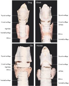
There is only one thyroid gland per animal, not right and left thyroids, as is frequently implied. In most mammals, the gland is divided into right and left lobes, connected ventrally by a variably developed isthmus, and species variation in shape exists In the horse, each lobe is distinctly ovoid and may be palpable in the live animal; certainly it is palpable when enlarged due to thyroid adenoma development. In goats the thyroid is somewhat flat and elongate.
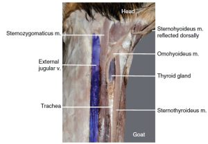
Observe: In the equine and ruminant, reflect the tissues over the dorsolateral margin of the cranial trachea, just caudal to the larynx, to locate the thyroid gland, typically a dark purple structure.
The thyroid gland of the pig differs considerably from the other species in that the two lobes are fused ventral to the trachea, and it is located nearer the thoracic inlet. Observe the porcine thyroid gland, still on the cadaver’s body, at the caudoventral aspect of the trachea.
Clinical Application: Thyroid Enlargement in the Horse
Benign, progressive, thyroid enlargement is not uncommon in the aging horse and can cause problems with swallowing and breathing due to external pressure on regional structures.
-
- Comparative thyroid glands. 7

-
- Ventral neck of the goat. 12

-
- Carotid sheath of the calf. 2
-
- Comparative thyroid glands of the different domestic animals. 7
-
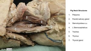
- Pig Neck Structures
-
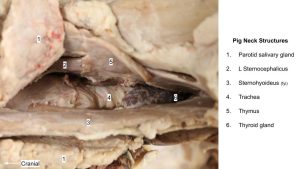
- Pig Neck Structures
Parathyroid Glands
Observe: Describe where the parathyroid glands are located, and spend a short amount of time trying to find them.
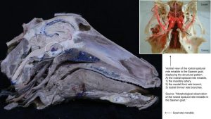
There are typically two pairs of parathyroid glands, the external parathyroids and the internal parathyroids. The internal parathyroids are typically very closely associated with or embedded in the thyroid gland. The external parathyroids may be in the vicinity of the thyroid gland (e.g. medial to the common carotid bifurcation in the ox) or some distance away e.g. in the horse, at the other end of the neck close to the caudal deep cervical lnn.!! Do not be too concerned with identification of these glands however do give a shout if you happen across any!
Review videos
Dog head veins – 1 min
Carnivore head arteries & thyroid gland, Walker – 11 min (watch until 16:30)
Carnivore head arteries & thyroid gland, Spradley – Watch until 12 min
Goat and horse head vasculature – 12 min
2025 video:
Comparative lymph nodes – 9 min
Terms
Vessels of the head |
|
| Features | Species (no calves) |
| Common carotid a | All. |
| Cranial thyroid a | Carnivore and horse |
| Internal carotid a | Dogs and equine (note remnant in goat and cat) |
| External carotid a | Carnivore and horse |
| Occipital a | Carnivore and horse |
| Linguofacial trunk | ID in Equine, also present in bovine and pig |
| Lingual a | ID in Carnivore, know that it is present in small ruminant. |
| Facial a | Carnivore and horse (on superficial side of horse only) |
| Caudal auricular a | Carnivore and horse |
| Superficial temporal a | Carnivore and horse |
| Transverse facial a. | Horse, Goat |
| Cornual a. | Goat |
| Maxillary a | Carnivore and horse |
| Inferior alveolar a | Carnivore and horse |
| Descending palatine a | Do not ID. Know is main channel rostral to maxillary. |
| Major palatine a | Carnivore and horse (in mouth) |
| Infraorbital a | Carnivore and horse (only rostrally as exits canal) |
| Maxillary rete mirabile (cat) | Cat |
| Epidural rete mirabile | Goat |
| Lingual v | All except calf |
| Facial v | All except calf |
| Linguofacial v | All except calf |
| Maxillary v | All except calf |
| External jugular v | All except calf |
| Transverse facial v | Equine |
| Deep facial v | Equine |
| Buccal v | Equine |
| Osteology | Species |
| Alar canal | Dog and horse |
| Mandibular foramen | Carnivore and horse. What artery enters. |
| Maxillary foramen | Carnivore and horse |
| Infraorbital canal | Carnivore and horse |
| Infraorbital foramen | Carnivore and horse |
| Major palatine foramen | Carnivore and horse |
| Major palatine canal | Carnivore and horse |
| Vessels of the brain |
|
| Features | Species |
| Arterial circle | All |
| Basilar a | All |
| Dorsal sagittal sinus | All |
| Cavernous sinus | All |
Structures of the head |
|
| Features | Species |
| Thyroid gland | All |
| External parathyroid | All. Do not ID. Know location. |
| Internal parathyroid | All. Do not ID. Know location. |
| Medial retropharyngeal ln | All |
| Lateral retropharyngeal ln | All except dog |
| Parotid ln | All |
| Mandibular ln | All |