Labs 10 and 11: Gluteal and Pelvic Limb Vasculature
Learning Objectives
- Identify the blood supply to the gluteal muscles.
- Identify main channel blood supply of the thigh and crus.
- Identify some arterial branches of the thigh and crus.
- Identify the saphenous venous system of the pelvic limb.
- Identify the popliteal lymph node in the carnivore and horse.
Vessels of the Carnivore Pelvic Limb
Branches of the Internal Iliac Artery
- Umbilical a. (covered in the pelvic cavity unit)
- Internal pudendal a. (covered in the pelvic cavity unit)
- Caudal gluteal a.
- Cranial gluteal a.
Perform the following dissection if the right limb has not been dissected to study the pelvic limb nerves:
Dissect: Remove the skin from the caudal part of the RIGHT trunk, pelvis, and thigh. Continue the midventral incision from the umbilicus to the root of the tail. In making this incision, closely encircle the external genital structures and the anus. Extend an incision distally on the medial surface of the thigh to the tarsus. Try to leave the subcutaneous vessels on the limb, taking special care on the medial aspect of the thigh. Encircle the tarsus with a skin incision. First, reflect the skin from the medial surface of the thigh and crus and then, starting at the tarsus, reflect the whole flap of skin from the lateral surface of the leg, stifle, thigh, pelvis, and abdomen to the mid-dorsal line. The cutaneous trunci m., if present, may be removed with the skin because it is more intimately attached to the skin than to the underlying structures.

Incise along red lines, following directions above (dashed red lines indicate incisions extended dorsally and/or laterally-aspects not directly seen in the figure).
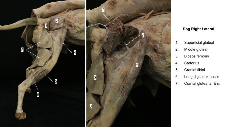
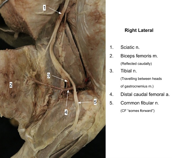
Transect and reflect the superficial and deep gluteal fasciae. For now, keep the medial fascia and fascia lata intact. Carefully, transect the biceps femoris midway between its origin and the stifle and reflect the transected muscle halves. The sciatic nerve and its branches lie directly underneath this muscle.
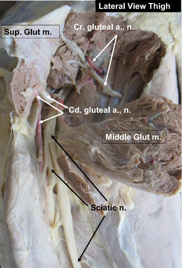
Expose the caudal insertion of the superficial gluteal muscle deep to the proximal edge of the biceps femoris m. Transect the insertion at this level and reflect the muscle dorsally. If possible, try not to break the caudal gluteal artery and nerve, and their branches, which supply this muscle.
Transect the middle gluteal muscle midway through its length. Reflect the cranial and caudal portions. Try not to break the cranial gluteal artery and nerve, or their branches, in doing so.
Observe: Observe the following arteries on the right pelvic limb, deep to the superficial and middle gluteal muscles and the biceps femoris muscle.
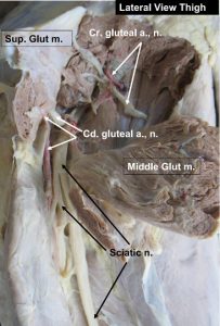
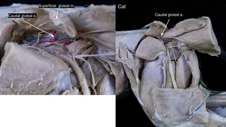
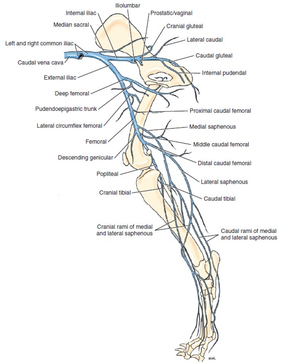
The caudal gluteal artery is the larger of the two terminal branches of the internal iliac artery. The internal iliac artery will be dissected during the urogenital unit. The caudal gluteal artery arises opposite the sacroiliac joint and passes caudally across the greater ischiatic notch and over the ischiatic spine lateral to the coccygeus muscle, parallel to the internal pudendal artery. Follow the caudal gluteal artery as it passes over the ischiatic spine with the sciatic nerve ventral to the sacrotuberous ligament. Remember: the cat lacks a sacrotuberous ligament. The artery supplies the superficial gluteal muscle (observe branches into this muscle), the rotators of the hip, and the adductor muscle. It divides into several branches, which supply the biceps femoris and the semitendinosus and semimembranosus muscles. Turn the biceps femoris caudally to expose the caudal gluteal artery lying deep to it, close to the sacrotuberous ligament and ischiatic tuberosity.
The cranial gluteal artery is a branch of the caudal gluteal artery in the dog. It passes across the cranial part of the greater ischiatic notch of the ilium and between the middle and deep gluteal muscles, which it supplies. Observe branches of the cranial gluteal artery as they enter the ventral side of these muscles. Note that in the cat, the cranial gluteal artery branches from the interal iliac artery. The caudal gluteal artery is a more distal branch of the internal iliac artery in the cat. This distinction will become more important when dissecting the internal pudendal artery, which is the continuation of the internal iliac artery once the caudal gluteal artery branches off.
-
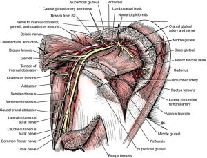
- Nerves, arteries, and muscles of the right hip, lateral aspect. 1

-
- Cranial gluteal a.
-
- Gluteal aa.
-

- Cat caudal gluteal a.
Femoral Artery and Branches
External iliac a. – main branch from abdominal aorta
Femoral a. – continuation of external iliac a. distal to the vascular lacuna
FYI: Superficial circumflex iliac a.
FYI: Lateral circumflex femoral a.
FYI: Proximal caudal femoral a.
Saphenous a.
FYI: Descending genicular a.
FYI: Middle caudal femoral a.
Distal caudal femoral a.
Popliteal a.
Caudal tibial – name change from popliteal to cranial tibial. No need to ID.
Cranial tibial a.
Dorsal pedal a.
Recognize that main channel arterial supply is on the plantar aspect of the pes.
Dissect: Carefully skin the right pelvic limb. Note/observe the following bolded arteries and structures, as described below.
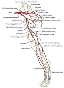
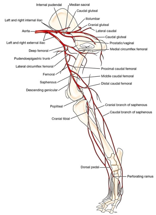
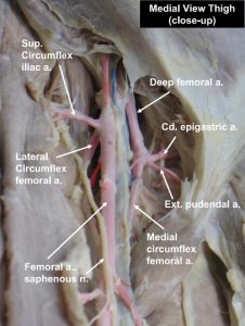
The external iliac artery arises from the aorta on a level with the sixth and seventh lumbar vertebrae and will be observed in the pelvic cavity unit. After passing through the abdominal wall, the external iliac becomes the femoral artery. The opening through which the external iliac artery passes is the vascular lacuna, located between the caudal border of the aponeurosis of the external abdominal oblique (inguinal ligament) and the pelvis.
The deep femoral artery is the only branch of the external iliac artery and arises inside the abdomen near the vascular lacuna and courses caudally. Two vessels leave the ventral surface of the deep femoral artery within the abdomen by a short pudendoepigastric trunk. These are the external pudendal and caudal epigastric arteries. The external pudendal artery passes through the inguinal canal. The inguinal canal will be discussed during the urogenital unit. The caudal epigastric artery arises from the pudendoepigastric trunk and passes cranially on the dorsal surface of the rectus abdominis. It supplies the caudal half of the rectus abdominis and the ventral parts of the oblique and transverse muscles.
NOTE: Typically in the cat, there is no pudendoepigastric trunk; the caudal epigastric a. and external pudendal a. each arise separately from the deep femoral a. Sometimes, this is the case in the canine as well.
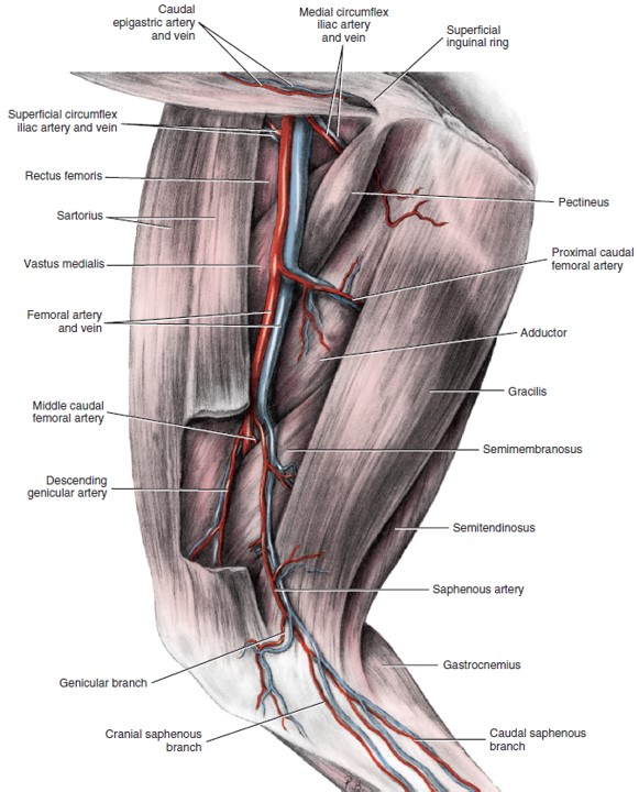
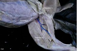
Observe the femoral triangle, which is bounded cranially by the sartorius, laterally by the vastus medialis and rectus femoris, and caudally by the pectineus and adductor. Its dorsal border is the abdominal wall, at the location of the vascular lacuna. The neurovascular bundle readily identified here consists of the femoral artery and vein and the saphenous nerve.
Dissect carefully on the proximal aspect of the medial thigh where the femoral artery emerges beyond the abdominal wall. Locate the deep femoral a. (image) as it branches from the external iliac a. Avoid dissecting more proximally as other inguinal structures will be damaged. If needed to see the PE trunk (image), transect the pectineus m. at its origin and reflect it distally (or remove a chunk entirely).
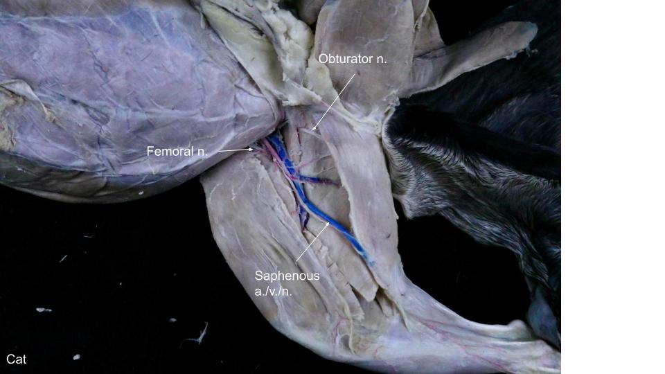
The continuation of the deep femoral artery after the PE trunk has branched off (or when the external pudendal branches if the PE trunk is absent) is called the medial circumflex femoral artery. To view the beginning of the medial circumflex femoral artery, transect the gracilis muscle halfway along its length and reflect the portions proximally and distally. Try not to break the obturator nerve as it courses along the lateral (deep) aspect of the gracilis m. Parts of the origin of the adductor may need to be removed to view this artery, but don’t follow it very far.
As the medial circumflex femoral artery approaches the large adductor muscle, it gives off branches that supply the adductor muscle, vastus medialis muscles, obturator muscles and semimembranosus muscle.
-
- Arteries of right pelvic limb, schematic medial view. 1
-

- Arteries and veins of the thigh, superficial dissection, medial aspect. 1
-
-
Vasculature of hindlimb of the cat, medial view with satellite
veins (not labeled) and saphenous system (labeled). 4

-
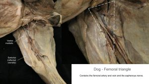
- Femoral triangle

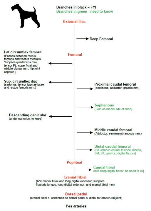
Femoral Artery and Branches
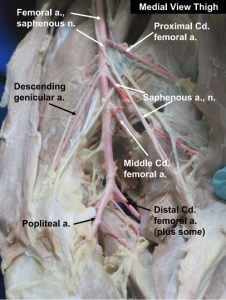
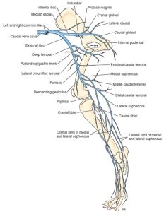
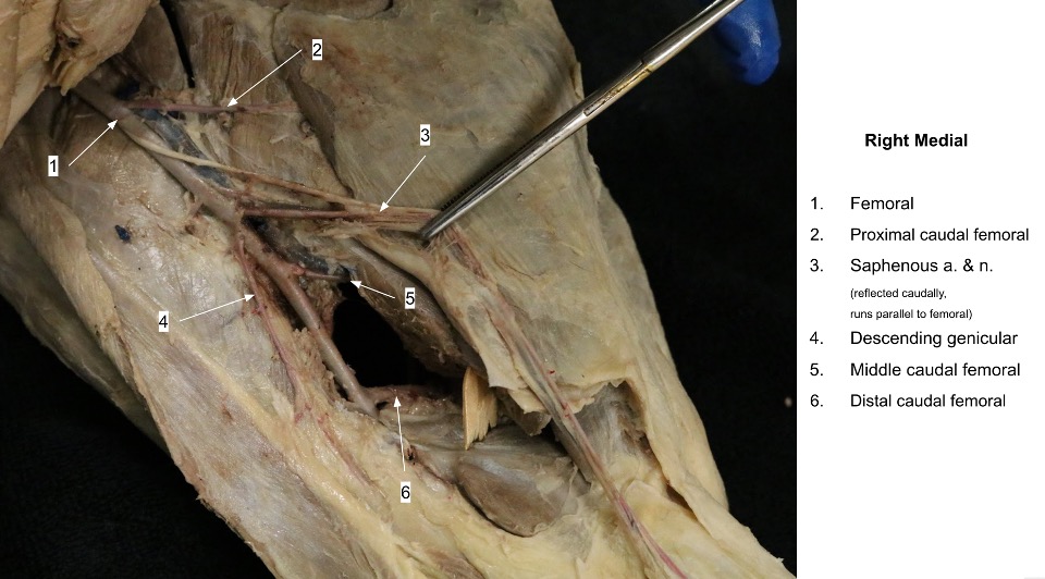
The femoral artery is the continuation of the external iliac artery beyond the level of the vascular lacuna (similar to how the subclavian artery name changes to axillary as it rounds the first rib). The branches of the femoral artery in the order that they typically arise are superficial circumflex iliac, lateral circumflex femoral, proximal caudal femoral, saphenous, descending genicular, and middle and distal caudal femoral arteries.
Observe: You may observe the following branches of the femoral artery as reference only. They are FYI for our purposes; however, identifying these will help prevent confusing them for the saphenous or distal caudal femoral aa., which are need-to-know.
FYI: The superficial circumflex iliac artery is a small branch that arises from the lateral side of the femoral artery near or with the lateral circumflex femoral artery. The superficial circumflex iliac artery courses cranially and supplies both parts of the sartorius, the tensor fasciae latae, and the rectus femoris. It becomes superficial at the cranial ventral iliac spine of the tuber coxae.
FYI: The lateral circumflex femoral artery is a large branch that passes between the rectus femoris and the vastus medialis. Although most of the vessel arborizes in the quadriceps, it also supplies the tensor fasciae latae, the superficial and middle gluteals, and the hip joint capsule.
FYI: The proximal caudal femoral artery leaves the caudal surface of the femoral artery distal to the origin of the lateral circumflex femoral in the mid-thigh region. It extends distocaudally over the pectineus and adductor muscles, which it supplies, and enters the deep surface of the gracilis.
-
- Dog

-
- Dog

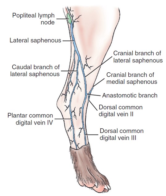
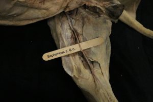
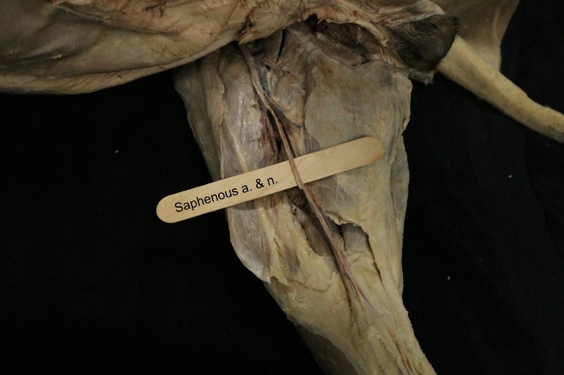
Dissect: Clean and observe the saphenous artery as it arises from the femoral artery, proximal to the stifle. It travels superficially, while the femoral artery travels deeper at this point. Note the saphenous nerve and medial saphenous vein, both of which run in the same neurovascular bundle as the saphenous artery. Also identify the lateral saphenous vein at this time.
The saphenous artery and nerve and the medial saphenous vein continue distally as a neurovascular bundle between the converging borders of the caudal part of the sartorius and gracilis muscles. The saphenous artery supplies the skin on the medial side of the stifle and terminates in a cranial and a caudal branch. The cranial branch of the saphenous artery arises opposite the proximal end of the tibia and terminates as the dorsal common digital arteries. The caudal branch of the saphenous artery arises at the proximal end of the tibia. It lies between the medial head of the gastrocnemius and the tibia. In the metatarsus it supplies the paw by branches that give rise to the plantar common digital arteries. It also gives off deep branches that contribute to a deep plantar arch that is the source of plantar metatarsal arteries.
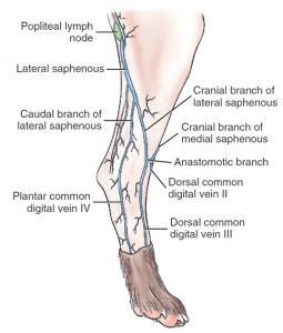
In some specimens the saphenous veins may be latexed enough to be identified. The medial saphenous vein is similar to the saphenous artery in its origin in the paw and termination in the femoral vein. The lateral saphenous vein has no comparable saphenous artery. It is formed by cranial and caudal branches in the leg that arise from venous arcades in the paw. It terminates in the distal caudal femoral vein. The cranial branch of the lateral saphenous vein is often used for venipuncture in the dog. It arises from an anastomosis with the cranial branch of the medial saphenous vein on the dorsal aspect of the tarsus and courses proximocaudally across the lateral surface of the leg.
-
- Veins of the pelvic limb, medial view. 1
-
- Superficial veins of the right hind limb, lateral aspect. 1
-
-
Vasculature of hindlimb of the cat, medial view with satellite
veins (not labeled) and saphenous system (labeled). 4
-
- Dog saphenous a.
-
- Cat saphenous a.
Clinical Application: Pelvic Limb Venipuncture in the Carnivore
In the cat, the medial saphenous vein is relatively larger than is the lateral saphenous vein. In the canine, the lateral saphenous vein is larger than its medial counterpart. Hence, venipuncture more often is performed in the medial saphenous vein in the cat and in the cranial branch of the lateral saphenous vein in the dog. Both veins can be used in either species, when needed.
FYI: The descending genicular artery arises from the femoral distal to the origin of the saphenous and supplies the medial surface of the stifle.
FYI: The middle caudal femoral artery arises distal to the descending genicular and saphenous arteries after the femoral artery passes lateral to the semimembranosus. It ramifies in the distal parts of the adductor and semimembranosus muscles.
The distal caudal femoral artery is a large vessel that arises from the caudal surface of the last centimeter of the femoral
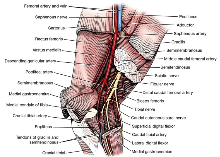
Dissect: To observe the distal caudal femoral a. transect the gracilis muscle at its distal attachment and reflect it. Transect the semimembranosus and semitendinosus muscles to uncover the medial head of the gastrocnemius muscle. This will expose the distal caudal femoral artery and its branches, which supply the biceps femoris, the semimembranosus, the semitendinosus, the gastrocnemius, and the digital flexors.
-
- Arteries and veins of the thigh, superficial dissection, medial aspect. 1
-

- Arteries and nerves of right popliteal region, medial view. 1
-
-
Vasculature of hindlimb of the cat, medial view with satellite
veins (not labeled) and saphenous system (labeled). 4
-

- Dog medial thigh
-

- Dog lateral thigh
Popliteal artery and lymph node; Cranital Tibial and Dorsal Pedal Arteries
The popliteal artery, a continuation of the femoral artery, is the main channel artery at the region of the knee. It passes between the two heads of the gastrocnemius muscle, crosses the medial surface of the superficial digital flexor muscle, and courses over the flexor surface of the stifle and through the popliteal notch of the tibia. It inclines laterally under the popliteus muscle and perforates the lateral digital flexor to reach the interosseous space. The popliteal artery supplies the stifle, gastrocnemius, and popliteus muscles and terminates as cranial and caudal tibial arteries. The caudal tibial artery is a small vessel that leaves the caudal surface of the popliteal in the interosseous space. It need not be observed.
Dissect: The lymph node lying in the fat directly caudal to the stifle is the popliteal lymph node. Clean the fat around the popliteal lymph node and identify the structure.
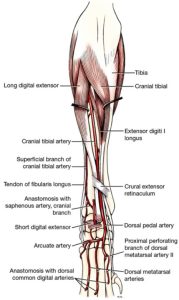
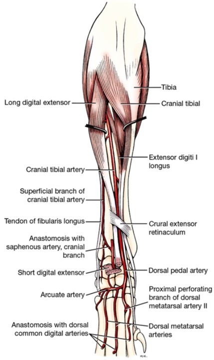
The cranial tibial artery is the continuation of the main channel distal to the point where the caudal tibial artery branches from the popliteal artery. Observe the cranial tibial artery as it passes between the tibia and the fibula.
Dissect: Reflect the fascia on the cranial aspect of the stifle and leg where it serves for the insertion of the biceps femoris. Separate the cranial tibial and long digital extensor muscles throughout their length. Observe the cranial tibial artery between these two muscles. It supplies the fibularis longus, long digital extensor, and cranial tibial muscles. The caudal tibial artery need not be identified, but know that it is the name change artery from popliteal to cranial tibial.
The cranial tibial artery is continued opposite the talocrural joint as the dorsal pedal artery. Branches supply the tarsus, and the dorsal pedal artery terminates in the arcuate artery at the level of the tarsometatarsal joint. Dissect down to this point. The arteries distal to the dorsal pedal a. need not be dissected.
-
- Arteries and nerves of right popliteal region, medial view. 1
-
- Cranial tibial artery of right limb, cranial view. 1
-
- Arteries of right hindpaw of the dog, plantar view. 1
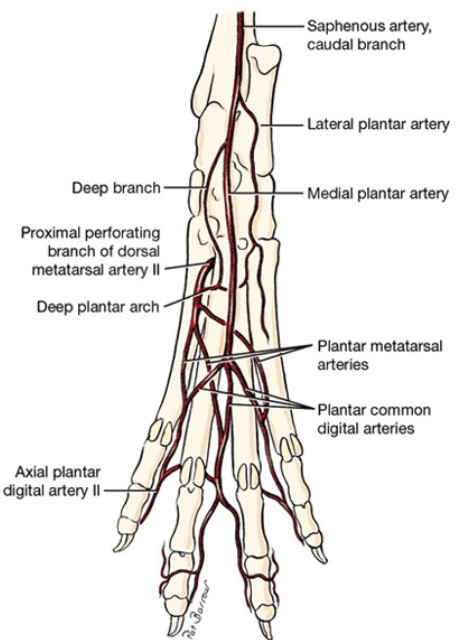
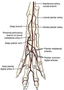
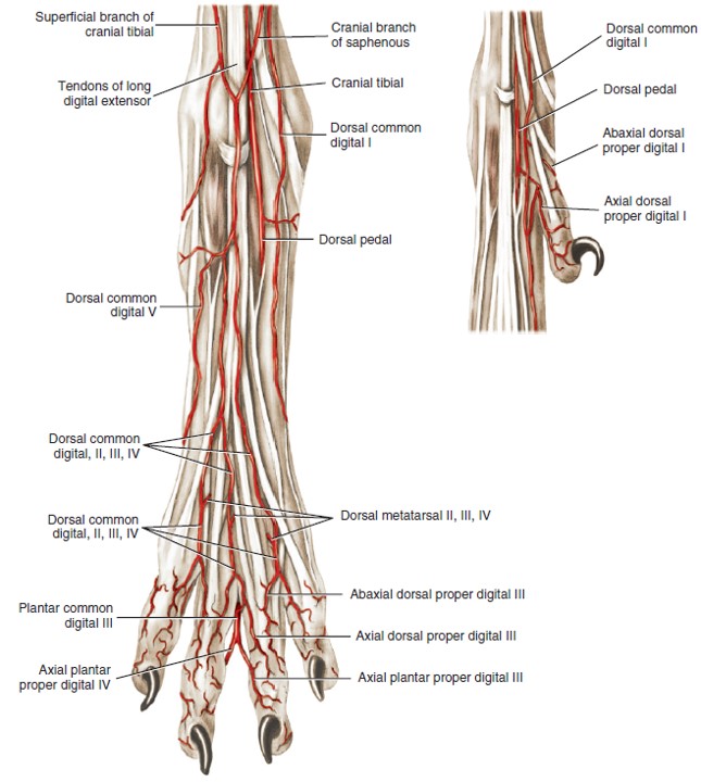
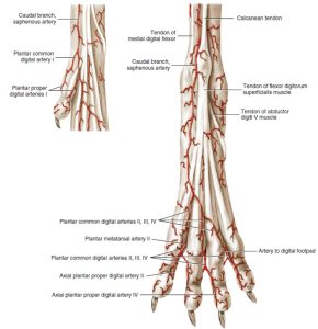
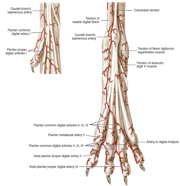
Observe: Use diagrams and lab models to describe the vasculature of the pes, as explained below. Need to know: Each digit is vascularized by four arteries.
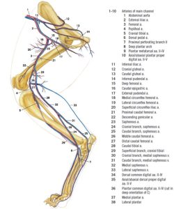
NEED TO KNOW: The main channel of blood supply to the pelvic limb and paw is via: external iliac a., femoral a., popliteal a., cranial tibial a., dorsal pedal a. From here, know that the main channel blood supply is on the plantar aspect of the pes. The main channel distal to the dorsal pedal artery is FYI: perforating branch, deep plantar arch, plantar metatarsal aa., and axial/abaxial plantar proper digital aa.
Similar to the manus, the arterial supply to the pes involves anastomosing arches fed by several branches. The dorsal and plantar aspects of the metatarsal region are vascularized by two layers of arteries each, as is the metacarpal region. The names of these arteries are FYI.
On the dorsal side, the most superficial layer is comprised of the FYI: dorsal common digital arteries. The deep layer is made up of the FYI: dorsal metatarsal arteries.
On the plantar side, the most superficial layer (closest to the ground) is comprised of the FYI: plantar common digital arteries while the deeper layer is formed by the FYI: plantar metatarsal arteries.
At the metatarsophalangeal joint, the dorsal metatarsal arteries anastomose with the dorsal common digital arteries and continue as the dorsal common digital arteries. The same thing occurs on the plantar surface, i.e. the plantar metatarsal arteries combine with the plantar common digital arteries and continue as the plantar common digital arteries.
Then each of the common digital arteries bifurcates to become FYI: axial/abaxial dorsal/plantar proper digital arteries.
Need to know: Each digit is vascularized by four arteries. FYI: These arteries are the axial and abaxial dorsal proper digital arteries and the axial and abaxial plantar proper digital arteries.
-

- Arteries of the right hindpaw and first digit, dorsal aspect. 1
-
- Superficial arteries of the right hindpaw, plantar aspect. 1
-

- Arteries of the pes of the cat. 4
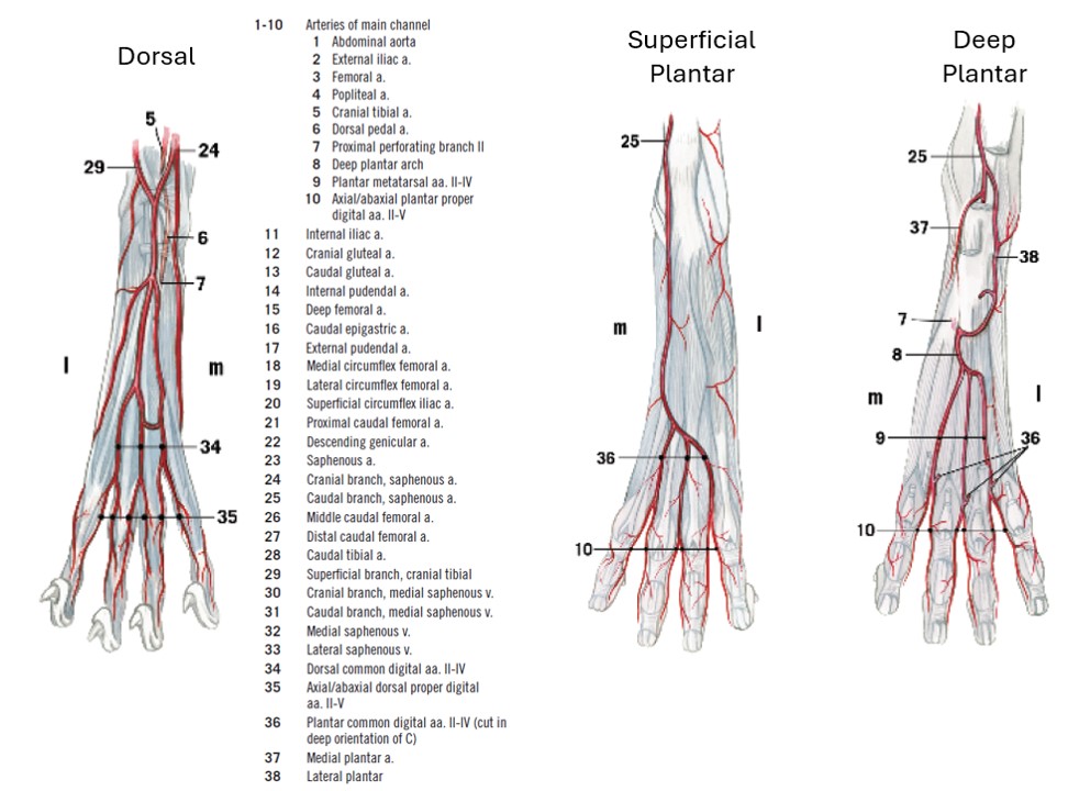
NEED TO KNOW: The main channel of blood supply to the pelvic limb and paw is via: external iliac a., femoral a., popliteal a., cranial tibial a., dorsal pedal a. From here, know that the main channel blood supply is on the plantar aspect of the pes. The main channel distal to the dorsal pedal artery is FYI: perforating branch deep plantar arch, plantar metatarsal aa., and axial/abaxial plantar proper digital aa.
Vessels and Lymph Nodes of the Ungulate Pelvic Limb
Tail
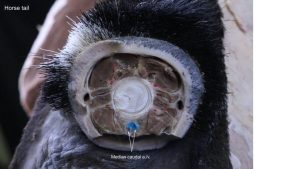
Observe: Observe the median caudal artery and vein in the transected tails of the equine and bovine cadavers.
FYI – hemal arches (bony bridges on ventral aspect of vertebra) may occur on the proximal caudal vertebrae. These arches can obstruct path of a needle intended to reach the median caudal v. in the ox.
Clinical Applications
Some common clinical implications of tail structures include:
- Cuff placement for blood pressure monitoring
- “Tail setting” operation in gaited show horses
- Tail amputation for pathologic reasons and animal husbandry reasons
- Caudal epidural anesthesia – this is performed in the horse and ox for obstetric procedures and for standing surgery in the perineal and pararectal regions e.g. repair of a rectovaginal fistula or perineal laceration. The injection is usually made between caudal vertebrae 1 and 2 (i.e. intercoccygeal). Raising (pumping) the tail identifies a typical depression over the dorsal interspace of the caudal vertebrae, where the needle is inserted. In small ruminants, calves, and the pig, epidural anesthesia is often performed at the lumbosacral space (i.e. cranial epidural anesthesia), partly because it is an easier space to identify.
-
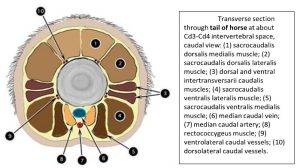
- Transverse section through the tail of the horse. 1
-
- Horse tail

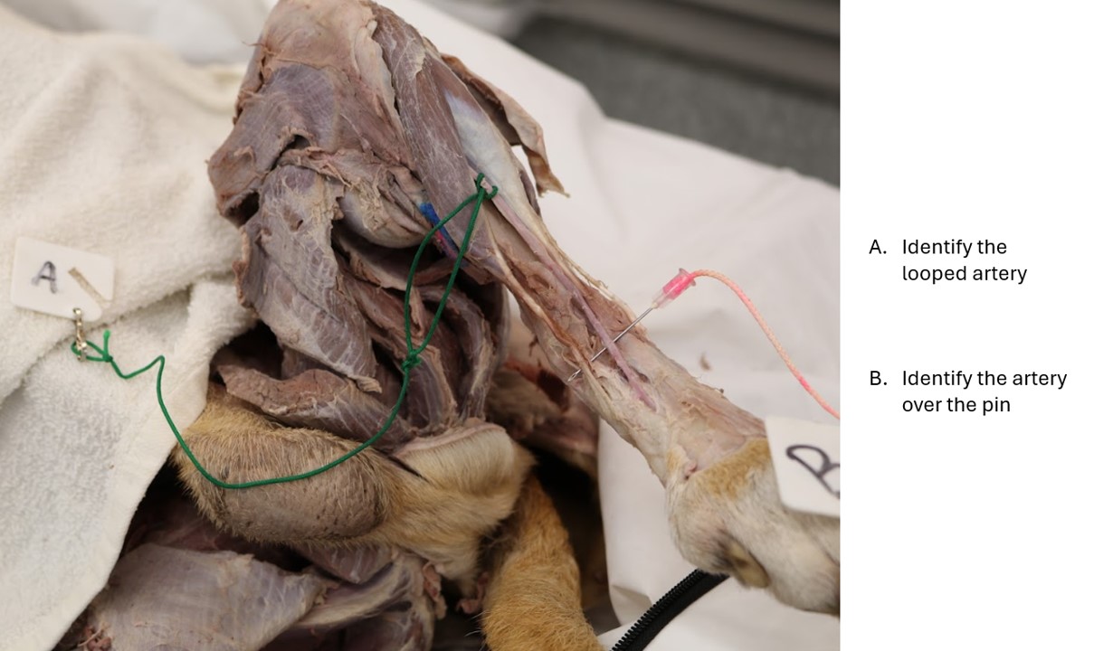
Ungulate Thigh Region Vessels
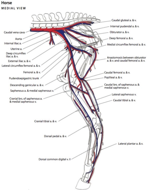
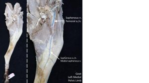
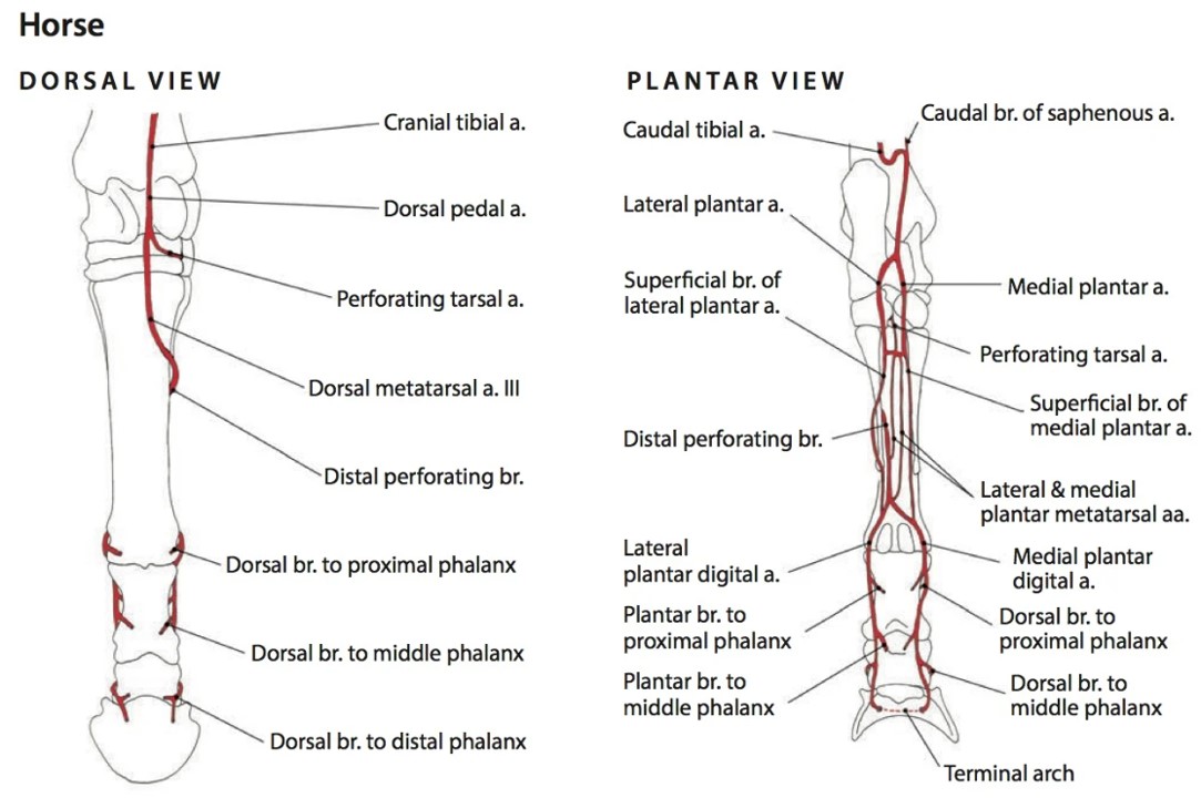
Main Channel Arteries of the Horse: Femoral, popliteal, cranial tibial a., to dorsal pedal a., to dorsal metatarsal a. 3, to medial and lateral digital aa.; on the plantar side (not main channel) are the medial and lateral plantar aa. In the horse and goat, identify the femoral artery and vein in the femoral triangle. Recall the borders of the femoral triangle. Note that in the ruminant, the femoral a/v pass between the two parts of the divided origin of this muscle.
Observe: Identify the medial saphenous v. and saphenous a. (large vein and smaller artery) and saphenous n., passing over the distal insertion of the gracilis m. and medial stifle.
Clinical Relevance
The saphenous a. can be used to palpate the pulse in ruminants.
-
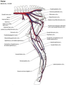
- Vessels of the pelvic limb of the horse, medial view. 5
-
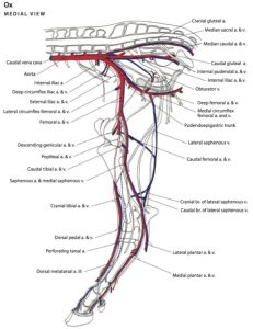
- Vessels of the pelvic limb of the ox, medial view. 5
-
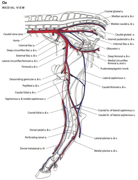
- Major branches of internal and external iliac arteries of cow, medial view. 2

-
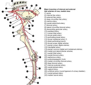
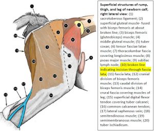
- Superficial structures of thigh and leg of newborn calf, medial view.2

-
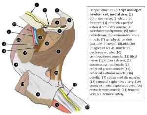
- Deeper structures of the thigh and leg of the newborn calf, medial view. 2

-
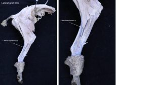
- Goat lateral saphenous v.

-
- Goat saphenous a.

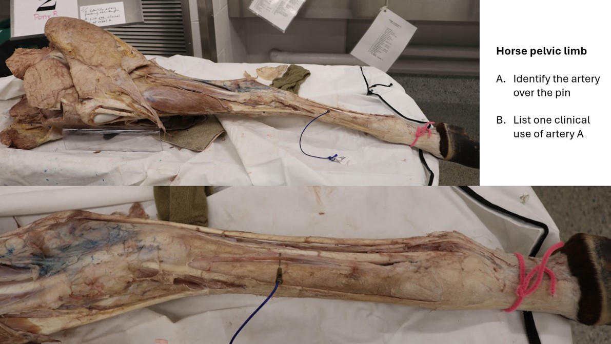
Vasculature of the Ungulate Crus
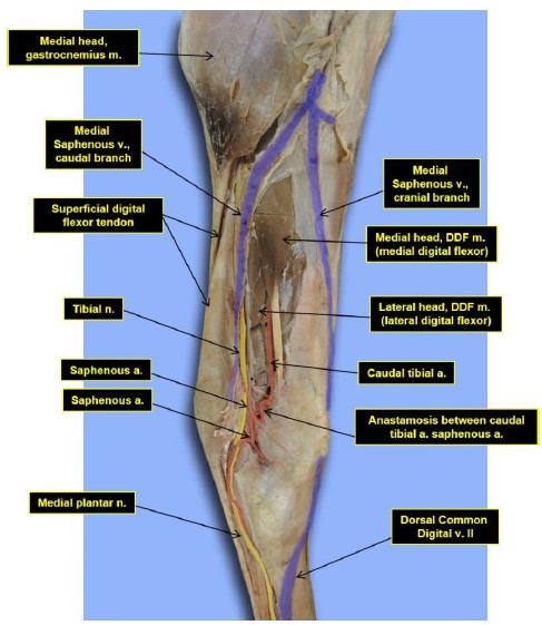
Observe: Observe the relatively large subcutaneous lateral saphenous vein on the distal lateral crus of the goat and horse. In the goat, identify the cranial tibial a. on the surface of the craniolateral tibia by looking between the fibularis tertius and long digital extensor mm. at the distal crus. In the horse, observe the intermuscular septum between the long and lateral digital extensor mm., about 10 cm above the tarsocrural joint. Locate the relatively shallow superficial fibular n. caudal to to this septum, and the more deeply located deep fibular n. and cranial tibial a. cranial to this septum.
-

- Medial crus and tarsus of the horse. 16
-
- Superficial structures of thigh and leg of newborn calf, lateral view. 2
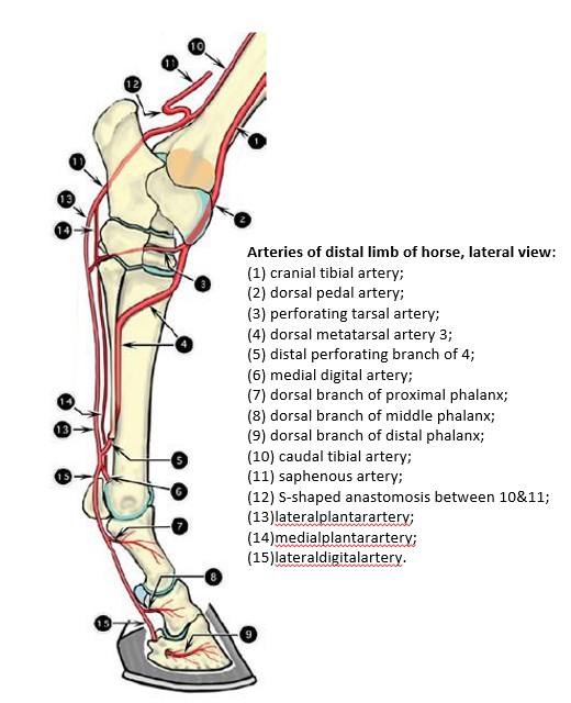
Observe: In the horse and goat, locate the cranial tibial a in the distal crus. This main channel artery continues distally across the dorsal tarsus, as the dorsal pedal a., and then on into the metatarsal region as the dorsal metatarsal a. 3. The dorsal metatarsal a. 3 lies in the groove between the 3rd metatarsus (MT3) and lateral splint bone (MT4) and is readily located there. Distally the dorsal metatarsal a. 3 passes under the lateral splint bone to the plantar side and divides to become the M/L digital aa. at the fetlock. On the plantar side of the metatarsus appreciate (but no need to dissect) that M/L plantar aa. travel with the M/L plantar nn. In the horse, because of the predominant MT3, rather than using traditional nomenclature for naming vessels and nerves in the metatarsus we simply refer to them as being medial or lateral to keep it simple clinically.
Clinical Relevance
Palpation of arterial pulses at fetlock, or catheterization of dorsal MT a. 3 for arterial blood pressure and blood gas sampling; intravenous regional limb perfusion of antimicrobials or local anesthetics, for example, using the M/L digital vv.
-

- Arteries of the distal limb of the horse, lateral view. 2
-
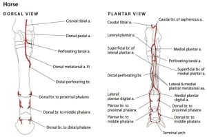
- Arteries of the distal left pelvic limb of the horse, dorsal and plantar views. 5
-
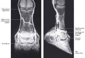
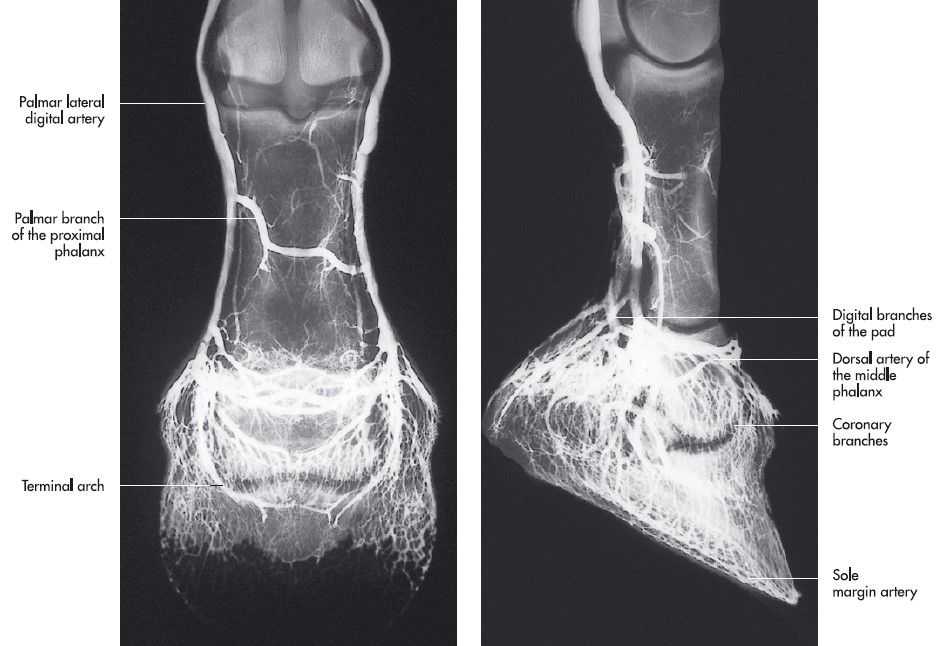
- Arteriogram of the foot of a horse, dorsopalmar (left) and lateromedial (right) projection. 7
-

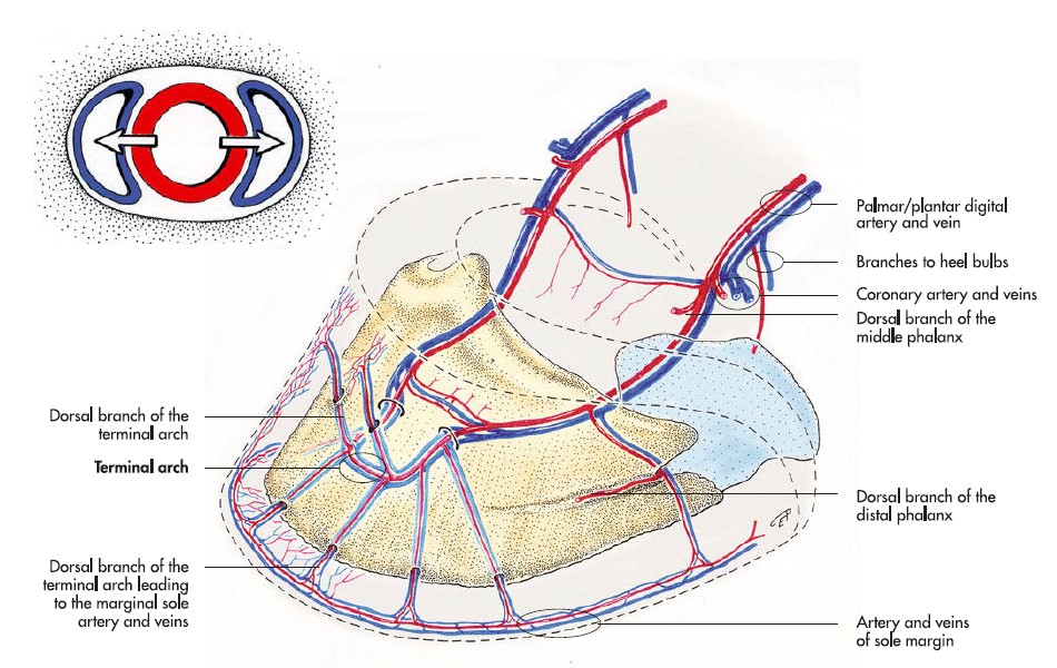
- Blood vessels of the distal phalanx of the horse. 7
Review videos
Dog vasculature (and nerves) – 23 min (much of this content has been cut and is FYI)
Goat main channel vasculature – 3 min
Terms List
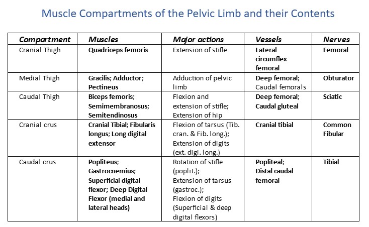
| Structures of the Pelvic Limb
|
|
| Features | Species (not in calf) |
| Popliteal ln | All |
| Cranial gluteal a/v | Carnivore |
| Caudal gluteal a/v | Carnivore |
| Deep femoral artery | ID in carnivore only. It will be dissected in the ungulate during the reproductive unit. |
| Pudendepigastric trunk | ID in carnivore only. It will be dissected in the ungulate during the reproductive unit. |
| Caudal epigastric a. | ID in carnivore only. It will be dissected in the ungulate during the reproductive unit. |
| External pudendal a/v | ID in carnivore only. It will be dissected in the ungulate during the reproductive unit. |
| Femoral a/v | All |
| Femoral triangle (identify, know borders, and know structures therein) | All |
| Vascular lacuna | All |
| Saphenous a | All |
| Medial saphenous v | All |
| Lateral saphenous v | All |
| Distal caudal femoral a/v | Carnivore |
| Popliteal a/v | All |
| Caudal tibial artery | Do not ID. Know that it is name change from popliteal to cranial tibial a. |
| Cranial tibial a/v | All, main channel picking up from popliteal a. |
| Dorsal pedal a/v | All (continuation of cranial tibial at level of the tarsus) |
| Vasculature on plantar aspect of pes | Know main channel is on plantar aspect of pes for carnivores – do not need to ID or name |
| Dorsal metatarsal a. III | Ungulate |
| Medial and lateral plantar aa. | Horse, associated with M/L plantar nn. in plantar metatarsus |
| Medial and lateral (plantar) digital aa. | Horse, main channel |
| Medial and lateral (plantar) digital vv. | Horse |
| Median caudal a/v | Tail transection horse and ox; vein may be doubled in ox |