Lab 1A: Upper Respiratory Tract 1 – Nose, nasal cavity, nasopharynx, paranasal sinuses (carnivore), hyoid.
Learning Objectives
- Identify the boundaries and structures of the nasal cavity across species
- Name the 3 divisions of the pharynx
- Identify the boundaries and structures of the nasopharynx and recognize species differences
- Identify and name the carnivore paranasal sinuses
- Review and identify the hyoid apparatus and associated muscles
- Associate the normal anatomy with clinical conditions and procedures.
Lab Instructions
Refer to the sectioned cadaver heads and focus on the medial (midline) surface. Much of the following anatomy is learnt by observation of the specimens. Once the anatomy of the carnivore is considered, move to the ungulate specimens to learn comparative features.
Nose and Nasal Cavity
Observe: Read the following text describing the carnivore anatomy and identify the bolded features by referring to images and studying the medial surface and rostral end of the sectioned head specimens.
Dissect: The medial surface view of the head specimens will vary, based on the plane of sectioning. It may be necessary to remove the nasal septum or its remnants to allow visualization of all nasal cavity structures. Remove the septum, after studying it, by using a scalpel or robust scissor to cut around its perimeter.
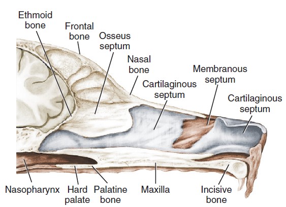
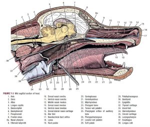
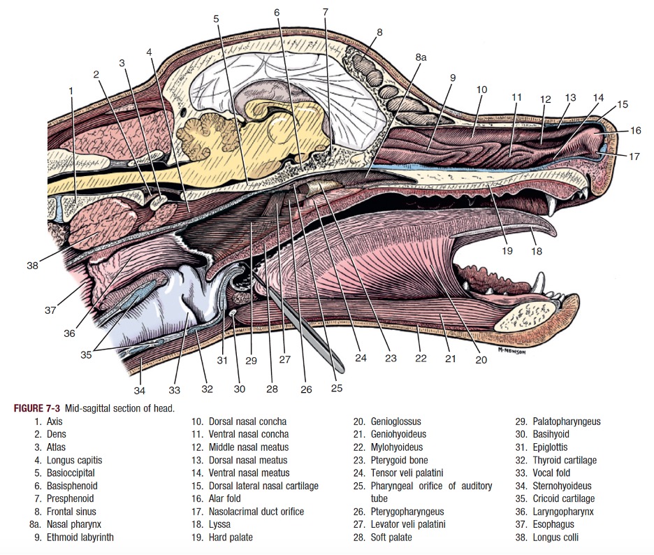
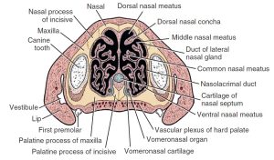
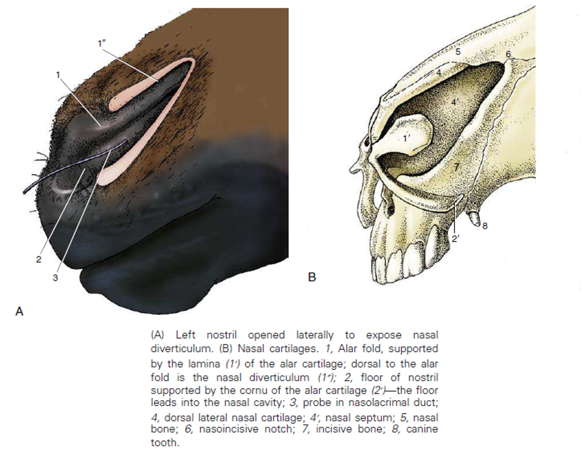
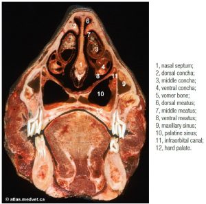
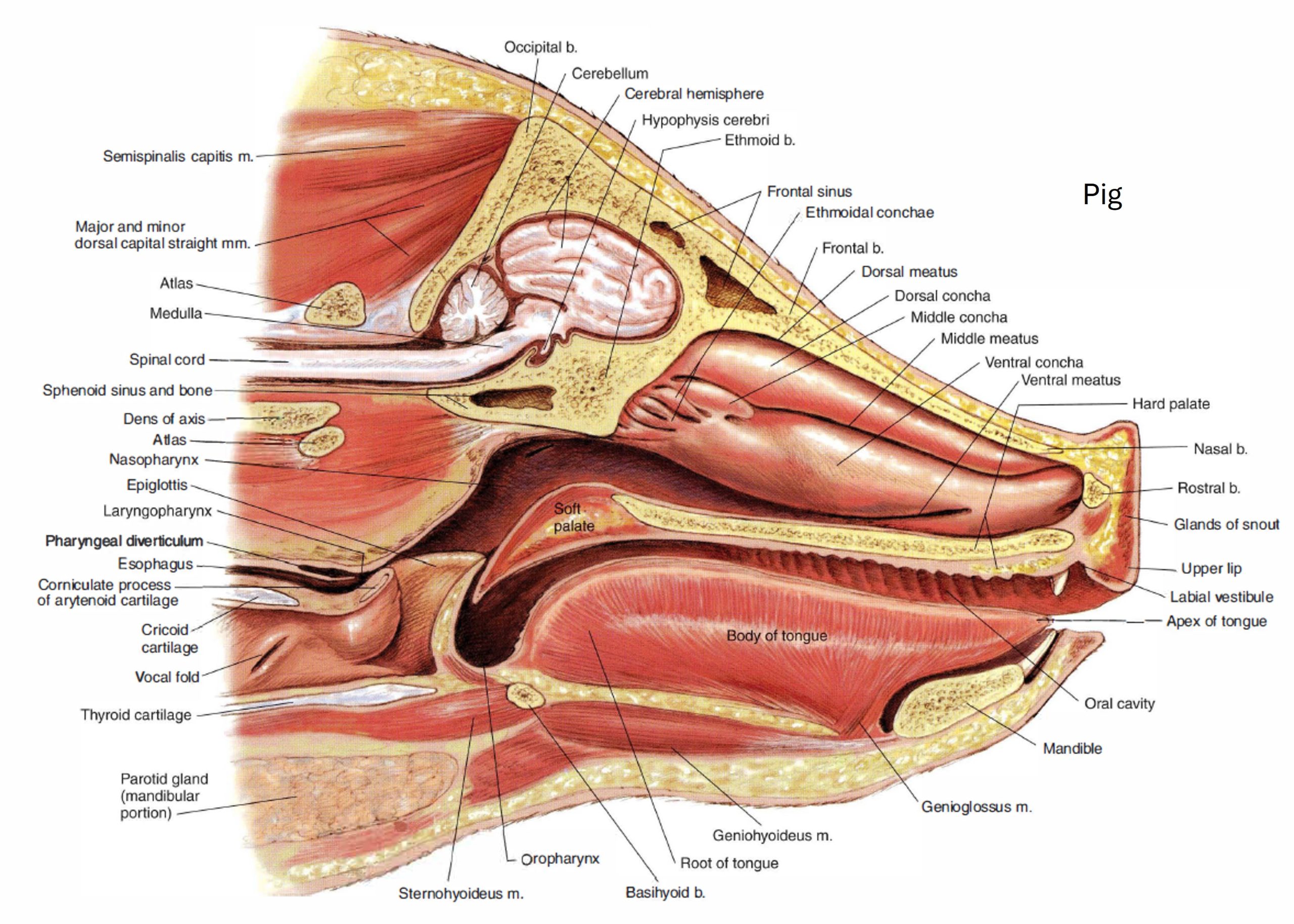
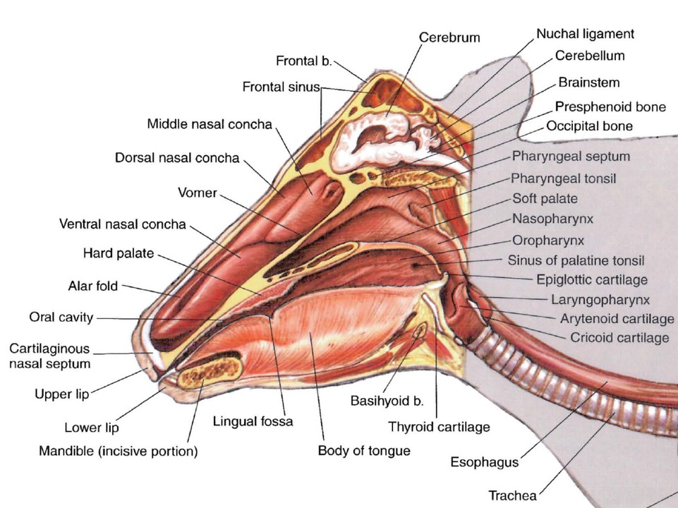
The external nose was studied in the Integument System (Semester 1). The nose is a construct of cartilages, modified epidermis, dense connective tissues and associated attached facial muscles. The openings in the nose are the nostrils (nares, singular naris) and these provide access to the nasal cavity. The vertical groove between the two nostrils and superior lip is the philtrum. The nasal cavity is separated on midline into left and right sides by the nasal septum. The nasal septum is primarily cartilaginous for its rostral 2/3rds, with central membranous and caudal osseous parts. Ventrally, the nasal septum fits into a groove formed by the vomer (single, V-shaped bone of the skull studied in the Musculoskeletal System); dorsally, it articulates with the nasal bones where they meet at the midline.
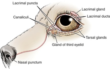
Once through the nostrils, the nasal cavity begins as the short nasal vestibule (vestibulum nasi). The nasal vestibule is lined by stratified squamous epithelium (skin) and a line demarcates this epithelium from the continuing respiratory epithelium of the rest of the nasal cavity. An orifice on the floor of the nasal vestibule, typically close to the line of demarcation, represents the opening of the nasolacrimal duct. Recall from the eye anatomy content in Semester 1, that this duct, part of the tear pathway, drains tears from the lacrimal sac located near the medial commissure (or canthus) of the eye. The nasolacrimal duct travels rostrally through the face to open in the nasal vestibule. Another duct related to the rostral nasal cavity is the incisive duct, which provides a connection between the oral and nasal cavities. Extending caudally from the incisive duct, close to its entrance into the nasal cavity, is the vomeronasal organ. This tubular structure, about 2 cm long, lies at the base of the nasal septum dorsal to the hard palate, and is an olfactory receptor of sexual stimuli. It can sometimes be seen on the sagittal section of the head if the cut was made slightly to one side of the midline.
The left and right sides of the nasal cavity end caudally at the common nasopharynx (studied further below). The choana (plural choanae) is the site of anatomical transition from the nasal cavity to the nasopharynx. It is akin to passing from one space to the next, going through a door-less opening, and without a raised perimeter to the opening to be able to readily say “that’s it, right there”. Instructors can help explain and identify the site of the choanae on the cadaver.
-
- Sagittal section of the dog nose, showing nasal septum. 1
-
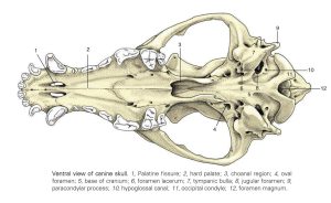
- Ventral view of dog skull. 8

-
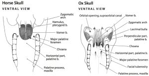
- Horse and ox ventral views of the skull, showing the choanae. 6
-

- Opening of the nasolacrimal duct in the dog. 1
Observe: Read the following descriptions and on the sectioned head specimens and on skulls separated along the median plane, study the soft tissue and bony features of the nasal conchae and nasal septum. Identify the bolded structures.
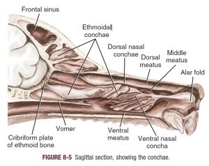
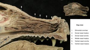
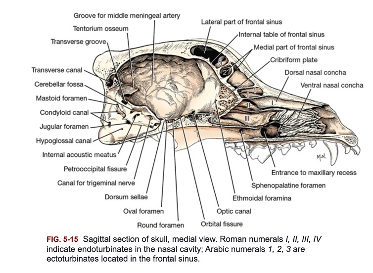
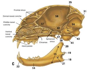
The thin bony scrolls of nasal conchae project into each side of the nasal cavity and, with their thick vascular mucosa, act as baffles to warm and cleanse inspired air. Their caudal portions also contain neuronal olfactory receptors, whose axons course to the olfactory bulbs of the brain through the cribriform plate of the ethmoid bone complex (studied in the Nervous System).
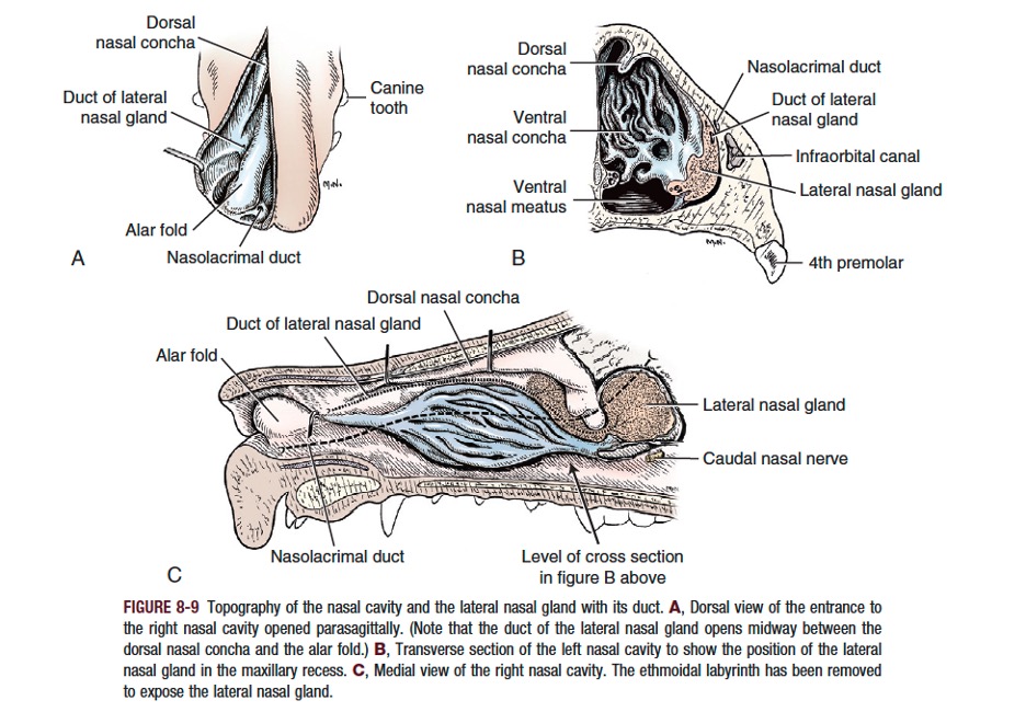
The dorsal nasal concha originates as the most dorsal scroll on the cribriform plate and extends rostrally as a shelf attached along the medial surface of the nasal bone.
The ventral nasal concha consists of several elongated scrolls that attach to the medial surface of the maxilla. It lies in the middle of the nasal cavity. A fold of mucosa, the alar fold, extends rostrally from the ventral nasal concha to the internal surface of the nose.
The ethmoidal conchae are a closely packed array of many delicate scrolls that attach to the cribriform plate caudally and occupy the fundus (caudal extent) of each nasal cavity. Their structure is reminiscent of a complex maze, and ethmoidal labyrinth is a commonly adopted term. The mucosa covering the ethmoidal conchae is rich with olfactory receptors for the special sense of smell. Dorsally, the scrolls extend as ectoturbinates into the rostral portion of the frontal sinus. Ventrally, as endoturbinates, the scrolls attach to the vomer and, in the dog, usually extend into the presphenoid bone (rostral part of the sphenoid bone complex). The ethmoidal conchae are part of the ethmoid bone complex. This central bone structure of the head, located between the cranium and the facial part of the skull, also includes the cribriform plate caudally, the median bony perpendicular plate forming the osseous part of the nasal septum, and the orbital laminae laterally. The ethmoid bone complex is surrounded by the frontal bone dorsally, the maxilla laterally, and the vomer and the palatine ventrally.
-
- Canine sagittal nasal cavity.3

-
- Canine midline section of head.1
-

- Canine nasal cavity topograph and lateral nasal gland.1
-
- Dog nose

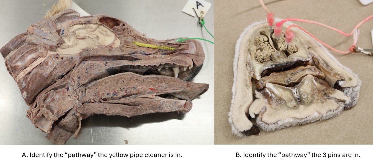
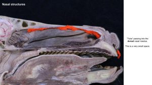
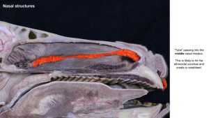
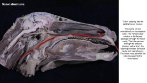
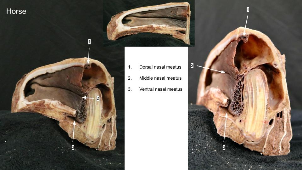
In each nasal cavity, the shelf-like dorsal nasal concha and the scrolls of the ventral nasal concha divide the cavity into four primary passages known as meatuses. The dorsal nasal meatus lies between the nasal bone and the dorsal nasal concha. The small middle nasal meatus lies between the dorsal nasal concha and the ventral nasal concha, whereas the ventral nasal meatus is between the ventral nasal concha and the floor of the nasal cavity. Because the conchae do not normally reach medially to contact the nasal septum, a sagittal vertical space, the common nasal meatus, is present on each side of the nasal septum. This space extends from the nares to the choanae in a longitudinal direction, and vertically, from the dorsal wall to the ventral floor of the nasal cavity.
-
- Transverse section of nasal cavity of the dog. 1
-
- Canine sagittal nasal cavity.3
Clinical Application
The ventral nasal meatus is the pathway along which to pass a nasopharyngeal, nasotracheal, nasoesophageal, or nasogastric tube. It is the largest meatus and is continued caudally and directly, as the tube passes through the choana, by the space of the nasopharynx.
Nasopharynx – a part of the pharynx
Observe: Read the following description of the pharynx and identify the bolded structures. Cadavers and wet and dry lab specimens should be examined.
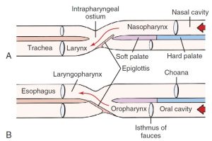
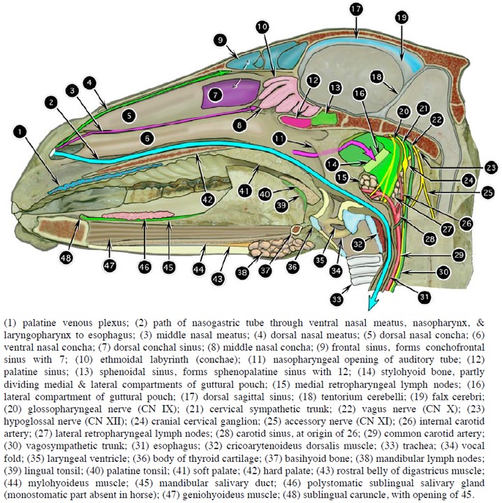
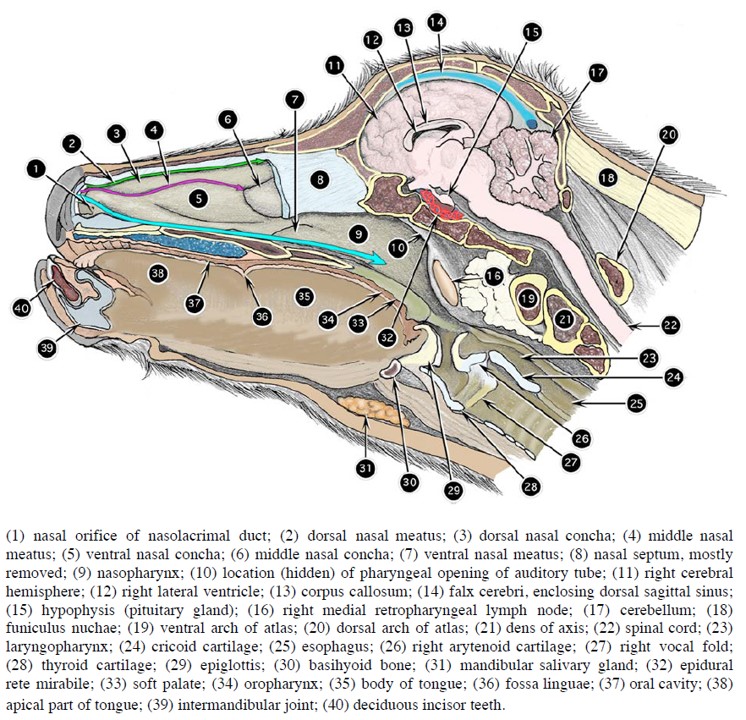
The pharynx is a muscular and mucosal-lined structure and space that has parts in both the upper respiratory and digestive systems. For context, all three parts of the pharynx are named here: nasopharynx, oropharynx, laryngopharynx. It forms the cross-roads of the intersecting respiratory and digestive tracts, allowing for food and liquids to be swallowed into the esophagus and air to be breathed into the larynx without compromise to the other tract.
The oropharynx and laryngopharynx are associated with the digestive system and will be considered in detail in that unit of study. The nasopharynx, as previously noted, continues the nasal cavity and extends caudally to be continued by the laryngopharynx and larynx. The floor of the nasopharynx is formed by the hard and soft palates. The nasopharynx extends from the choanae to the margin of the palatopharyngeal arches and the caudal border of the soft palate. A palatopharyngeal arch is a fold of mucosa that covers the palatopharyngeus muscle; there is a left and right arch. The arches pass caudolaterally along the walls of the pharynx, one each side, extending from the caudal border of the soft palate. The arches meet on dorsal midline. Therefore, the arches and the soft palate form a complete ring of soft tissue, creating an opening between the space of the nasopharynx and the space of the laryngopharynx. This opening is called the intrapharyngeal ostium, and we will focus on it more in the horse, for clinical reasons. On the lateral wall of the nasopharynx, dorsal to the middle of the soft palate, is an oblique, slitlike opening, the pharyngeal opening of the auditory tube. In the caudodorsal wall of the nasopharynx lymphoid tissue accumulates as the pharyngeal tonsils.
-
- Canine midline section of head.1
-
-
Diagram of the pharyngeal chiasma. A, During respiration.
B, During swallowing. 1
-

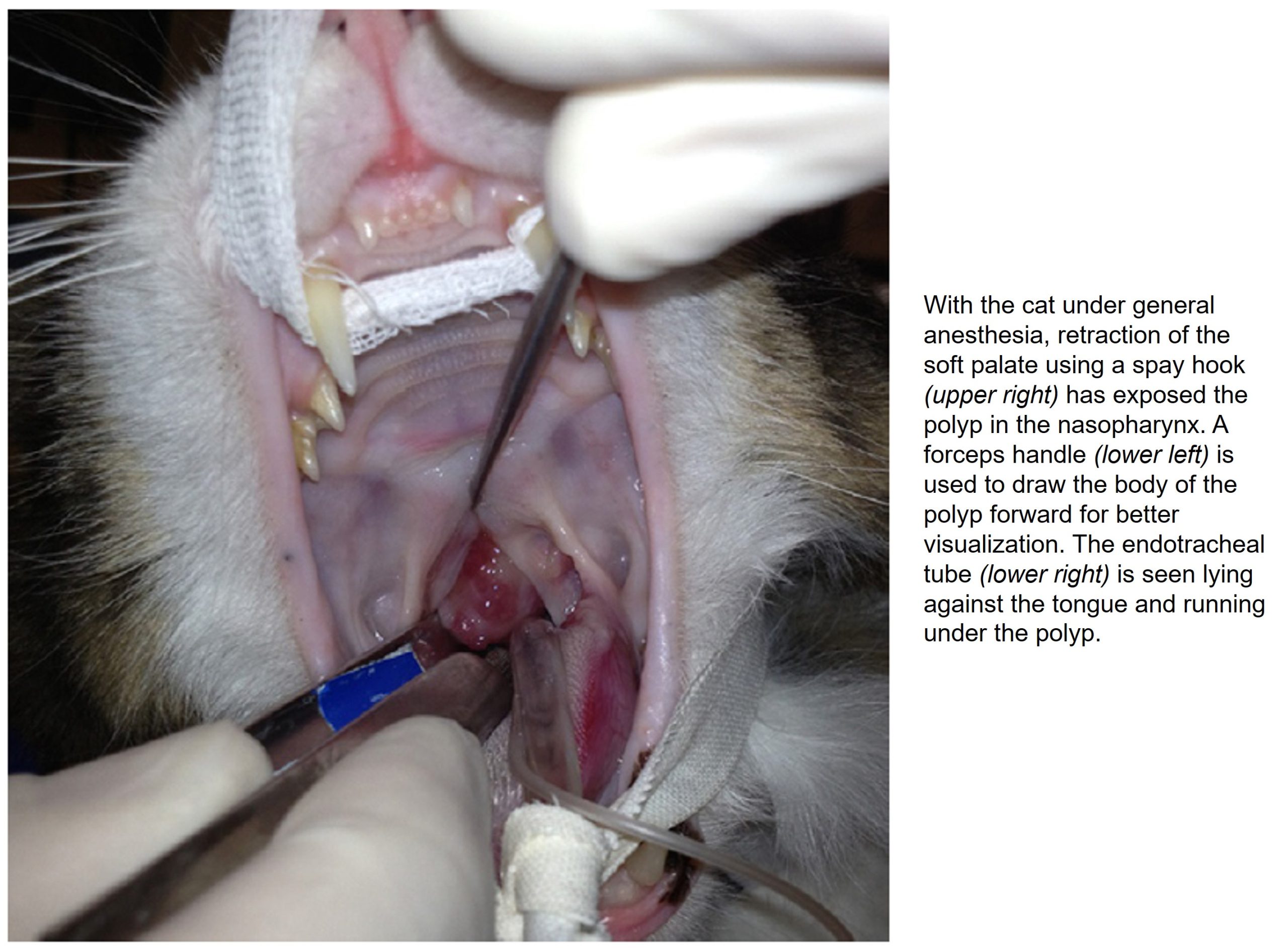
- A nasopharyngeal polyp in a cat. 9
-
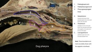
- Dog pharynx

-
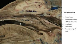
- Dog nasopharynx

Ventral to the soft palate, and therefore ventral to the nasopharynx, lies the oropharynx. The laryngopharynx is dorsal to the larynx, and is the common pharyngeal continuation of the nasopharynx and oropharynx. Functionally, it primarily belongs to the digestive tract. It extends from the previously named palatopharyngeal arches, plus the base of the epiglottis, to the beginning of the esophagus.
A series of pharyngeal muscles affects significant actions of the pharynx, including the complex, highly integrated function of swallowing. A few of these muscles will be considered when studying the digestive tract.
Regarding the nasopharynx, muscles of the soft palate and nasopharyngeal walls are very important for function. In depth study of the function of these muscles has been performed in the horse, because of diseases that result in collapse and narrowing of the nasopharynx or displacement of the soft palate (see comparative ungulate content in this lab). These diseases are performance limiting problems in the equine athlete due to subsequent airway obstruction. These muscles are not dissected and are listed here for interest and completeness only:
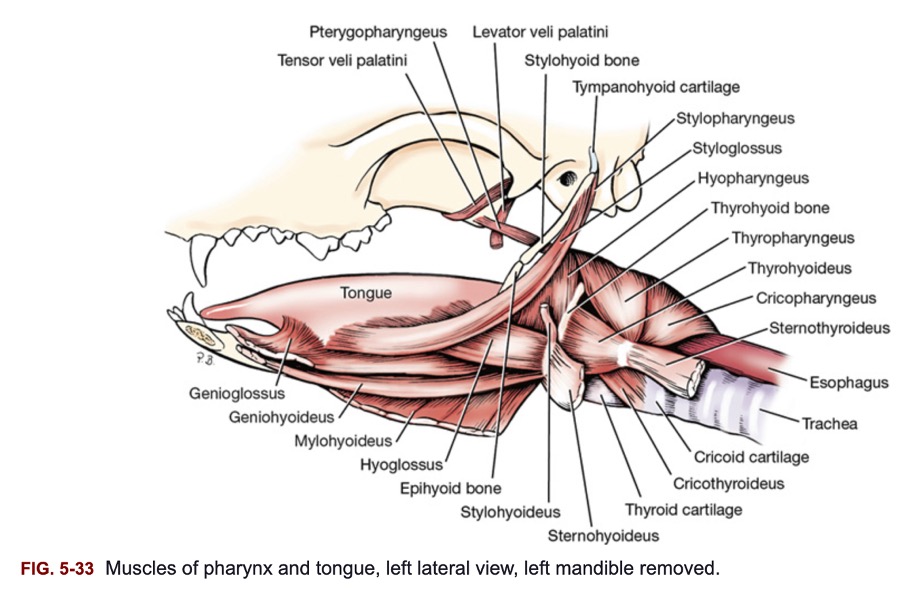
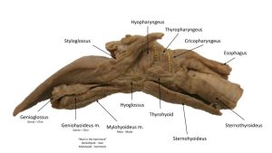
- The palatopharyngeus m. passes from the soft palate into the lateral and dorsal wall of the pharynx. Its border is in the palatopharyngeal arch. This muscle constricts and shortens the pharynx.
- The pterygopharyngeus m. arises from the pterygoid bone, passes caudally, and is inserted in the dorsal wall of the pharynx. This muscle constricts and shortens the pharynx.
- The stylopharyngeus m. arises from the stylohyoid bone and passes caudolaterally deep to the hyopharyngeus and thyropharyngeus mm. to be inserted in the dorsolateral wall of the pharynx. It acts to dilate the pharynx.
- The levator veli palatini m. arises from the tympanic part of the temporal bone and passes ventrally to enter the soft palate caudal to the pterygoid bone. It raises the caudal end of the soft palate.
- The tensor veli palatini m. arises mainly from the cartilaginous wall of the auditory tube and is inserted on the pterygoid bone and medially on the soft palate.
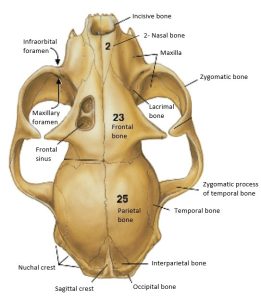
Paranasal sinuses – carnivore
Observe: Use available skulls and cadavers to identify the following bolded structures.
Paranasal sinuses are air-filled spaces between outer and inner cortices of bones of the skull. Put another way, the bone has been pneumatized, making it lighter in construct. The paranasal sinuses are lined with respiratory mucosa and they drain directly or indirectly into the nasal cavity. The functions of the paranasal sinuses include protection of the deeper located structures of the head, a reduction in the weight of the head, and immune defense. Paranasal sinuses are present in all the species we are studying and may be the focus of disease conditions.
In the canine, there is a lateral, rostral and medial frontal sinus located between the outer and inner tables of the frontal bone. The feline (like the horse), on the other hand, has a singular frontal sinus. The lateral frontal sinus in the canine is much larger than the others. Its size and shape vary with the type of skull. It occupies the zygomatic process and extends caudally, bounded laterally by the temporal line and medially by the median septum. It may be partially divided by bony septa extending into it. Ethmoidal ectoturbinates project into the rostral floor of the sinus. The rostral frontal sinus is small and lies between the median plane and the orbit. The ethmoid labyrinth bulges into this sinus. The medial frontal sinus lies between the median septum and the walls of the other two sinuses. It is very small and may be absent. As previously mentioned the sinuses communicate with the nasal cavity for drainage.
Cats also have paired sphenoid sinuses within the sphenoid bone complex, separated by a septum. These sinuses, which are not found in dogs because they are filled by endoturbinates (see ethmoidal conchae above), are the most caudal of the paranasal sinuses and can generally be found along the cut, medial edge of the split heads. The other sinuses may need to be observed in bony models in which these sinuses are specifically opened.
Observe: For the next two structures below – The maxillary recess is observed on a skull specimen and the lateral nasal gland is not identified. Discern the location of the lateral nasal gland and describe its function.
The maxillary recess of carnivores is a sinus in function but it is not called a sinus because it is not an enclosed space of the maxilla. The walls of the maxillary recess are formed laterally and ventrally by the maxilla, palatine and lacrimal bones and medially by the orbital lamina of the ethmoid bone, with a narrowed opening to this functional cavity. The recess lies in a plane expanding the upper 4th premolar to last molar tooth. The recess communicates with the nasal cavity. The lateral nasal gland occupies the rostral portion of the maxillary recess. This gland’s duct opens rostrally into the dorsolateral aspect of the nasal vestibule, and its serous secretion prevents rostral nasal desiccation caused by nasal panting.
-

- Canine sagittal skull section.1
-
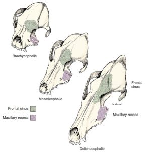
- Paranasal sinuses in three types of skulls of the dog. 1
-
- Canine nasal cavity topograph and lateral nasal gland.1
-
- Cat skull with mandible, medial view.4
-
- Cat skull, dorsal view.4

Hyoid apparatus (revisited) and related muscles
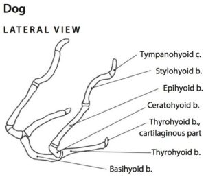
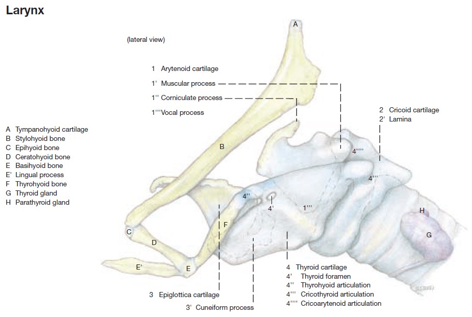
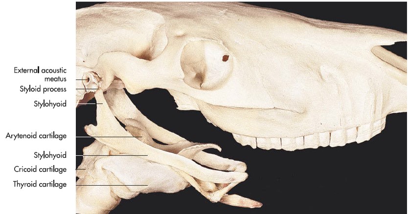
The hyoid apparatus is a construct of bones that attach the root of the tongue and larynx to the base of the skull. ‘Hyoid’ is from the root words ‘hyo’-[Gr] related to U, the letter Upsilon, and ‘-eidos’ [Gr] shape or form. Therefore the hyoid is the U-shaped set of bones. All bones are paired, except the basihyoid. The hyoid is attached to the skull via the tympanohyoid cartilage, which connects the stylohyoid to the mastoid process of the petrous temporal bone in carnivores. At the larynx, the thyrohyoid bones attach to the rostral cornua of the thyroid cartilage of the larynx, via a short cartilaginous end. Remaining hyoid bones include the epihyoid and ceratohyoid. The hyoid apparatus is flexible, moving with swallowing, for example. This structure was studied in the Musculoskeletal System (as part of the axial skeleton) and returns here because of its close relationship to the larynx and the equine guttural pouch. It may show up again because of its association with the Alimentary System!
Observe: Review specimens available in the lab and recall the bolded names of the bones of the hyoid apparatus. Portions of the bones of the hyoid are also visible on the sectioned cadaver heads.
-
- Dog hyoid apparatus. 6
-
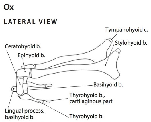
- Ox hyoid apparatus. 6
-
- Horse hyoid apparatus. 6
The hyoid muscles have an attachment to the hyoid apparatus, which suspends the larynx and anchors the tongue. They function in swallowing, lolling, lapping, and retching, causing movements of the tongue and larynx. All muscles of this group have names with the suffix –hyoideus. The prefixes of the hyoid muscles designate the bone or part from which they arise.
Observe/dissect as needed: Review specimens available in the lab to identify and study the bolded muscles of the hyoid apparatus. Also isolate the muscles to be able to identify them on the sectioned cadaver head. The sternohyoideus m. stumps will be present.
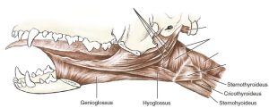
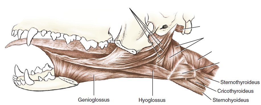
The sternohyoideus m., studied in the MSK system, is the ventrally located neck strap muscle, attached at the sternum caudally and to the basihyoid bone cranially. Recall, it travels with the sternothyroideus m. much of its pathway and then these two muscles diverge cranially to their respective insertions. These muscles draw the larynx caudally. The thyrohyoideus m. is a short muscle that lies dorsal to the sternohyoideus. It extends from the thyroid cartilage of the larynx to the thyrohyoid bone. The sternohyoideus and thyrohyoideus muscles are innervated by ventral branches of cervical spinal nerves and the hypoglossal nerve (CN XII).
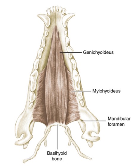
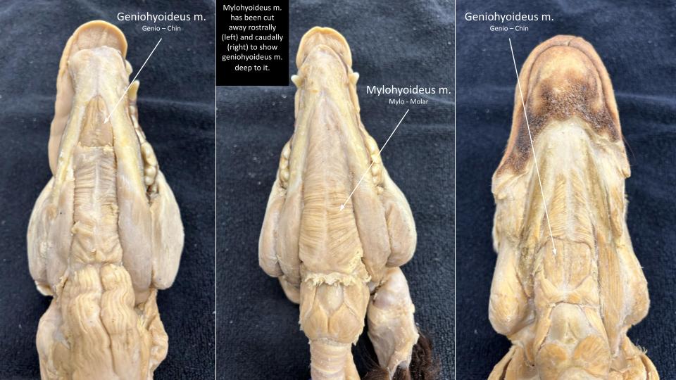
The mylohyoideus m. spans the intermandibular space. It arises as a thin sheet of transverse fibers from the medial surface of the body of the mandible. It is inserted on its fellow muscle at the midventral raphe. Caudally, it inserts on the basihyoid. It forms a sling that aids in the support of the tongue. Recall from the Nervous System, the similarly named mylohyoid n. (from mandibular n. of CN V) innervates the mylohyoid m.
The geniohyoideus m. lies deep to the mylohyoideus. It is a muscular strap that arises on and adjacent to the intermandibular articulation. It parallels its fellow along the median plane and attaches to the basihyoid. Contraction of the geniohyoideus draws the hyoid apparatus and larynx rostrally. It is innervated by the hypoglossal nerve.
-
- Muscles of the tongue and pharynx of the dog, lateral aspect. 1
-
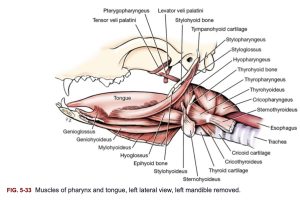
- Muscles of pharynx and tongue, left lateral view, left mandible removed, canine.1
-

- Hyoid muscles canine dorsal view of mandible.1
-
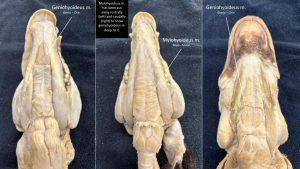
- Dog mylohyoideus and geniohyoideus mm.
-
- Dog hyoid mm.

COMPARATIVE UNGULATE ANATOMY
Observe: Apply your knowledge of the carnivore anatomy to identify similar and unique features in our ungulate species. Study the prosected and sectioned head specimens to learn the anatomy.
Nose and Nasal cavity
The external anatomy of the ungulate nose has been considered in the integument unit. Recall the nasal plane of the small ruminant (similar to the carnivore), the expansive nasolabial plane of the ox, and the rostral plane of the pig snout-disk. The horse has great capacity to dilate its nostrils for maximal intake of air when exercising strenuously.
Clinical Application
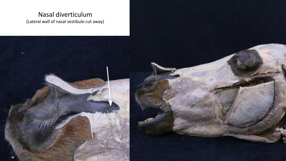
A unique feature of the nasal vestibule in the horse is the presence of a blind-ended cutaneous pouch, the nasal diverticulum, aka the false nostril. This pouch is located caudodorsolaterally over the region of the nasoincisive notch (skull feature) and may assist with funneling air into the nasal cavity. A tube to be passed via the nasal cavity could be misdirected into the nasal diverticulum.
Observe in horse: Insert a finger caudally into the dorsolateral part of the nostril and define a blind-ended cutaneous pouch. This is the nasal diverticulum.
-

- Opening of the nasolacrimal duct in the horse. 7
-
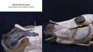
- Horse nasal diverticulum
Clinical Application – nasal diverticulum
 Q1: This horse presented with a slowly expanding soft swelling as shown in the image. The horse elicited no pain to palpation of the swelling and it did not feel to have increased heat. Airway sounds were normal and airflow was normal from both nostrils. There has been no nasal discharge. What is your most likely diagnosis?
A – Abscess secondary to foreign body
B – Atheroma
C – Squamous cell carcinoma
Observe: examine the medial surfaces of the sectioned head specimens to identify the bolded comparative structures of the nasal cavity in the ungulates. Refer to labeled images for assistance.
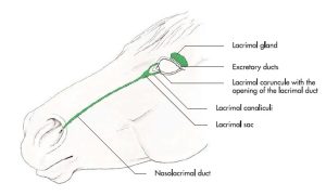
The opening of the nasolacrimal duct is readily identifiable in the horse at the mucocutaneous junction of the nasal vestibule, where skin meets respiratory mucosa. The opening is ~2mm diameter and usually lies on the cutaneous side of the junction, on the floor of the vestibule.
-
- The lacrimal apparatus of the horse. 7

-
- Opening of the nasolacrimal duct in the horse. 7
Clinical Application – nasolacrimal duct
The thick nasal septum partitions the nasal cavity into left and right sides. In the horse, the dorsal nasal concha and ventral nasal concha each have rostral scrolled parts and caudal sinus parts, separated by a conchal septum. The caudal conchal sinus regions are considered part of the paranasal sinuses complex (see Lab 3A). The rostral conchal scrolls contain air spaces called bullae, that are subdivided further into cells by transverse septa. Spelunking may be a good way to think of exploring the cavities of the equine conchae. Spelunking? From the [L] spelunca, from the [Gk] spelynx, which means “cave”. Some cave-exploring enthusiasts are evidently good with the less obscure terms “caver and caving.”
-
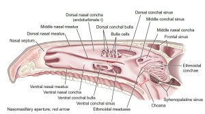
- The right side of the equine nasal cavity, with the nasal septum removed. 9
-
- Sagittal section of horse head. 2
-
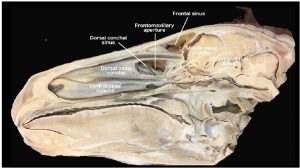
- Sagittally sectioned equine skull showing the structures of the right nasal passage and their association with the paranasal sinuses. 9

-
- Horse nasal structures

The dorsal nasal concha of ruminants and swine is relative simple, compared to the horse. The ventral nasal concha of ruminants and swine is slightly more complex in that it has a dorsal and ventral scroll rolling from a common central origin.
-

- Sagittal section of newborn calf head. 2
-
- Sagittal section of a prosected Ox head. 9

-
- Transverse section of an Ox prosection at the level of the last premolars. 9
Clinical Application – nasal conchae, nasal septum
Beyond the physiological function of filtering and humidifying inspired air the nasal conchae are not uncommonly a focus of upper respiratory tract disease management, particularly infections. Infections of the nasal conchae can be complicated to treat because of the many ‘nooks and crannies’ for organisms to colonize and for exudate to accumulate, and the consequent challenges in effectively flushing and draining these spaces. Innovative techniques have been described in the horse as the intricate anatomy of these conchal tissues has been carefully explored and mapped.
Deviation of the nasal septum may cause respiratory obstruction and limit performance in the sport horse. Nasal septum resection is a surgical treatment. One can imagine the hemorrhage related to such a surgery and the need to be prepared, to limit blood loss intra- and postoperatively.
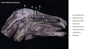
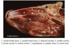
In the ethmoid region, the most prominent concha observed is the middle nasal concha. This concha is short, only extending the length of surrounding ethmoidal conchae in the horse, but extending much further in ruminants. This concha also contains a conchal sinus space within its walls. The middle nasal concha is readily observed in the horse and ruminant.
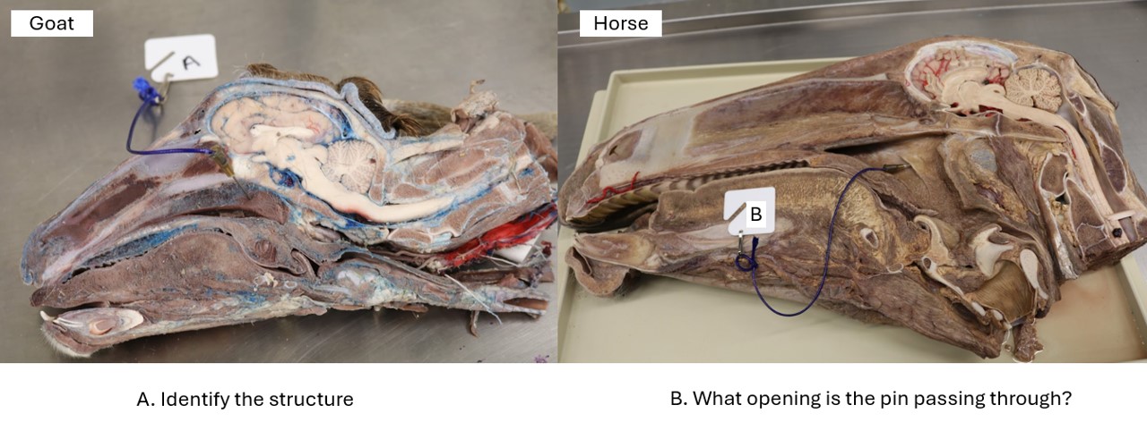
Similar to the carnivore, dorsal, middle, ventral and common nasal meatuses are present. Ethmoidal meatuses are also present between adjacent ethmoidal conchae. A fissure-like opening in the horse, the nasomaxillary opening, is located at the caudal extent of the middle nasal meatus, bound by the adjacent walls of the dorsal and ventral nasal conchae. This opening represents the communication between the paranasal sinuses and the nasal cavity and will be studied in Lab 3A. The nasal cavity ends caudally at the choanae, which mark the rostral beginning of the nasopharynx. The choanae represent the transition (an analogy is to think of passing between two rooms connected by a wide opening) from the nasal cavity space into the nasopharynx space.
-
- The right side of the equine nasal cavity, with the nasal septum removed. 9
-
- Sagittal section of horse head. 2
-
- Sagittal section of newborn calf head. 2
-
- Horse and ox ventral views of the skull, showing the choanae. 6
-
- Dorsal nasal meatus

-
- Middle nasal meatus

-
- Ventral nasal meatus

-

- Horse nasal meatus
Clinical Application
-
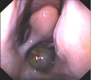
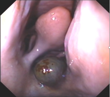
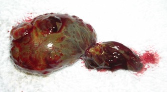
- A. Endoscopic view of ethmoid hematoma in nasal cavity, ventral to middle nasal concha.
-
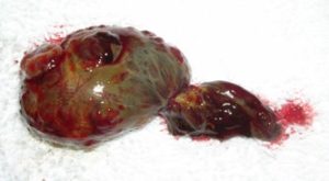
- B. Ethmoid hematoma in A. removed from nasal cavity by snaring.
Nasopharynx – a part of the pharynx
Observe: Structures of the pharynx are again best identified on the medial surface of the sectioned head. Refer to figures to help identify the bolded structures.
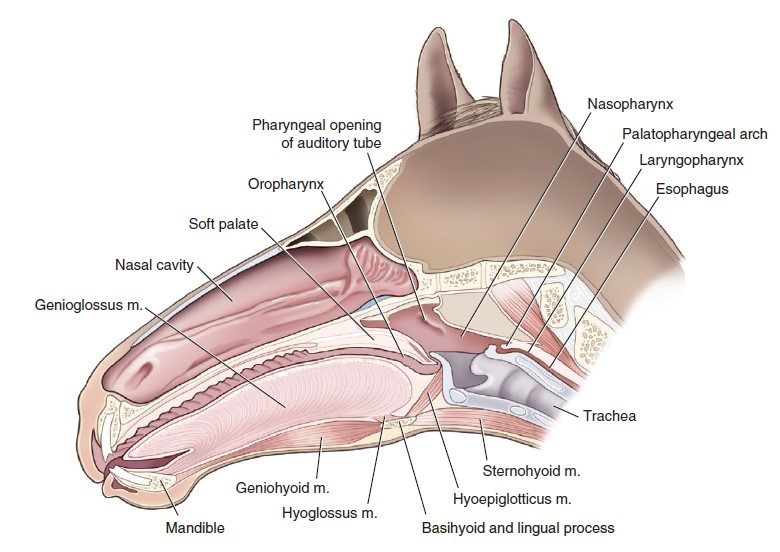
As for the carnivore, the pharynx consists of 3 main regions: nasopharynx, oropharynx, laryngopharynx. The nasopharynx continues the nasal cavity at the choanae and lies dorsal to the hard and soft palate. The nasopharynx ends caudally at the palatopharyngeal arches and the caudal border of the soft palate. The arches and the soft palate form a complete ring of soft tissue, creating an opening between the space of the nasopharynx and the space of the laryngopharynx. This opening is called the intrapharyngeal ostium, and the rostral aspect of the larynx projects through this opening into the nasopharynx space, in the normal horse. This anatomy will be considered in more detail in Lab 2A, when the larynx is studied. The pharyngeal opening of the auditory tube (left and right sides) is seen as a large cartilaginous flap in the horse, and a relatively small, tight opening in the ruminant and pig. The (dorsal) pharyngeal recess is a blind-ended niche in the caudodorsal region of the nasopharynx in ungulates. This recess is not present in the carnivore. In the horse, uncommonly, the pharyngeal recess may be extended further caudally by an epithelial tubular pouch, an inconstant vestige that can also occur as an independent evagination of the nasopharynx. In the donkey, the pharyngeal recess is normally a much deeper tubular cavity compared to the horse.
FYI for now – the oropharynx continues the oral cavity and is ventral to the soft palate. The oropharynx ends caudally at the base of the epiglottis. The laryngopharynx continues the nasopharynx and oropharynx. The laryngopharynx occupies the space lateral to the entrance of the larynx (this part of the laryngopharynx forms the piriform recesses) and then continues dorsal to the larynx to connect with the esophagus. These parts of the pharynx will be considered in detail in the Alimentary System unit.
-

- The 3 parts of the equine pharynx: nasopharynx, orophraynx, and laryngopharynx.9
-
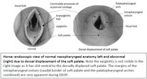
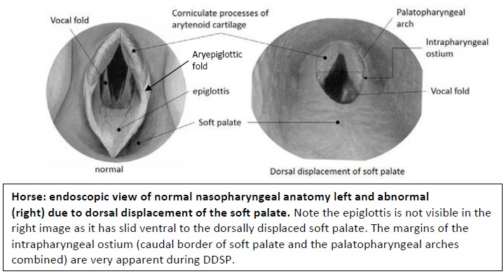
- Horse: endoscopic view of normal nasopharyngeal anatomy left and abnormal (right) due to dorsal displacement of the soft palate.
-
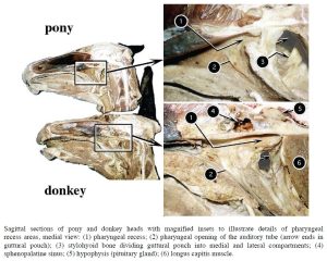
- Sagittal sections of pony and donkey heads with magnified insets to illustrate details of pharyngeal recess areas, medial view.2

Clinical Application – nasopharynx
Dynamic collapse of the nasopharynx in the horse occurs at high intensity exercise. The lumen of the nasopharynx will narrow on inspiration, likely due to neuromuscular dysfunction. This is a performance limiting problem. Scarring of the nasopharynx can also narrow the lumen. Scarring is secondary to inflammation and the condition is referred to as nasopharyngeal cicatrix. Dorsal displacement of the soft palate ie DDSP, occurs in the horse when the soft palate, which is the floor of the nasopharynx, flips up and lies on top of the epiglottis, causing partial airway obstruction – this anatomical dysfunction will be better understood once the larynx is studied. The pharyngeal opening of the auditory tube provides a handy entrance into the guttural pouch in the horse and we are often driving an endoscope into the pouch to examine it (see future lab). A condition of young, maturing horses (and other species) is pharyngeal lymphoid hyperplasia (PLH), diagnosed on endoscopic exam. Lymphoid tissue may swell to the point of appearing like a cluster of grapes in the pharyngeal recess – related to the young animal’s exposure to a novel environment and the consequent immune response. PLH is usually self limiting but may require medical treatments to help ‘settle things down’.
Pharyngeal collapse in a horse – 1 min
Observe: The unique pharyngeal diverticulum in the pig is identified on the sectioned head specimen.
Unique to the pig, the nasopharyngeal mucosa forms a median caudal blind pouch, the pharyngeal diverticulum, directly dorsal to the laryngopharynx and esophagus.
-

- Head of the pig, showing the pharyngeal diverticulum.30
Clinical Application – pharyngeal diverticulum
Observe: The unique pharyngeal septum in the pig and ruminant is identified on sectioned head specimens, especially those that were sectioned just off midline.
Unique in the ruminant and pig, a vertical midline tissue septum, the pharyngeal septum, provides partial (ruminant) or complete (pig) division of the nasopharynx in its rostral portion. The pharyngeal septum is a direct continuation of the nasal septum. Pharyngeal tonsils are located on the pharyngeal septum.
-
- Pharyngeal septum of the ox. 30
Paranasal sinuses
This complex system of sinuses in the ungulate will be examined in Lab 3A (phew!).
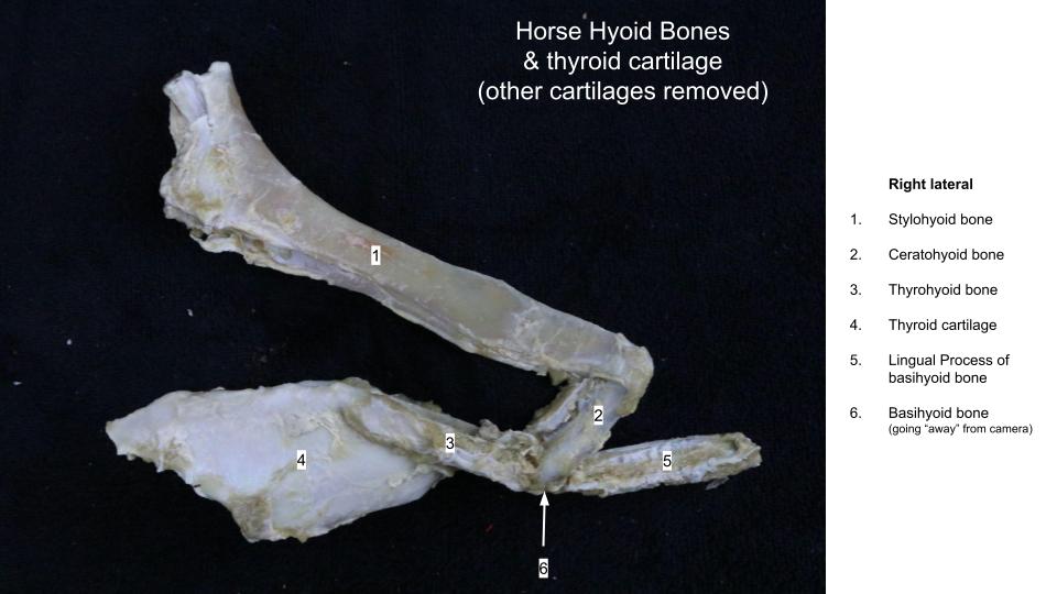
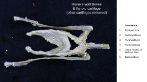
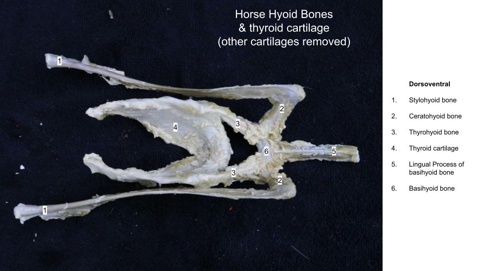
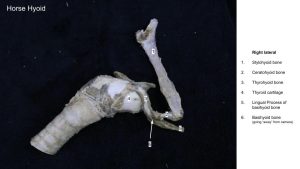
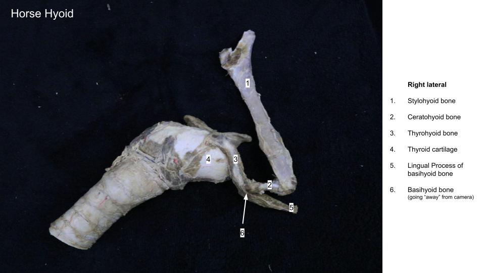
Hyoid apparatus – horse only
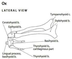
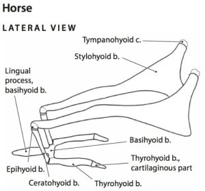
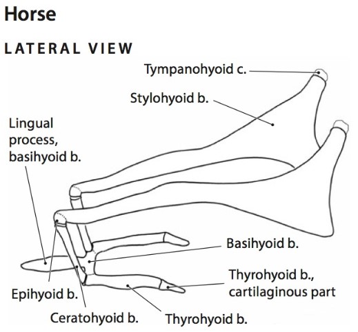
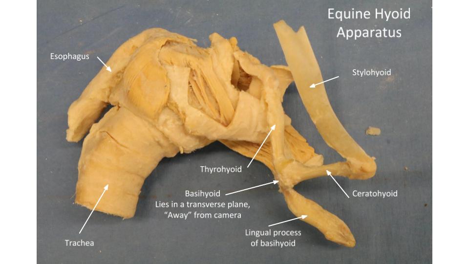
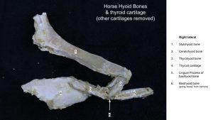
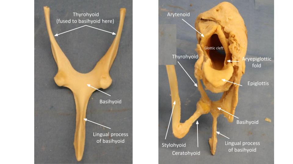
The horse hyoid apparatus differs from carnivores. Firstly, the epihyoid is fused to the stylohyoid, and it is not identified as a separate bone. Therefore the ceratohyoid connects the stylohyoid to the basihyoid. Secondily, the horse has a prominent lingual process that extends rostrally, from the midpoint of the basihyoid, into the root of the tongue [FYI – the ruminant hyoid apparatus has a lingual process too, shorter and rounded, compared to the elongate, pointed equine lingual process]. Another feature of the horse is that the basihyoid fuses to the thyrohyoid bones at their articulation, creating a large ‘wishbone’ structure.
As for the carnivore, all bones are paired, except the basihyoid. The hyoid is attached to the skull via the tympanohyoid cartilage, which connects the stylohyoid to the styloid process of the petrous temporal bone in the horse and ruminants (mastoid process of the petrous temporal bone in carnivores). At the larynx, the thyrohyoid attaches to the rostral cornu of the thyroid cartilage of the larynx, via a short cartilaginous end.
Muscles with attachments to the hyoid apparatus in the ungulate are similar to the carnivore, and do not need to be identified.
Observe: Study the horse hyoid apparatus to recall the bolded bones previously learnt in the Musculoskeletal System. Refer to images to aid in bone identification. There is no need to identify muscles of the hyoid apparatus in the horse, in part because clinically we are more concerned with the bones.
-
- Horse hyoid apparatus. 6
-

- Horse hyoid apparatus. 14
-

- Skull of a horse with hyoid bone and larynx. 7
-

- Horse hyoid
-
- Horse hyoid
-
- Horse hyoid
-
- Horse hyoid
-

- Horse lingual process of basihyoid
Clinical application – hyoid apparatus
The horse hyoid apparatus is not uncommonly a focus of surgical and medical attention. Temporohyoid osteoarthropathy (THO) is a painful inflammatory/infectious condition of the articulation of the hyoid to the skull (can you recall from the Nervous System the CN signs related to this disease and why?). Fusion of the normally movable temporohyoid joint may result from THO. The ceratohyoid on the affected side is removed to manage THO – why might this be helpful? And there are other surgical techniques for THO management.
The basihyoid and lingual process are used to help anchor suture in the laryngeal tie-forward surgery to manage dorsal displacement of the soft palate.
Review videos
Dog nasal structures, Spradley – 5 min, watch until 32:30
Dog nasal structures, Walker – 5 min, watch until 5:20
Dog nasopharynx, Spradley – 2 min, watch until 4:00
Dog nasopharynx, Walker – 2 min, watch until 19:00
Horse hyoid – Watch until 2 min
Horse, pig nasal cavity, nasopharynx – watch until 8:30
Pig nasopharynx – 1 min, watch until 12:30
Calf and pig pharyngeal septum and auditory tube – 2 min
2025 videos:
Nasal cavity vs nasopharynx – 1 min
Dog nasal cavity – 4 min
Comparative nasopharynx – 7 min
Hyoid apparatus – 5 min
Terms
| Carnivore terms |
|
|---|---|
| Nose and Nasal Cavity |
|
| Nasal cavity | Dorsal nasal concha |
| Nostril | Ventral nasal concha |
| Nasal septum | Ethmoidal conchae |
| Nasal vestibule | Dorsal nasal meatus |
| Opening of nasolacrimal duct | Middle nasal meatus |
| Choana(e) | Ventral nasal meatus |
| Common nasal meatus | |
| Pharynx | Paranasal sinuses |
| Nasopharynx | Frontal sinus |
| Palatopharyngeal arches | Maxillary recess |
| Pharyngeal opening of auditory tube | Lateral nasal gland – be aware of presence, not identified. |
| Intrapharyngeal ostium | Sphenoid sinus (cat) |
| Soft palate | |
| Hyoid apparatus bones |
Hyoid muscle |
| Stylohyoid | Sternohyoideus m. |
| Epihyoid | Thyrohyoideus m. |
| Ceratohyoid | Mylohyoideus m. |
| Basihyoid | Geniohyoideus m. |
| Thyrohyoid | |
| Ungulate terms |
|
|---|---|
| Nose and Nasal cavity | Identify in all ungulates unless specified |
| Nasal cavity | |
| Nostril | |
| Nasal vestibule | |
| Dorsal nasal concha | |
| Ventral nasal concha | |
| Middle nasal concha | horse and ruminant |
| Ethmoidal conchae | |
| Dorsal nasal meatus | |
| Middle nasal meatus | |
| Ventral nasal meatus | |
| Common nasal meatus | |
| Nasal septum | |
| Nasal diverticulum | Horse = false nostril |
| Choana(e) | Opening(s) of nasal cavity into nasopharynx |
| Opening of nasolacrimal duct | Horse only |
| Nasopharynx – ungulates |
|
| Pharyngeal opening of auditory tube | |
| Palatopharyngeal arch | |
| Soft palate | |
| (Dorsal) pharyngeal recess | |
| Intrapharyngeal ostium | |
| Pharyngeal septum | Ruminant, pig |
| Pharyngeal diverticulum | Pig |
| Hyoid apparatus – horse only |
Comments |
| Stylohyoid | Attaches to skull via tympanohyoid cartilage |
| Ceratohyoid | |
| Basihyoid | |
| Lingual process (of basihyoid) | |
| Thyrohyoid | Attaches to thyroid cartilage of larynx |