Lab 6A: The Abdominal Cavity of the Carnivore (pt. 2)
Learning Objectives
- Apply the embryology of gut and peritoneum development to identify and understand the location and function of the abdominal viscera and peritoneum in the cat and dog.
- Identify the different sections of the small intestine and their mesenteries.
- Identify the different parts of the pancreas.
- Identify the different sections and flexures of the large intestine.
Lab Instructions
The Small Intestine and Pancreas
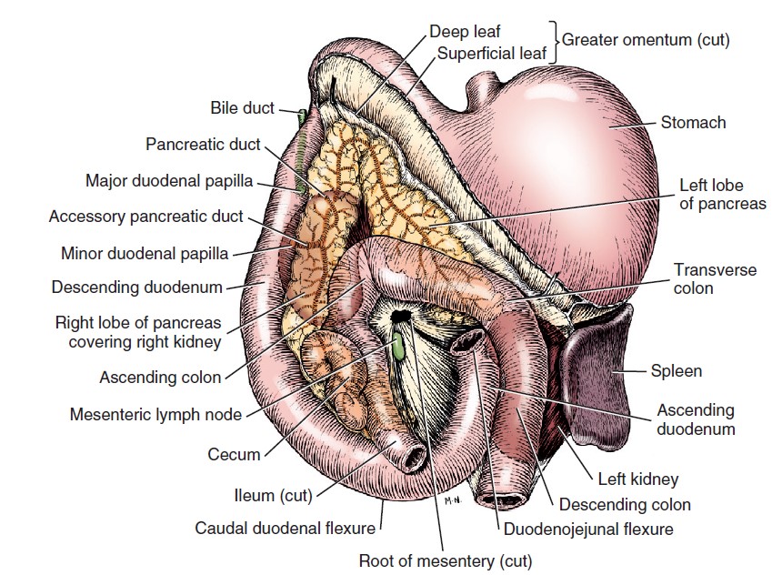
The duodenum is the most fixed part of the small intestine. It is suspended by the mesoduodenum. The mesoduodenum originates at the dorsal abdominal wall and the root of the mesentery and extends to the duodenum. On the right side it passes to the descending duodenum and encloses the right lobe of the pancreas between its layers (the left lobe of the pancreas is covered by the deep leaf of the greater omentum). Cranially, it is continuous with the greater omentum across the ventral surface of the portal vein. Caudally, the mesoduodenum passes from the root of the mesentery to the caudal flexure of the duodenum. On the left, it is attached to the ascending duodenum, and at the duodenojejunal flexure, it is continuous with the mesentery of the jejunum. The ascending duodenum is secondarily attached to the mesocolon of the descending colon by the duodenocolic fold.
-

- Abdominal viscera of the dog, ventral aspect. 1
-
-
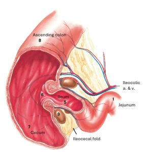
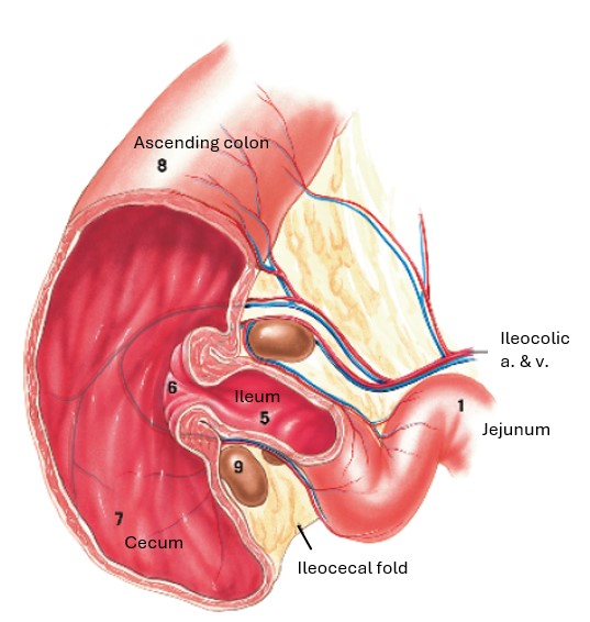
Dog small intestine in situ and its relationship to surrounding abdominal organs. Cutaways: ileocolic junction and a cross section of a
segment of jejunum with the mesenteric vessels. 9
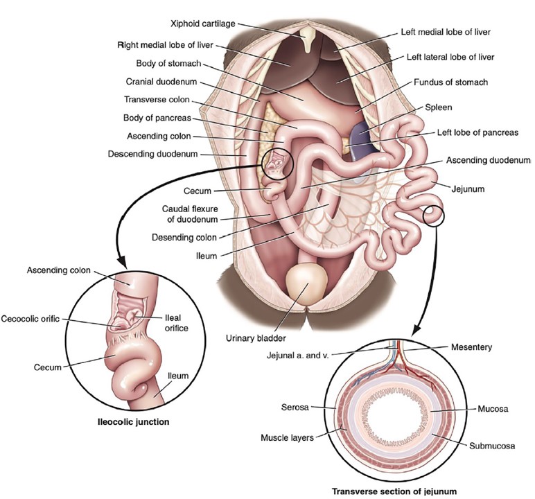
Observe: Reflect the greater omentum cranially and the jejunum to either side to expose the duodenum.
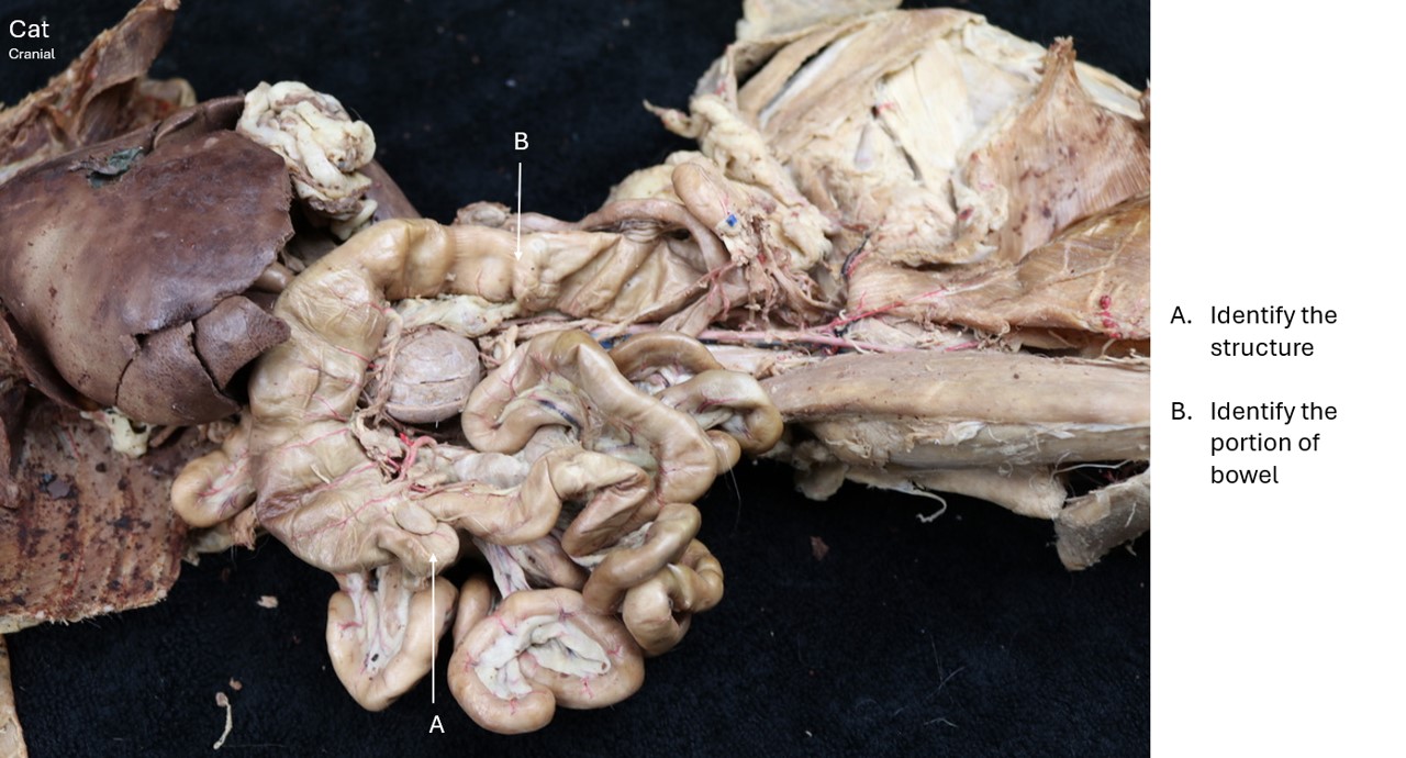
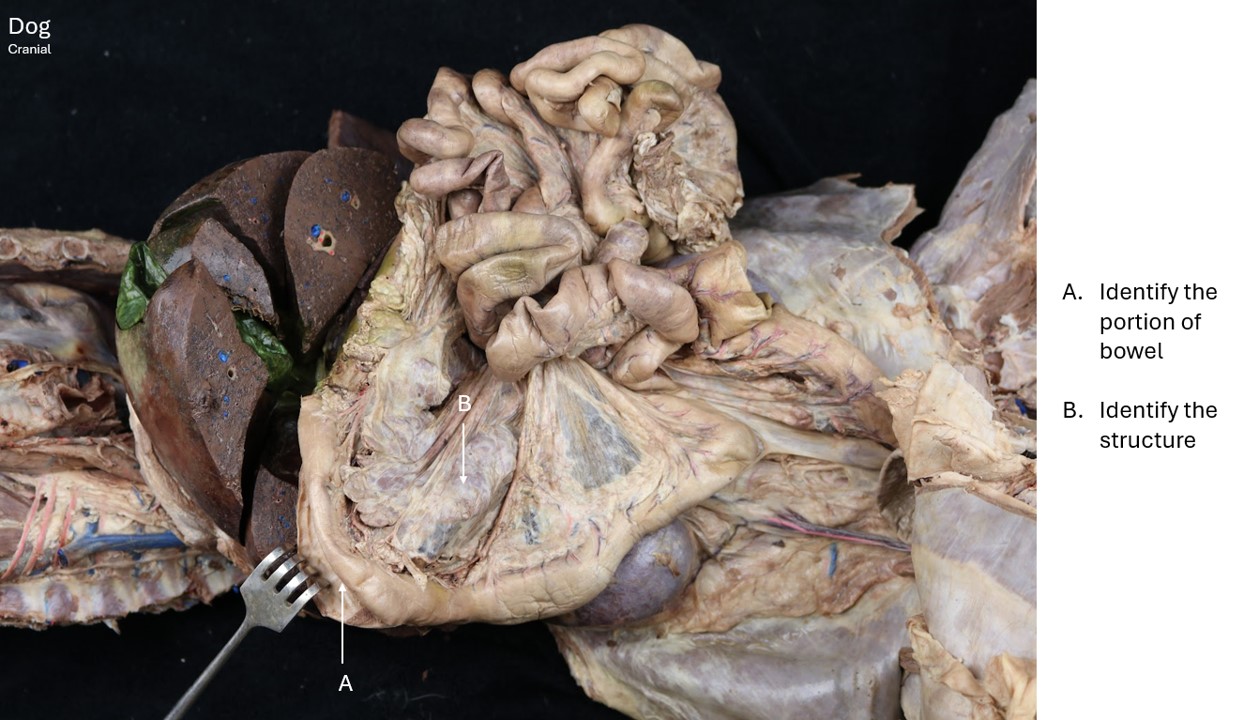
The jejunum forms the many coils of the small intestine, which occupy the ventrocaudal part of the abdominal cavity. They receive their nutrition from the cranial mesenteric artery, which is in the root of the mesentery. The root of the mesentery is the attachment of the mesentery of the jejunum and ileum (the mesojejunoileum) to the dorsal body wall. Vessels and nerves pass in the mesentery to supply the intestine and mesenteric lymph nodes lie along the vessels in the mesentery. The jejunum begins at the left of the root of the mesentery and is the longest portion of the small intestine. At the ileocolic junction, the mesojejunoileum is continuous with the mesocolon.
-
- Dog intestinal tract. 9

Observe: Trace the jejunum from the duodenojejunal flexure on the left to its termination at the ileum on the right side of the abdomen.
The ileum is the terminal portion of the small intestine. It is short and passes cranially on the right side of the root of the mesentery and joins the ascending colon at the ileocolic orifice. This narrow orifice is surrounded by a sphincter. There is no clear demarcation between jejunum and ileum.
Observe: Note the vessel (the antimesenteric ileal a.) that courses on the antimesenteric side of the ileum from the cecum toward the jejunum. This vessel approximates the length of the ileum (~10 cm in the canine, ~2.5 cm in the feline).
-
- Abdominal viscera of the dog, ventral aspect. 1
-
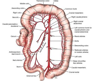
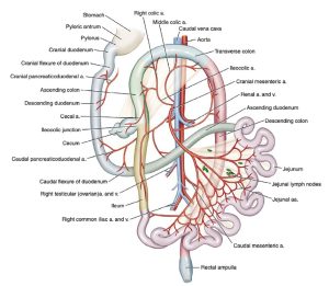
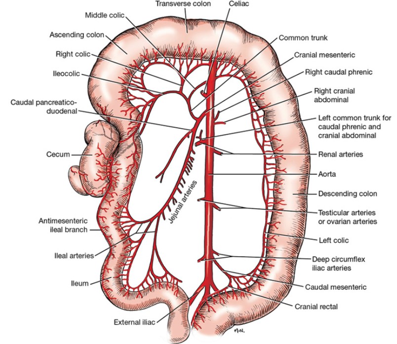
- Branches of cranial and caudal mesenteric arteries, ventral aspect. 1
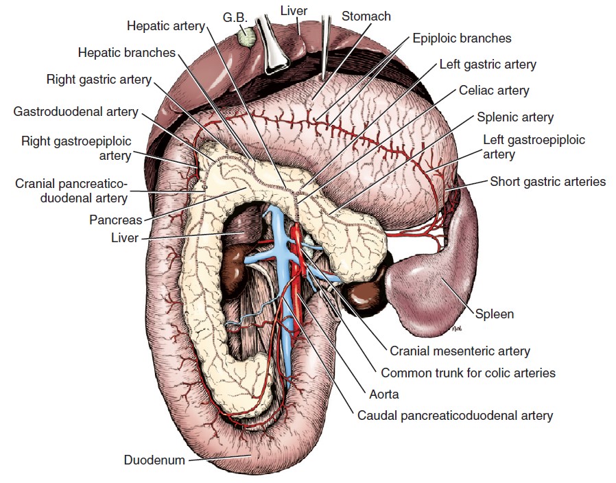
The pancreas is lobulated and is composed of a body and two lobes. The body lies adjacent to the pylorus of the stomach. The right lobe lies dorsomedial to the descending duodenum enclosed by the mesoduodenum. It is ventral to the right kidney.
Observe: Pull the descending duodenum ventrally and to the left to expose this right lobe of the pancreas in the mesoduodenum.
The left lobe of the pancreas lies between the peritoneal layers that form the deep leaf of the greater omentum. It is caudal to the stomach and liver and cranial to the transverse colon.
-
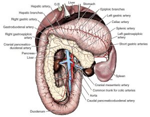
- Celiac and cranial mesenteric arteries, ventral aspect. (Stomach reflected cranially.) 1
-

- Pancreas of the cat, ventral view. 4
-
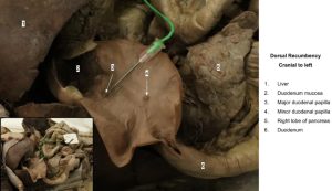
- Duodenal papilla

Observe: Reflect the greater omentum cranially and the small intestine and transverse colon caudally to observe the left lobe of the pancreas.
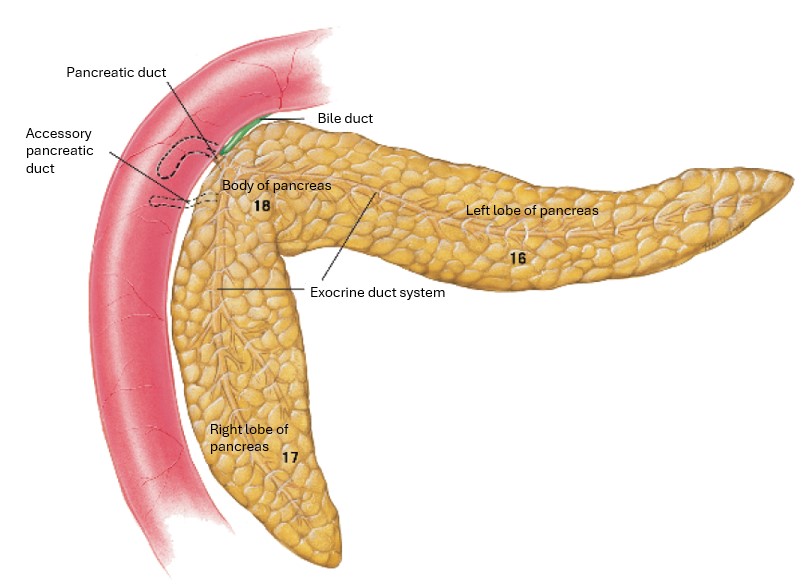
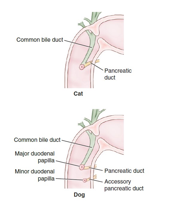
The pancreatic duct system is variable. Most dogs have two ducts; these open separately in the descending duodenum but communicate in the pancreas. The pancreatic duct is the smaller of the two ducts and is sometimes absent. It opens close to (but separate from) the bile duct on the major duodenal papilla.
Dissect: Make a 2-3 in long incision through the free border of the cranial part of the descending duodenum. Firmly scrape away the mucosa with the scalpel handle and identify the major duodenal papilla on the side the mesoduodenum attaches.
The larger accessory pancreatic duct in the dog opens into the duodenum on the minor duodenal papilla 2 to 5 cm caudal to the major papilla. Approximately 20% of cats will also have an accessory pancreatic duct, and thus a minor duodenal papilla.
-
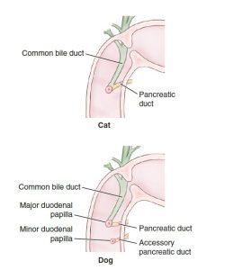
- Pancreatic and bile ducts of the dog and cat. 9
-
- Pancreas of the cat, ventral view. 4
-
- Duodenal papilla
Observe: Identify the accessory duct in the mesoduodenum between the right lobe of the pancreas and the descending duodenum. Identify the minor duodenal papilla, if it has been located, a few centimeters aboral to the major duodenal papilla.
The Large Intestine
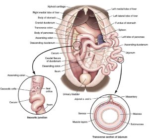
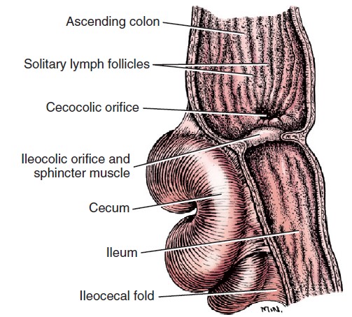
The cecum, a part of the large intestine, is an S-shaped, blind tube located to the right of the median plane at the junction of the ileum and colon. It is ventral to the caudal extremity of the right kidney, dorsal to the small intestine, and medial to the descending duodenum. The cecum communicates with the ascending colon at the cecocolic orifice. The cecum is connected to the ileum by the ileocecal fold – a triangular connecting peritoneum between these two bowel segments.
-
-
Dog small intestine in situ and its relationship to surrounding abdominal organs. Cutaways: ileocolic junction and a cross section of a
segment of jejunum with the mesenteric vessels. 9
-

- Longitudinal section through ileocolic orifice, ventral aspect. 1
-
- Junction of small and large intestine of the cat, ventral view. 4
-

- Dog cecum
-

- Cat colon
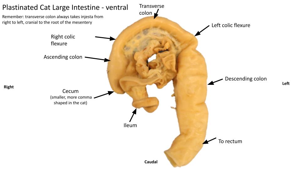
Observe: Observe the ileocolic and cecocolic orifices on lab models. Note that the cecum of the cat is proportionally (and absolutely) smaller than that of the dog.
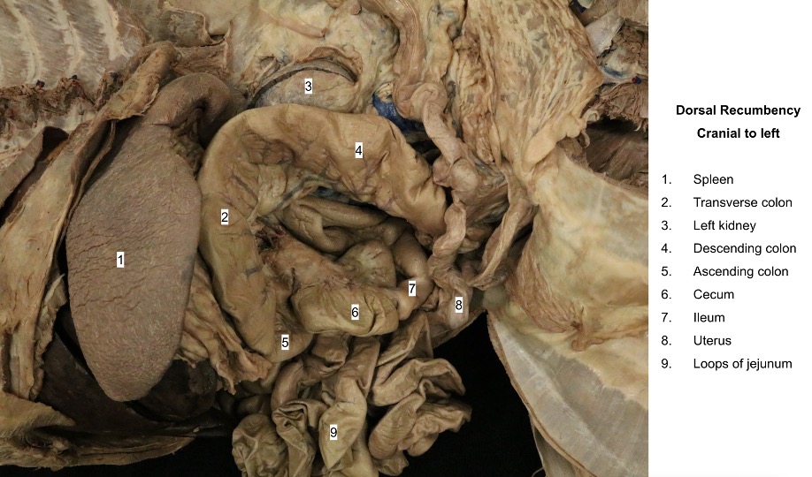
The colon is located dorsally in the abdomen, suspended by its connecting peritoneum called the mesocolon. The mesocolon is one continuous mesentery attached to each part of the colon.
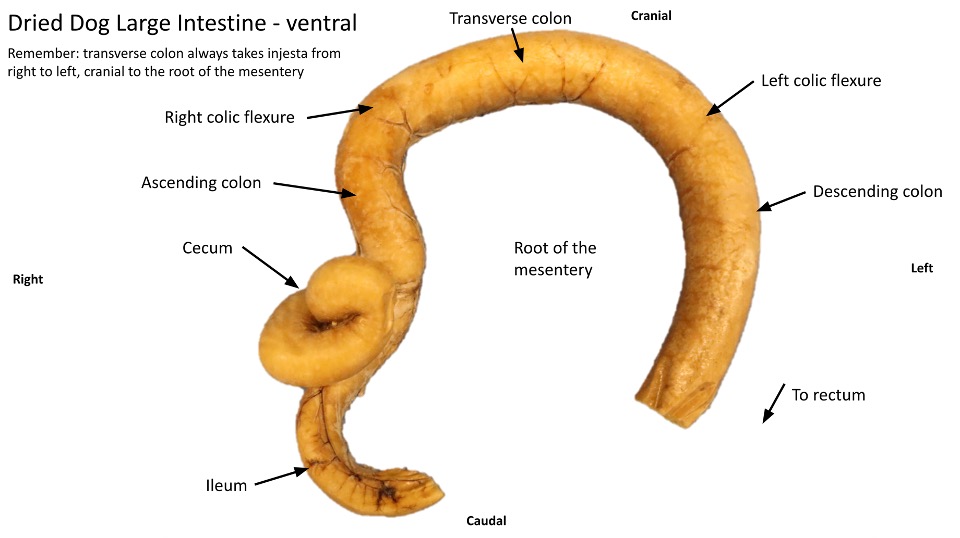
The colon is divided into a short ascending colon, which lies on the right of the root of the mesentery; a transverse colon, which lies cranial to the root of the mesentery; and a long descending colon, which lies at its beginning on the left of the root of the mesentery. The bend between the ascending and transverse colons is known as the right colic flexure, and that between the transverse and descending colons is known as the left colic flexure. The descending colon terminates at a transverse plane through the pelvic inlet. It is continued by the rectum (which is primarily located in the pelvic cavity and will be studied in the urogenital unit).
-
- Branches of cranial and caudal mesenteric arteries, ventral aspect. 1
-
- Dog cecum
-

- Dog colon
Review videos
Dog Duodenal papilla, small intestine, cecum, large intestine, pancreas – 8 min, watch until 24:30
Cat abdominal cavity – 15 min, watch until 17
Terms
| Term | Notes (This lab is exclusively focused on the carnivore) |
| Duodenum | |
| Mesoduodenum | Mesentery of the duodenum |
| Descending duodenum | Oral section of the duodenum |
| Ascending duodenum | Aboral section of the duodenum |
| Duodenojejunal flexure | Where duodenum transitions to jejunum |
| Duodenocolic fold | Connecting mesentery b/n duodenum and descending colon |
| Major duodenal papilla | Found inside lumen of duodenum; where bile and pancreatic ducts secrete |
| Minor duodenal papilla | Found inside lumen of duodenum; where accessory pancreatic duct secretes |
| Jejunum | |
| Mesojejunum | Mesentery of the jejunum |
| Mesenteric lymph nodes | Typically found within mesojejunum |
| Ileum | |
| Ileocolic orifice | Opening between ileum and large intestine |
| Antimesenteric ileal artery | Found on antimesenteric side of ileum; useful indicator for length of ileum (very short in feline) |
| Pancreas | |
| Left lobe | |
| Right lobe | |
| Pancreatic duct | Do not identify; know that it dumps into duodenum via major duodenal papilla |
| Accessory pancreatic duct | Do not identify; know that it dumps into duodenum via minor duodenal papilla |
| Cecum | |
| Cecocolic orifice | Opening between cecum and ascending colon |
| Ileocecal fold | Connecting mesentery between ileum and cecum |
| Colon | |
| Mesocolon | Mesentery of colon |
| Ascending colon | Most oral section of colon; found on right side of root of mesentery |
| Right colic flexure | Transition from ascending colon to transverse colon |
| Transverse colon | |
| Left colic flexure | Transition from transverse colon to descending colon |
| Descending colon | Found of left side of root of mesentery |