Lab 10A: Peritoneum and Stomach of the Ungulates
Learning Objectives
- Describe the topography of the abdominal viscera as viewed from left and right sides and correlate this to physical examination findings when percussing and auscultating.
- Review the anatomy of peritoneum and how it relates to the embryological development of the gut.
- Identify the different parts of the greater omentum in the ruminant.
- Describe the ruminant stomach, identifying both external and internal structures of the four chambers.
- Identify the different parts of the horse stomach.
- Identify the different parts of the pig stomach.
Lab Instructions
For this lab, you will be studying instructor prosections and wet and dry specimens around the lab. Please refer to these materials when reading this guide and be sure to use a diversity of materials in your studies. Because these materials are limited, please feel free to jump to different sections of this lab guide to make the most of your time in lab. This guide is organized by species, allowing you to quickly scroll from section to section rather than having to read through text regarding a species that you may not be standing at.
External Topography and Landmarks
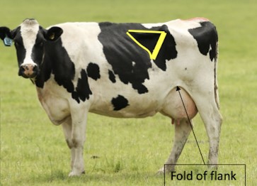
The lateral part of the abdominal wall that is not protected by the rib cage, pelvis, or thigh is called the flank. In the horse, the last rib is very close to the tuber coxae, leaving only about one hand’s-width space in the upper flank. Surgical intervention in this area is therefore limited in the horse, however laparoscopy is readily performed via the flank. The ruminant has a large flank area, and this allows for convenient access to the abdominal cavity.
Paralumbar Fossa: This triangular depression in the upper part of the flank is especially prominent in the standing domestic ruminant, in which it is an important surgical area. It is bound dorsally by the transverse processes of lumbar vertebrae L2 to L5, cranially by the last rib, and caudoventrally by the tense band or ridge of the ventral part of the internal abdominal oblique (IAO) m., created by the weight of the abdominal viscera. In the living horse and pig, the paralumbar fossa is small in size and often not a fossa at all, so it is less apparent.
The flank fold is a skin fold that extends from the caudoventral abdominal wall to the craniomedial aspect of the thigh near the stifle. The cutaneus trunci m. is contained within this fold, which accounts for the active twitching of the fold, especially in the horse (remember the cutaneous trunci m.??). The fold can also be used as a handhold in the restraint of calves and small ruminants- hence, the term “flanking a calf down.”
-
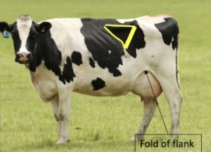
- Paralumbar fossa and fold of the flank. M. Gerard
-
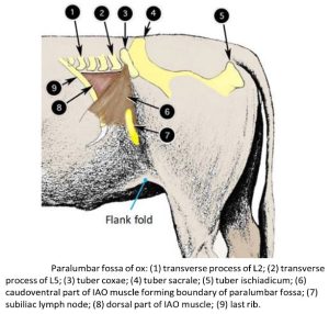
- Paralumbar fossa of the ox. 2

-

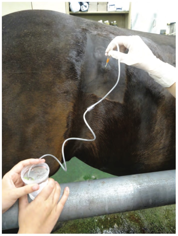
- Percutaneous cecal trocarization through the right paralumbar fossa of the horse. AAEP
Observe: Identify the flank, paralumbar fossa, and flank fold in the calf and goat cadavers.
Clinical application
Review of Peritoneum
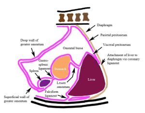
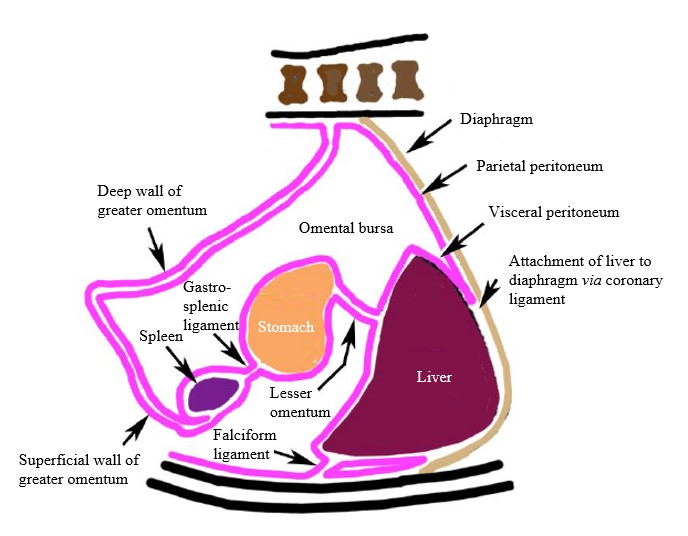
The peritoneum is the serous membrane that lines the walls of the abdominal cavity, extends partially into the pelvic cavity, envelops the descended testes of the male, completely or partially covers all organs in the abdominal cavity, and forms multiple connections among structures. Note that the word is singular- it is one continuous membrane that does all the above. Anatomists apply regional names to specific parts of this membrane. Here is the way Dr. Smallwood “outlines” the peritoneum:
- Parietal Peritoneum– lines the walls of the abdominal cavity; extends into the pelvic cavity, and outside the cavity in the male (and bitch) through the inguinal canals (derives from somatic mesoderm).
- Visceral Peritoneum– closely invests an organ’s surface; adherent to that surface (derives from splanchnic mesoderm).
- Connecting Peritoneum– double folds of peritoneum that connect things:
- Mesentery – connects the bowel to the body wall (e.g., mesoduodenum, mesocolon); in the broader usage, connects some other organ to the body wall (e.g. mesovarium).
- Omentum – attaches the stomach to something; the greater omentum connects the stomach to the body wall (embryologically it was the dorsal mesogastrium), and the lesser omentum attaches the stomach to the liver (embryologically, part of ventral mesogastrium).
- Fold – connects different parts of the bowel or another organ to one another (e.g., ileocecal fold, genital fold).
- Ligament – connects organs other than the bowel to the body wall or to the bowel (e.g., broad ligament, falciform ligament [liver to body wall, seen in calf], gastrosplenic ligament).
-
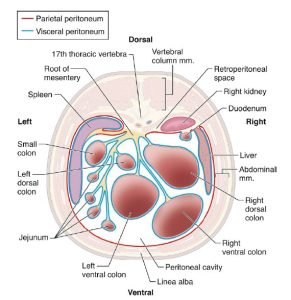
- Transverse section schematic defining the peritoneum and peritoneal cavity of the horse. 9
Review of the Embryological Development of the Gut
Embryologically, the stomach primordium and proximal duodenum are attached to the dorsal and ventral body walls by the dorsal and ventral mesogastria. The liver primordium grows ventrally from the duodenum and develops between the layers of the ventral mesogastrium. That part of the ventral mesogastrium between the liver and the body wall becomes the falciform ligament, and that part between the stomach and liver becomes the lesser omentum.
-
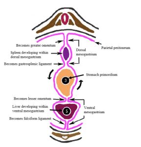
- Representation of embryonic attachments of developing stomach, transverse section, caudal view. 2
-
- Peritoneal attachments following embryonic rotation of stomach around a dorsoventral axis, sagittal section, right lateral view. 4
Observe: Look for the falciform ligament on the ventral body wall of the goat or calf cadaver. It will be invested with fat and should be in close association with the liver when the body wall is in its natural position.
The dorsal part of the stomach primordium grows faster than the ventral part, thus creating greater (dorsal) and lesser (ventral) curvatures of the stomach. As the greater curvature (dorsal part) rapidly expands, it becomes “top- heavy” and “flops over” to the left side, carrying with it the dorsal mesogastrium, which becomes the greater omentum. After it flops over, the greater curvature, which now faces to the left, continues to grow faster so that the stomach now rotates around a dorsoventral axis, carrying the pylorus to the right and cranially.
In the ruminant, continued expansion and modification along the proximal part of the greater curvature, now facing the left, results in the formation of the rumen and reticulum. Similarly, an outgrowth and modification near the lesser curvature results in formation of the omasum. Knowing this, it makes sense that, in the adult animal, the greater omentum is closely associated with the rumen and reticulum and greater curvature of the abomasum, whereas the lesser omentum is related to the omasum and lesser curvature of the abomasum. Conversely, the attachments of the greater and lesser omenta in the adult animal mark the embryonic greater and lesser curvatures of the stomach.
The spleen develops in the dorsal mesogastrium of the embryo. Thus, it makes sense that the spleen is associated with the greater curvature (and greater omentum) of the stomach in the adult. That part of the greater omentum between the stomach and the spleen is referred to as the gastrosplenic ligament. In the ruminant, the gastrosplenic ligament has been replaced by an area of direct attachment to the body wall. This is a consequence of the massive expansion and direct contact of the ruminant stomach (specifically the rumen and reticulum parts, commonly referred to as the ruminoreticulum) to the craniodorsal abdominal wall, such that the peritoneum receded back out of the area, leaving a direct adhesion instead of a mesenteric attachment. Also, as a result of the expansion of the ruminoreticulum, the spleen of the ruminants was separated away from its embryonic association with the greater omentum.
A few comments on the omenta: In those ungulates with a simple stomach (horse and pig), the omenta remain relatively simple, whereas in the ruminant, the stomach and the greater omentum are more complex. In the horse and the pig, the greater omentum, like that of the dog/cat, consists of a simple peritoneal fold that extends from the greater curvature of the stomach to the dorsal body wall. The superficial wall of the greater omentum extends caudally a variable distance from the greater curvature before turning back on itself as the deep wall, which continues cranially, then dorsally, to the body wall. Between the superficial and deep walls is an enclosed part of the peritoneal cavity known as the omental bursa. Commensurate with its small stomach, the greater omentum of the horse is not really so great (poorly developed) and does not typically extend back to cover the intestinal mass. It is interesting to note that, among our common domestic mammals, the horse has the most poorly developed greater omentum and is most susceptible to peritonitis. One recognized function of the greater omentum is that it quickly adheres to any area of peritoneal insult and seals it off. Its vessels then serve as an avenue of attack for inflammatory cells to combat any invasion of the peritoneal cavity by foreign material (e.g., bacteria, intestinal leakage). Have you surmised? Yes, ruminants, with their spectacularly robust greater omentum, also have relatively excellent ability to combat peritonitis and to “wall off” focal areas of insult.
OK, end of theory – let’s apply it, but not a bad idea to refer back frequently, particularly to the figures!
-
- Greater omentum of a ruminant, left lateral. 7

-

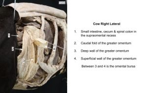
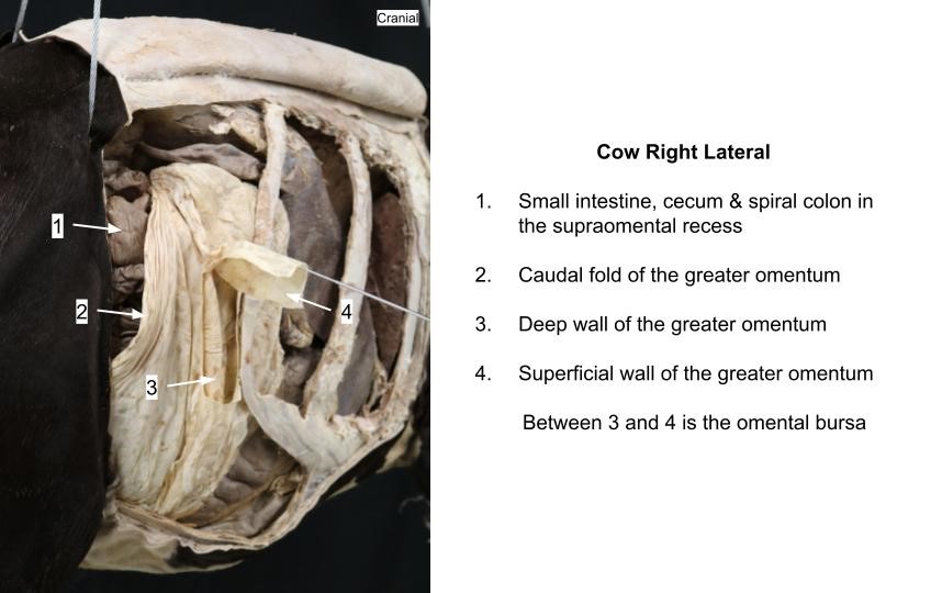
- Greater omentum of a ruminant, right lateral. 7
-
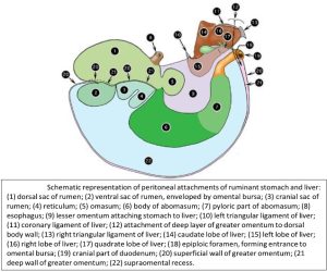
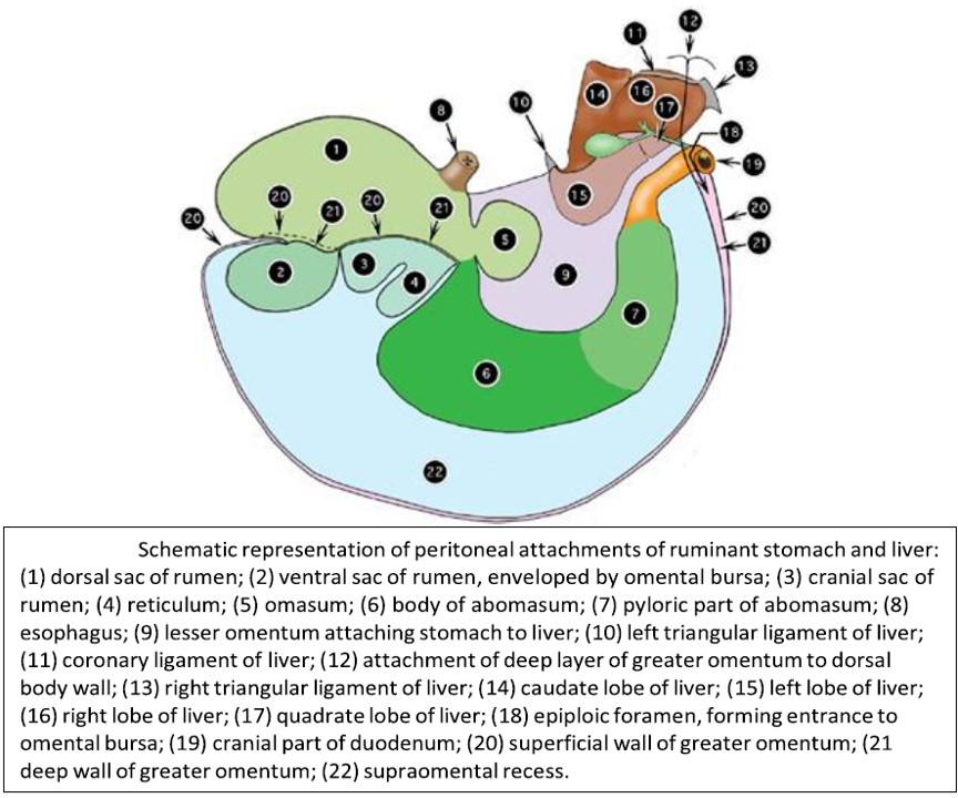
- Schematic representation of peritoneal attachments of ruminant stomach and liver. 2
-
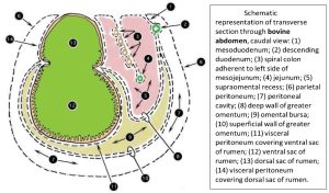
- Transverse section through bovine abdomen, caudal view. 2
-
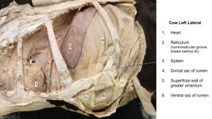
- Left cow topography

-
- Right omentum
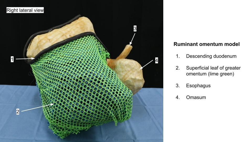
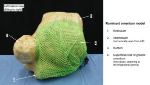
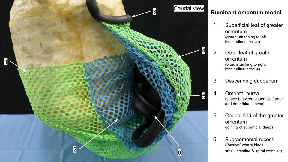
Lindsey and Dr. Gallenstein created this stunning chicken wire, paper mache, fiberglass and fishnet piece of art in 2018 after seeing students struggle with understanding the attachments and arrangement of the ruminant greater omentum. This model is intended to help you transfer that understanding to the cadavers, NOT to be testable material on this model.
-

- Right
-
- Left

-

- Caudal
Omentum and Associated Structures
Omentum of the Calf
The attachments of the greater omentum of the ruminant can best be understood by first studying that of the newborn calf.
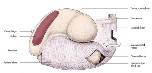
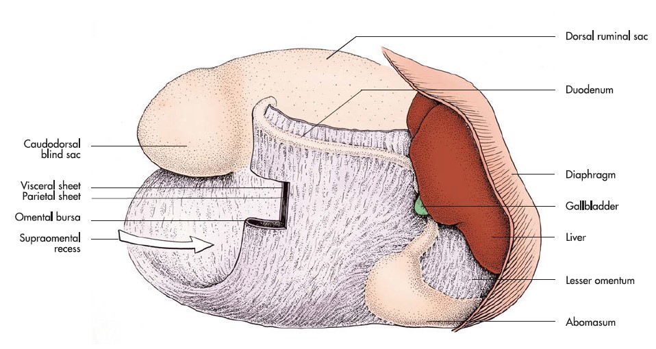
Observe: First, identify the caudal fold of the greater omentum on the right side. This free fold represents the line of reflection of the superficial wall back on itself to form the deep wall. Follow the free fold to the rumen, and note that it attaches along the caudal groove. Here the two walls separate; the superficial wall continues in the left longitudinal groove, whereas the deep wall continues in the right longitudinal groove.
Thus, the ventral sac of the rumen is enveloped by the omental bursa, but it is not within the bursa, because the bursa is a potential space between the two walls of the greater omentum. The ventral sac is covered by visceral peritoneum, which is continuous with the greater omentum along its line of attachment. The relationship of the ventral sac of the rumen to the omental bursa is analogous to the relationship of the heart to the pericardial sac. The deep and superficial walls come back together in the cranial groove of the rumen, and both walls continue along the greater curvature of the abomasum.
-
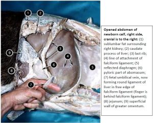
- Opened abdomen of newborn calf. 2

-

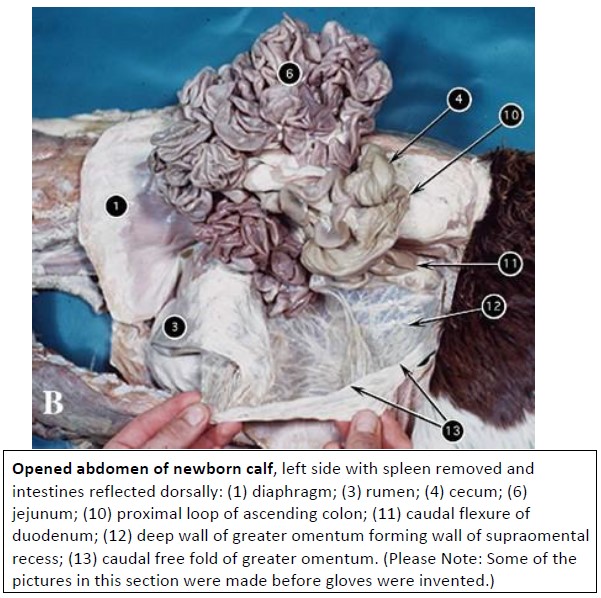
- Opened abdomen of newborn calf, left. 2
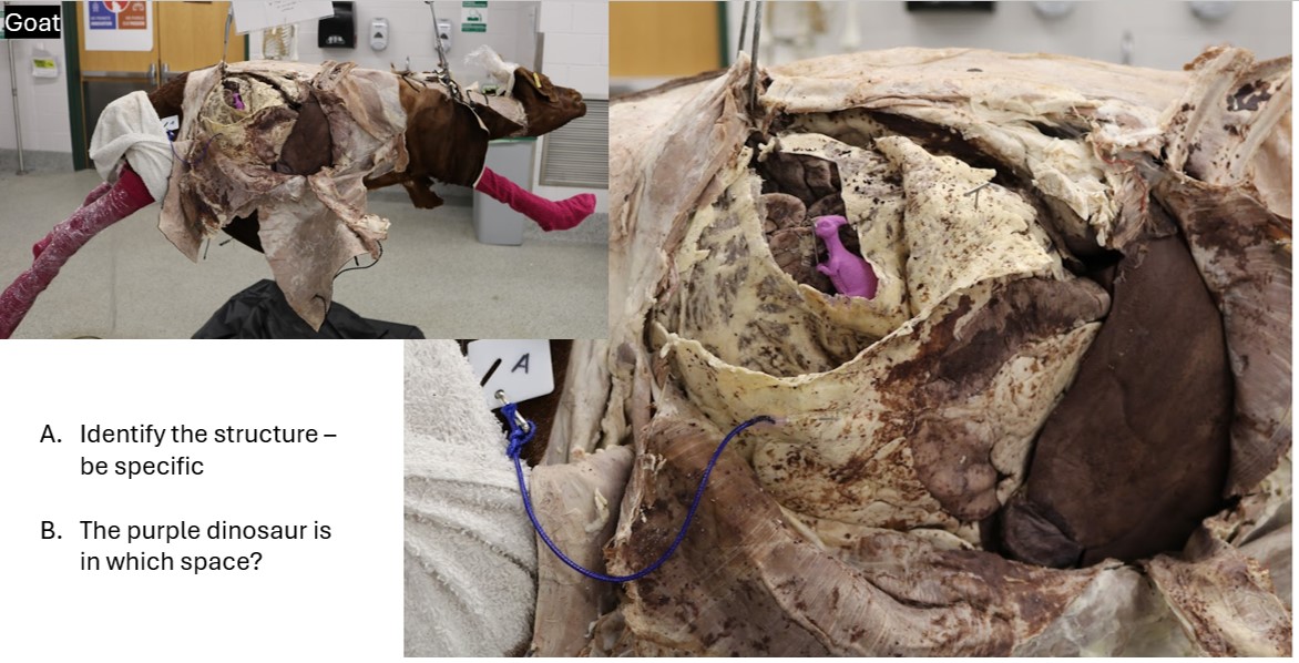
Observe: Return now to the caudal border, and reflect the intestinal mass dorsally out through the left flank. Follow the free border to the right side, and note that it attaches here to the duodenum at about the point of the caudal flexure (where the descending duodenum turns sharply cranially to become the ascending duodenum). Both the superficial and the deep walls attach along the antimesenteric border of the descending duodenum. More proximally, the deep wall detaches from the cranial part of the duodenum, and continues dorsally to the body wall. The greater omentum forms a double-walled “basket” around the intestinal mass, which is known as the supraomental recess.
From the right side, lift the liver craniodorsally, and locate the lesser omentum extending between the lesser curvature of the abomasum and the visceral surface of the liver. While holding the liver up, pass your hand dorsal to the descending duodenum (at its cranial end), and under the caudate lobe of the liver, heading towards midline. You should locate a slit-like, natural opening at the dorsal border of the lesser omentum, large enough to accommodate one or more fingers.
Congratulations, you have just found the epiploic foramen, and you now have your finger in the omental bursa. As you can see, the peritoneal cavity proper communicates with the omental bursa through this opening.
-
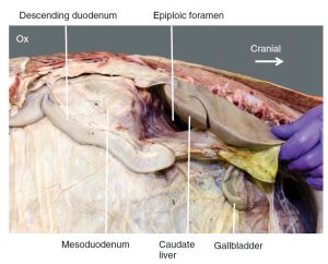
- Epiploic foramen of the ox. 30

Observe: In summary, before moving on make sure you have identified the following in the calf: greater omentum, caudal fold of greater omentum, superficial and deep walls of greater omentum, descending duodenum, caudal duodenal flexure, omental bursa, epiploic foramen, supraomental recess, greater curvature and lesser curvature of abomasum, lesser omentum.
Omentum of the Goat
Observew: On the right side, identify the superficial wall of the greater omentum, which attaches to the descending duodenum. Slide your hand caudodorsally along the superficial wall until you locate the caudal fold of the greater omentum, which extends from the caudal flexure of the duodenum on the right to the caudal groove of the rumen on the left.
Notice how nicely your hand fits around the caudal fold of the omentum; your fingertips are now in the supraomental recess and are resting on the deep wall of the omentum.
Surgeons manipulate this fold to allow the organ of interest (e.g., uterine horn) to slip out of the supraomental recess, and then release the fold to “sack up” and keep contained wandering loops of bowel.
On the right side, an incision was made about 5 cm ventral to the mid part of descending duodenum. Look through this window at the exposed deep wall. The omental bursa is opened when this window is created in the superficial wall.
Now move around to the left side, and note the attachment of the superficial wall along the left longitudinal groove of the rumen. Again, locate the caudal fold and trace it back toward the right side of the animal. A better understanding of the omental attachments can be gained by reference to a transverse schematic drawing.
-

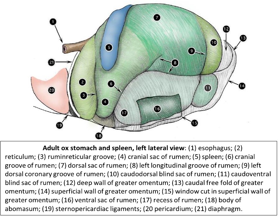
- Adult ox stomach and spleen, left lateral view. 2
-

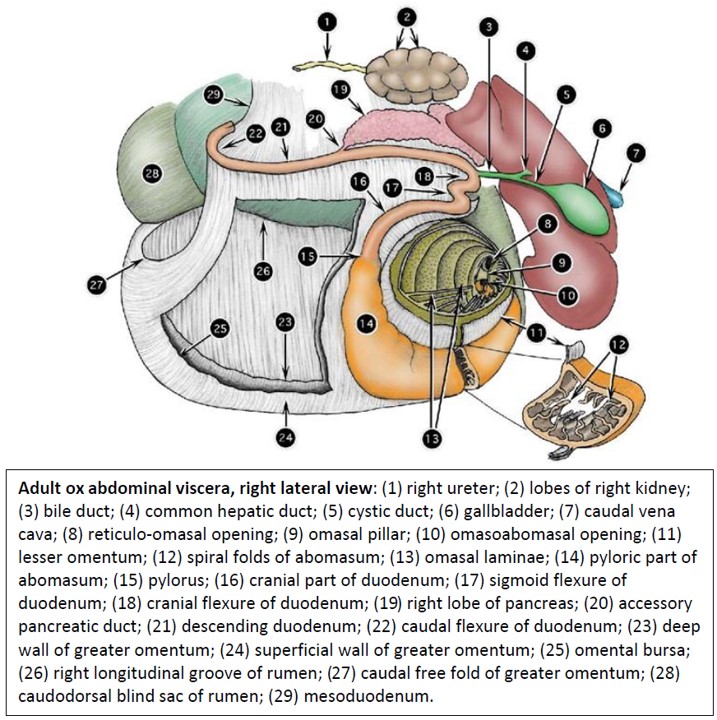
- Adult ox abdominal viscera, right lateral view. 2
-
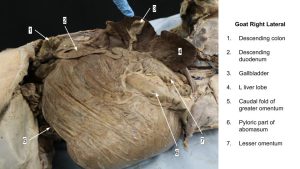
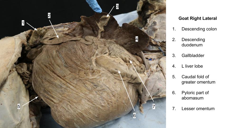
- Goat omentum, right side
Observe: In summary, before moving on make sure you have identified the following in the goat: greater omentum, caudal fold of greater omentum, superficial and deep walls of greater omentum, descending duodenum, caudal duodenal flexure, omental bursa, supraomental recess, greater curvature and lesser curvature of abomasum, lesser omentum.
The Stomach
In addition to the instructor prosections, please also refer to plastinated, dry and wet models of stomachs. You will be expected to be able to identify these structures in any of these contexts.
Based on what in the lab is available to you at the moment, feel free to jump to the section of any species and come back to the others when you can.
Ruminant Stomach (Calf and Goat)
The ruminoreticulum (i.e. the rumen and reticulum compartments), occupies the entire left half (and more) of the abdominal cavity of an adult ruminant. It is difficult to observe all the grooves and other features in a large animal, but this can easily be accomplished in the newborn calf. These potential ruminants (physiologically not fully developed to ruminate yet) have all the right parts, they just haven’t gotten big and bulky yet.
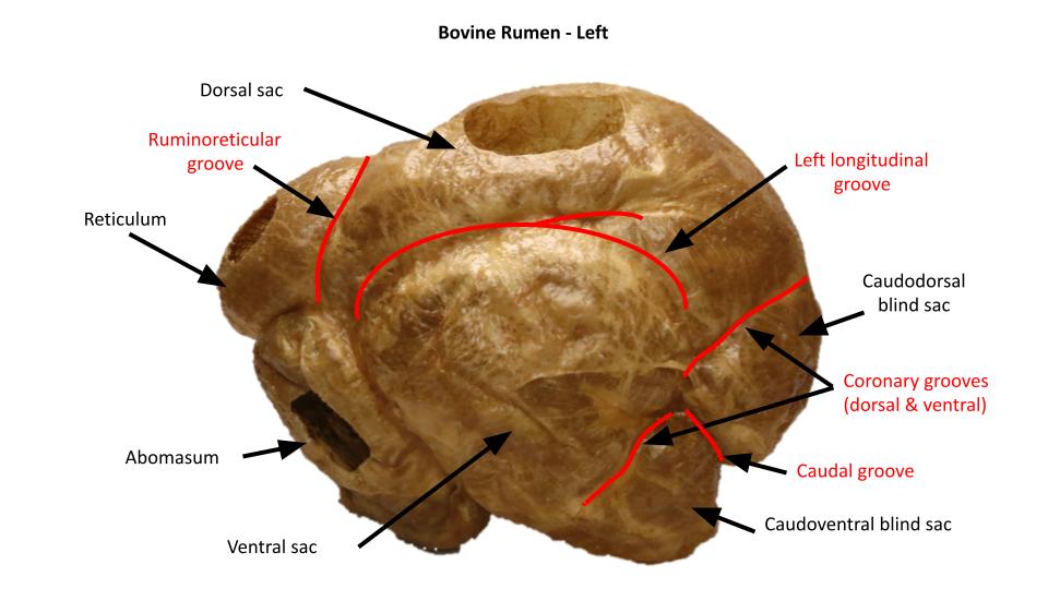
Observe: Examine the external surface of the ruminant stomach and initially identify the 4 major compartments (rumen, reticulum, omasum and abomasum) and then identify the grooves and sacs of the ruminoreticulum. Use the dry models to visualize this anatomy too.
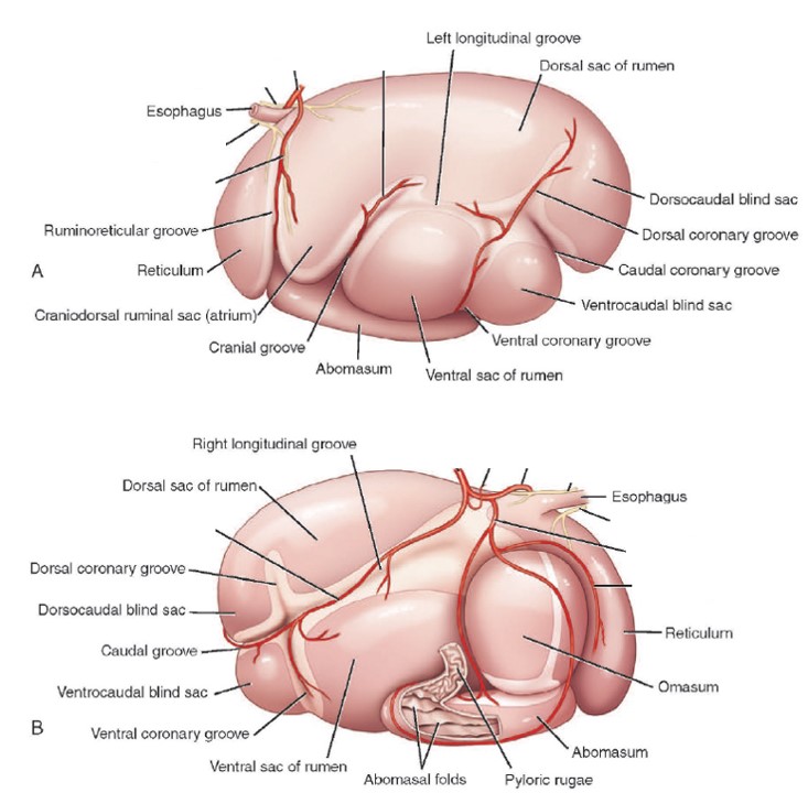
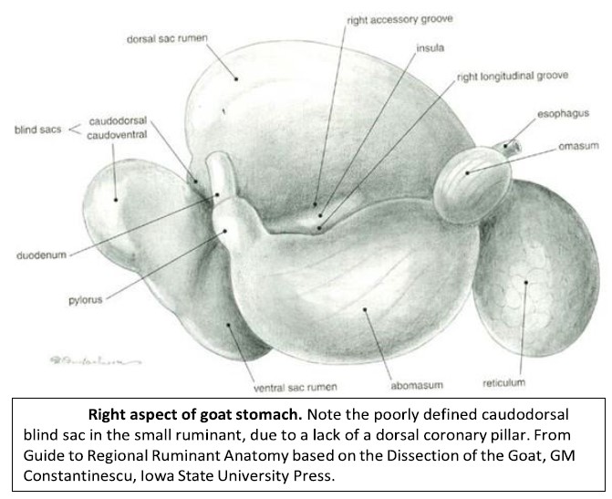
NOTE: The reticulum of the sheep and goat is proportionately smaller than that of the ox. The caudodorsal blind sac is also poorly defined.
-
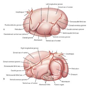
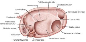
- Anatomy of the ruminant’s forestomach. A: left, B: Right. 9
-
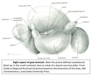
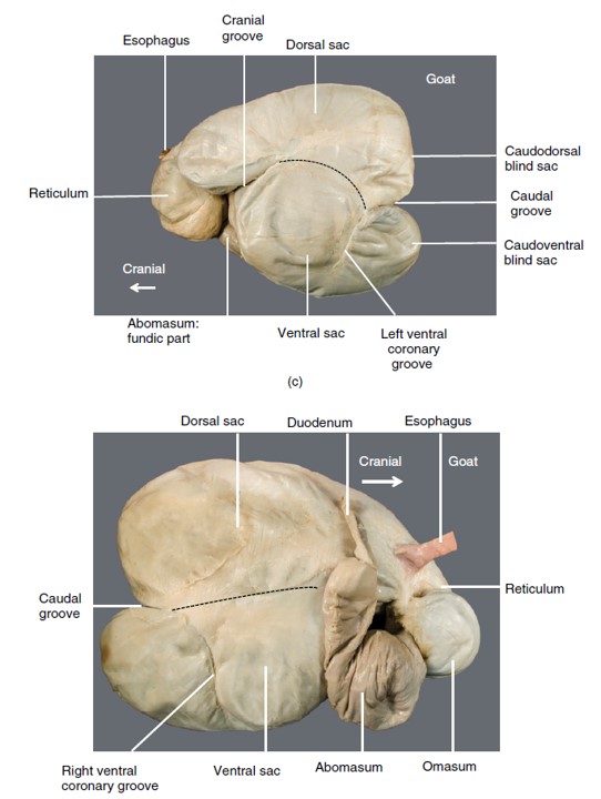
- Right aspect of the goat stomach.
-

-
Goat’s rumen. Note that the caudoventral blind sac extends more caudally than
the caudodorsal blind sac. 16
-
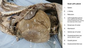
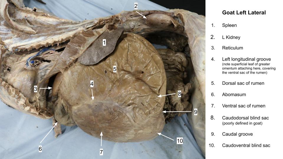
- Goat rumen
-
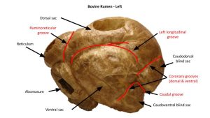
- Left ox stomach
-

- Right ox stomach
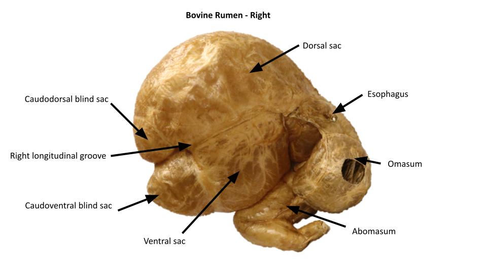
Observe: Using the images, make sure you identify all of the following on the external surface of the ruminant stomach: rumen, reticulum, omasum, abomasum, ruminoreticulum, dorsal, ventral, and cranial sacs of rumen; caudodorsal and caudoventral blind sacs of rumen; the cranial, left longitudinal, caudal, right longitudinal, dorsal coronary, ventral coronary, and ruminoreticular grooves.
The following structures described below are found inside the ruminant rumen.
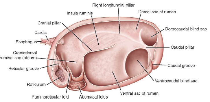
The grooves on the outside of the rumen correspond with the pillars on the inside, which carry the same designations. The ruminal pillars represent in-folding and thickening of the muscular wall.
Observe: Identify the pillars and sacs of the rumen, in the cadaver and view models too.
-
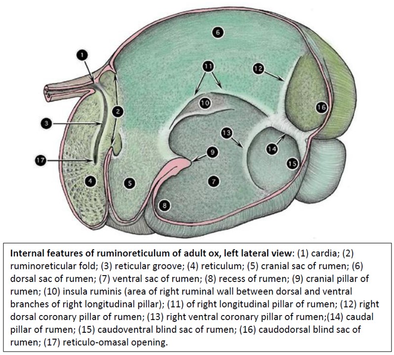
- Anatomy of the inside of the ruminant’s forestomach, left lateral. 9
-
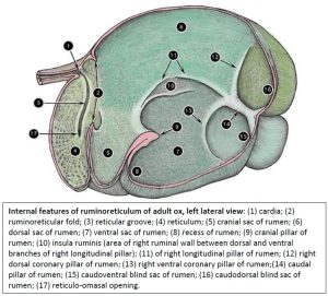
- Internal features of ruminoreticulum of adult ox, left lateral view. 2
The recess of the rumen is the part of the ventral sac below the cranial pillar. The cranial sac is relatively smaller in the sheep and the goat. Although a caudodorsal blind sac is usually described in small ruminants, the absence of dorsal coronary pillars in the sheep and the goat makes this a poorly defined area in these animals. It is because the ruminoreticular opening is so large that these two compartments are often considered together, both anatomically and physiologically, as the ruminoreticulum.
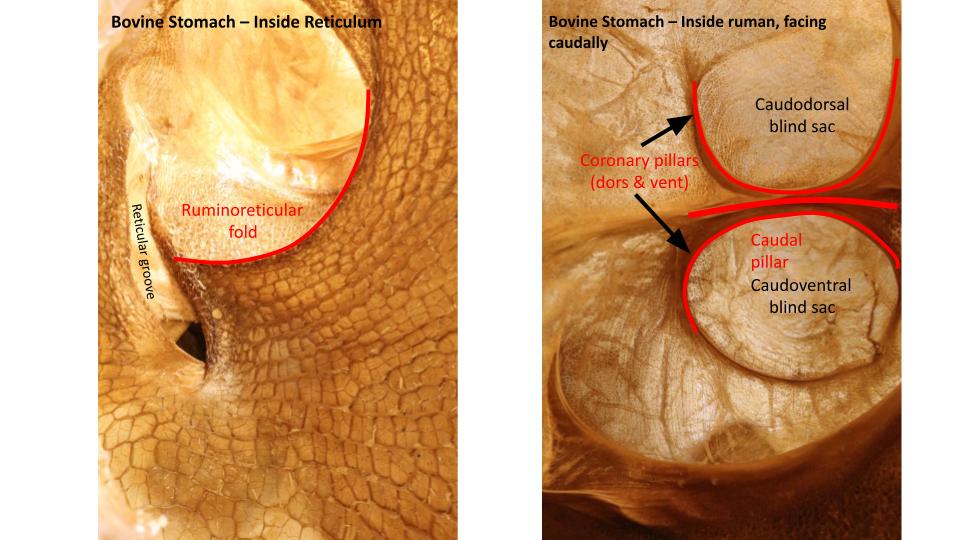
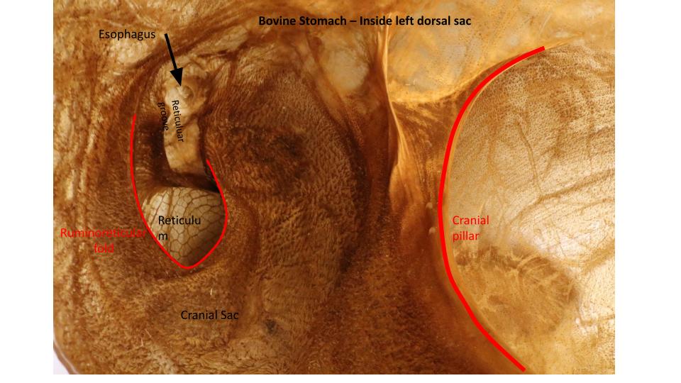
Observe: Identify all of the following in the rumen: internal appearance of dorsal, ventral, and cranial sacs, caudodorsal and caudoventral blind sacs of rumen, recess of the rumen; Pillars = cranial, left longitudinal, caudal, right longitudinal, dorsal coronary, and ventral coronary pillars; ruminoreticular fold, ruminoreticular opening.
Clinical Applications
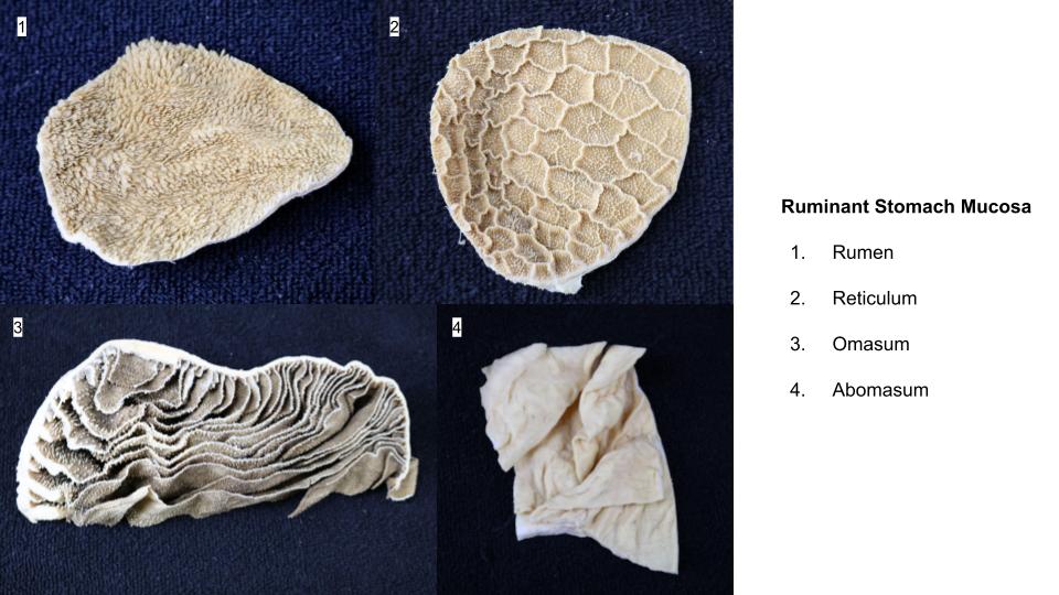
Observe: Look for a window incised into the reticulum and examine its characteristic honeycomb lining. Observe the mucosa of the rumen and reticulum.
The non-glandular, stratified squamous mucosa of the rumen is characterized by the presence of closely packed, tongue-shaped ruminal papillae, which give it the appearance of Astroturf (or “gastroturf”). The papillae vary in shape and number throughout regions of the rumen. The reticulum mucosa has a very distinct honey-comb pattern.
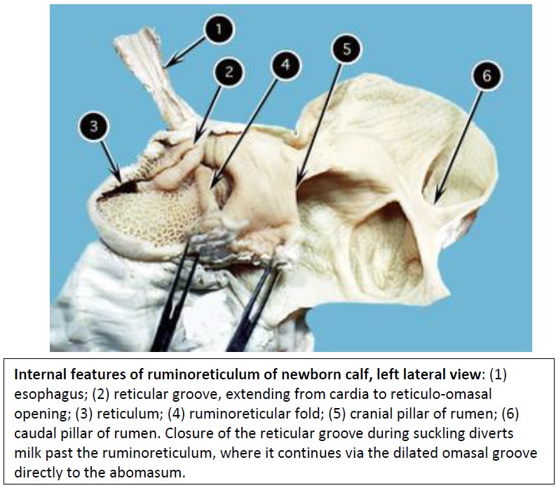
The cardia is located in the reticulum, not in the rumen, and therefore the esophagus actually enters the reticulum. The momentum of the coarse, light material that is propelled through the cardia with reasonable velocity undoubtedly clears the ruminoreticular fold and enters the rumen. On the other hand, heavy objects such as nails, spark plugs, or flashlight batteries more probably fall out of the cardia into the reticulum, where they may reside for life. Sharp objects may also penetrate the wall of the reticulum and cause traumatic reticulitis and may continue to migrate into liver, or through the diaphragm into lungs, pericardial sac and even the heart (traumatic reticulopericarditis).
Note the distinct groove, the reticular groove, beginning at the cardia and spiraling down the reticular wall, to lead to the reticulo-omasal opening. This groove is the first part of the gastric groove. Do not confuse the ruminoreticular groove, an external feature of the ruminant stomach, with the reticular groove, an important internal structure.
The reticular groove plays an important role in the suckling animal and thus the calf should receive special attention here. When the young ruminant suckles milk, it initiates a strong (vagal) reflex closure of the muscular lips of the reticular groove, converting it from a groove to a tube. Simultaneously, there is dilation of the reticulo-omasal opening and omasal groove so that, ideally, the milk completely bypasses the forestomach and is delivered directly to the abomasum, where the milk-digesting enzyme rennin is secreted.
-

- Internal features of ruminoreticulum of newborn calf, left lateral view. 2
-
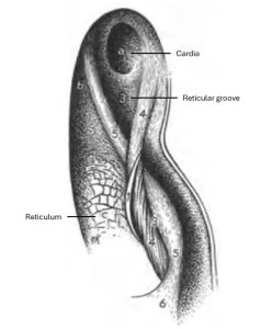
- Reticular groove of the ox, caudoventral aspect. 36

-
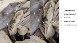
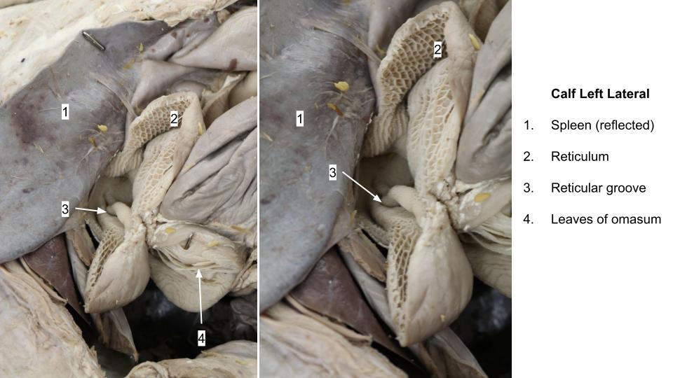
- Calf reticular groove
Observe: Make sure to identify the following in the ruminant: ruminal papillae, reticulum and its mucosa, cardia, reticular groove.
Observe: Examine the omasum internally and externally. NOTE: the omasum of the goat (and sheep) is more oval and proportionately smaller than that of the ox.
Identify the omasum and omasal laminae in the ruminant.
Note the normal position of the body of the abomasum and observe that it is not firmly fixed in this position (i.e. can readily be displaced). Then look for a removed section of the wall of the abomasum.
In the newborn calf, coagulation of the milk results from the action of the enzyme rennin, which accounts for a common name for the abomasum, the rennet. Calling it the “true stomach” is a tad ridiculous, because there is only one stomach per animal and the other parts are real too.
-
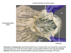
- Omasum and abomasum of the ox. 16

-
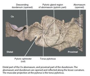
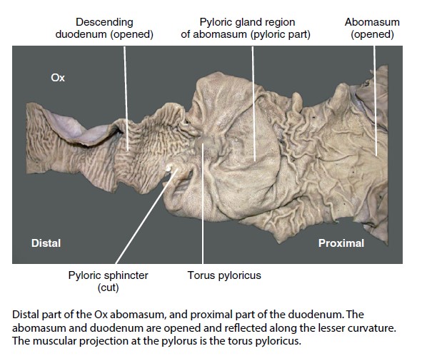
- Distal part of the Ox abomasum, and proximal part of the duodenum. 16
-

- Omasum and abomasum
-
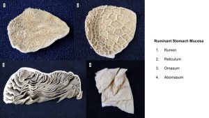
- Stomach mucosa types
Observe: Locate the knob-like torus pyloricus, (also present in the pig) which assists in closure of the pylorus.
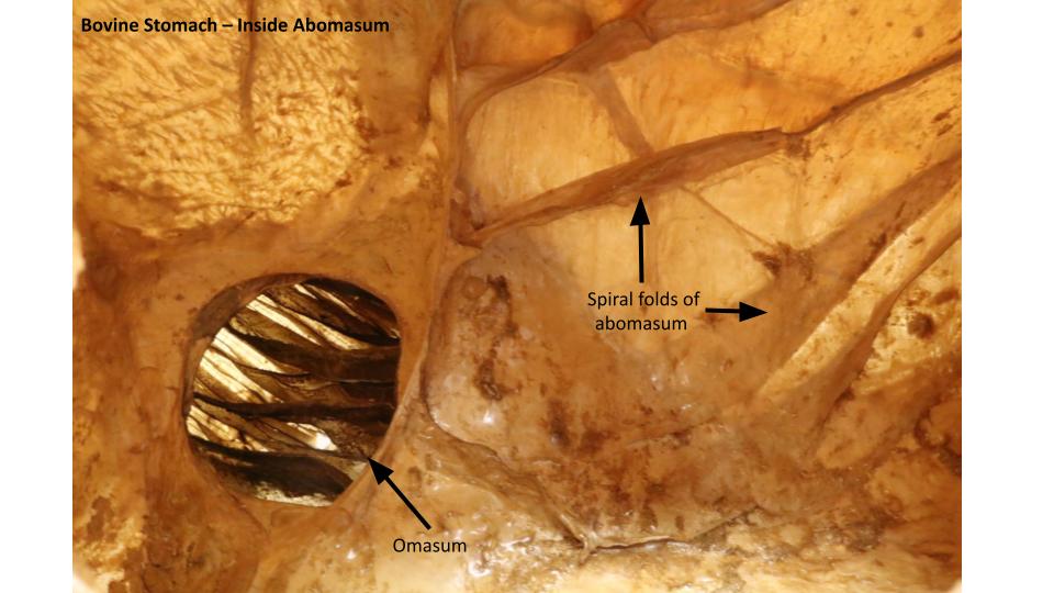
Note the mucosal elevations known as spiral folds, best developed in the body and fundic regions of the abomasum. The abomasum is the glandular region of the ruminant stomach.
Ruminant stomach clinical case
Ruminant emergency clinical case
Clinical Application
Horse Stomach
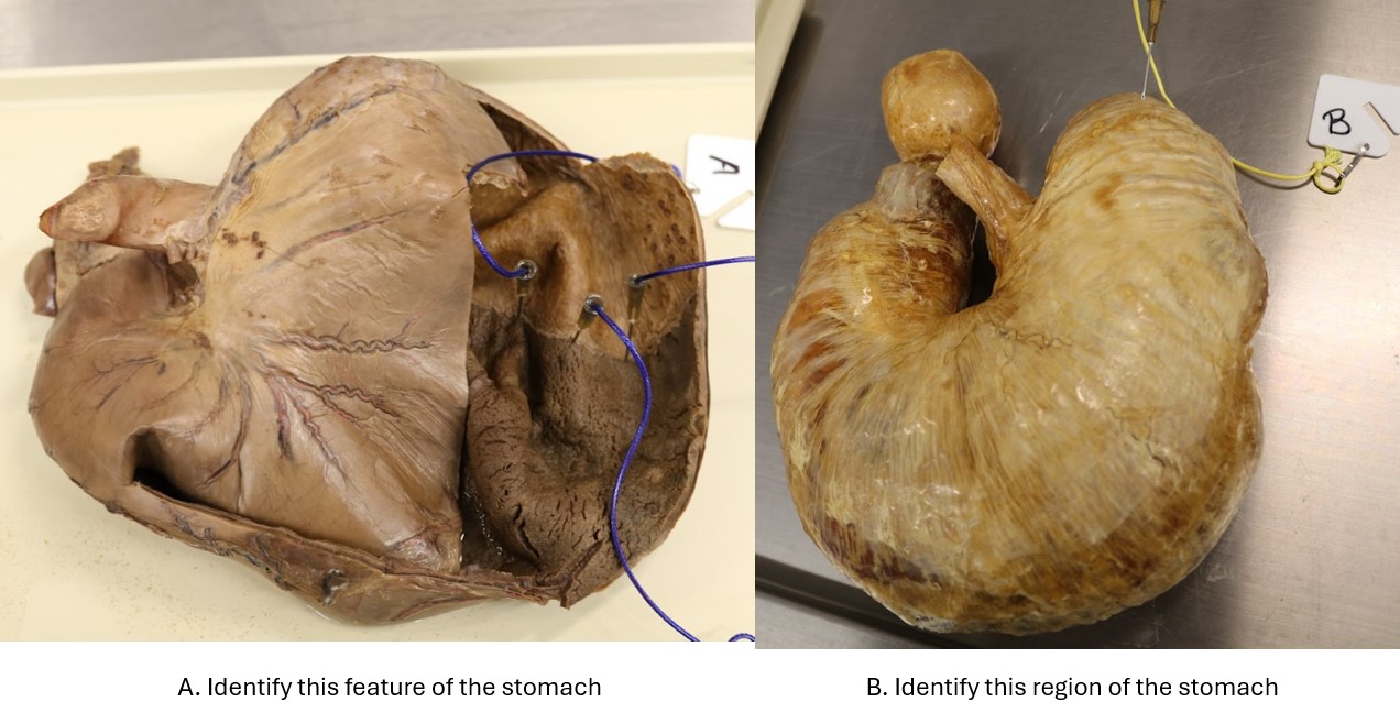
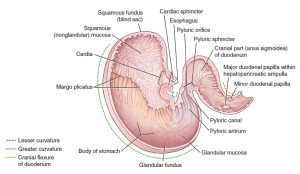
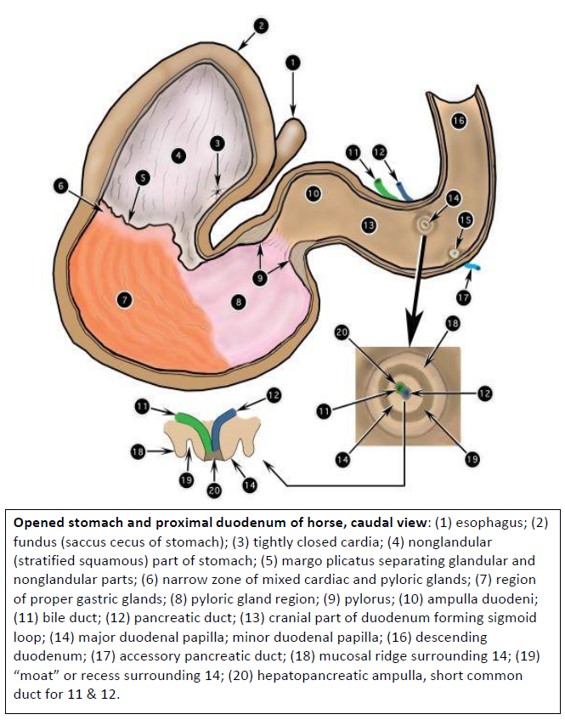
The fundus is enlarged to form a saccus cecus (blind sac), which is lined by a non-glandular, stratified squamous epithelium. The junction of the non-glandular (squamous) and glandular regions is marked by a ridge, the margo plicatus. The larvae of the common botfly, Gasterophilus intestinalis, may be found attached to the stomach wall. The glandular part of the stomach is divided into three regions.
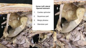
Observe: In a wet or cadaveric horse stomach, try to push your finger back up through the cardiac opening.
Not so easy, right? The horse has a very strong cardiac sphincter. This sphincter, combined with the strong muscular tunic of the more distal part of the esophagus, makes it practically impossible for the horse to vomit or even eructate (belch). Excessive, and quick, ingesta/gas accumulation in the equine stomach can cause complete rupture of the organ! (a life-threatening problem that often leads to death/euthanasia of the horse).
From the body of the stomach, the organ narrows distally to form the pyloric part of the stomach (first the pyloric antrum, which then leads to the pyloric canal). At the end of the pyloric part of the stomach, is the pyloric sphincter (aka pyloris), which is well developed in the horse.
-
- Cross-sectional anatomy of the horse stomach. 9

-
- The equine duodenum and its papillae. 9

-

- Opened stomach and proximal duodenum of horse, caudal view. 2
-
- Horse stomach

Observe: Before moving on, make sure you identify all of the following in the equine: fundus (= saccus cecus in horse), margo plicatus, squamous part of stomach, glandular part of stomach, cardiac sphincter, body of stomach, pyloric part, pyloric sphincter, greater curvature, lesser curvature.
Clinical Application
The Pig Stomach
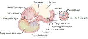
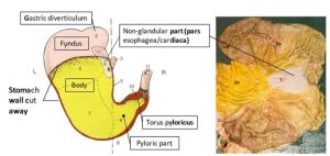
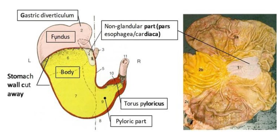
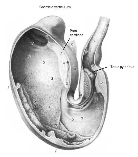
The most obvious modification of the pig stomach is the small out-pocket at the apex of the fundus, termed the gastric diverticulum.
The non-glandular part of the stomach is small and is represented by a narrow strip of stratified squamous epithelium that extends from around the cardia into the gastric diverticulum, anatomically the pars cardiaca, and most commonly called the pars esophagea in the literature/clinically. This non-glandular area is where gastric ulcers typically occur in pigs. Gastric ulcers are a common problem in the modern swine-production industry, and can result in significant economic loss to the producer. The glandular regions of the stomach are large and can be distinguished in part by mucosal color and prominence of mucosal folds.
Internally, at the pyloric sphincter note the musculofatty protuberance from the lesser curvature, the torus pyloricus. With the surrounding circular muscle fibers, the torus pyloricus forms a sort of ball-and-seat valve for more efficient, watertight closure of the pylorus (similar to ruminants).
-
- Porcine stomach. The very small squamous area is a common location for gastric ulcers in the pig. 8
-
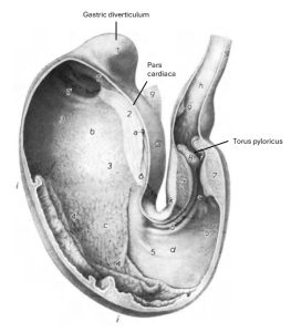
- Pig stomach. 36
-
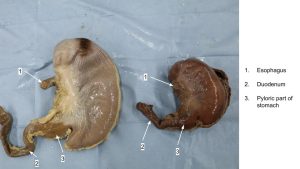
- Pig stomachs

-
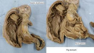
- Pig stomach

Observe: Make sure to identify all of the following in the pig: gastric diverticulum, fundus, cardiac sphincter, pars esophagea/cardiaca (= squamous or non-glandular region of stomach), glandular region of stomach, body, pyloric part, torus pyloricus, pyloric sphincter, greater curvature, lesser curvature.
Clinical Application
Clinical relevance: gastric ulcers, gastric ulcers, gastric ulcers of the pars esophagea.
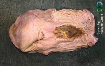
Refer to the image above of the porcine internal stomach. What region of the stomach is affected by a deep ulcer?
Review Videos
Horse and pig stomach – 8 min
Ruminant greater omentum – 3 min
Ruminant stomach (models) – 17 min
Ruminant stomach (cadaver & model) – 4 min
Key Terms
| Term | Species/Notes |
| Flank | Goat and ox |
| Paralumbar fossa | Goat and ox |
| Flank fold | Goat and ox |
| Peritoneum | Understand the different types of peritoneum on a conceptual level |
| Parietal peritoneum | |
| Visceral peritoneum | |
| Connecting peritoneum | |
| Falciform ligament | Identify on ventral body wall of goat and calf |
| Greater omentum | Ruminant |
| Superficial wall | |
| Deep wall | |
| Omental bursa | |
| Supraomental recess | |
| Caudal fold | |
| Epiploic foramen | All |
| Gastrosplenic ligament | All |
| Ruminant stomach | All of the following will be identified in the stomach of the ox and goat |
| Rumen | |
| Dorsal sac | |
| Ventral sac | |
| Cranial sac | |
| Caudodorsal blind sac | |
| Caudoventral blind sac | |
| Recess of rumen | |
| Cranial groove and pillar | Groove = external feature; pillar = internal feature |
| Caudal groove and pillar | |
| Left longitudinal groove and pillar | |
| Right longitudinal groove and pillar | |
| Dorsal coronary groove and pillar | |
| Ventral coronary groove and pillar | |
| Rumenoreticular groove and fold | Fold = internal structure associated with rumenoreticular groove |
| Ruminal papillae | Mucosal structure of the rumen |
| Reticulum | |
| Rumenoreticulum | Combined rumen and reticulum |
| Rumenoreticular opening | |
| Reticular groove | |
| Cardia | |
| Reticular mucosa | Characteristic “honeycomb” appearance |
| Omasum | |
| Omasal lamina | Mucosal structure of the omasum |
| Abomasum | |
| Greater curvature | |
| Lesser curvature | |
| Spiral folds | Mucosal structure of the abomasum |
| Torus pyloricus | |
| Equine stomach | All of the following will be identified in the horse’s stomach |
| Glandular and non-glandular (aka, “squamous”) portions | |
| Saccus cecus | = fundus in the horse stomach |
| Cardiac sphincter | |
| Margo plicatus | Ridge between glandular and non-glandular portions |
| Body | |
| Pyloric part | |
| Pyloric sphincter | |
| Greater curvature | |
| Lesser curvature | |
| Pig stomach | All of the following will be identified in the pig stomach |
| Gastric diverticulum | |
| Pars cardiaca/Pars esophagea | = squamous or non-glandular region of stomach |
| Body | |
| Fundus | |
| Torus pyloricus | |
| Pyloric sphincter | |
| Glandular region | |
| Greater curvature | |
| Lesser curvature |