LAB 2A: Alimentary Structures of the Head and Neck
Learning Objectives
- Identify the major extrinsic salivary glands and describe the locations of the opening of their ducts in the oral cavity.
- Name and identify the structures, boundaries, & subparts of the oral cavity.
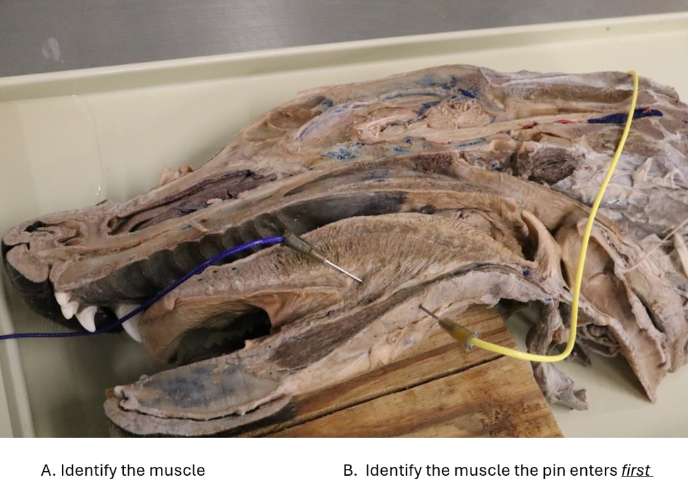
- Identify the muscles of mastication.
- Identify the lingual and hyoid muscles and name their basic attachments.
- Name and identify the boundaries and subparts of the pharynx.
- Identify muscles of the pharynx.
- Identify the esophagus and describe species differences amongst the ungulates.
Welcome to the gastrointestinal (GI) tract! Otherwise known as the “alimentary canal” (alimentum [L] = “nourishment”), this system is fundamentally one long tube stretching from the mouth to the anus. Along the way, this tube has modifications related to the specific demands of that region of the GI tract, as well as a host of accessory organs that contribute to the breakdown and absorption of ingesta.
Our approach to the labs in this unit will be to start at the oral cavity and work our way aborally (away from the mouth) towards the anus. With that in mind, let’s begin with your cadavers’ heads!
The Salivary Glands
Salivary Glands of the Carnivore
Observing the medial aspect of your bisected head, notice the tongue is attached to the floor of the oral cavity by a thin piece of tissue. This is a ventral median fold of mucosa, known as the lingual frenulum.
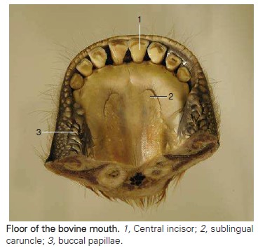
Observe: Turn the tongue medially and observe the slightly raised elevation of mucosa that is lateral to the rostral part of the frenulum and protrudes from the floor of the oral cavity. This is the sublingual caruncle.
Extending caudally from the caruncle is the sublingual fold. (This is easier to see in the live animal.) The mandibular duct and major sublingual duct are found in this fold. They course rostrally to open on or beside the sublingual caruncle, separately or through a common opening. *You do not need to expose the ducts, but do look for the caruncles.*
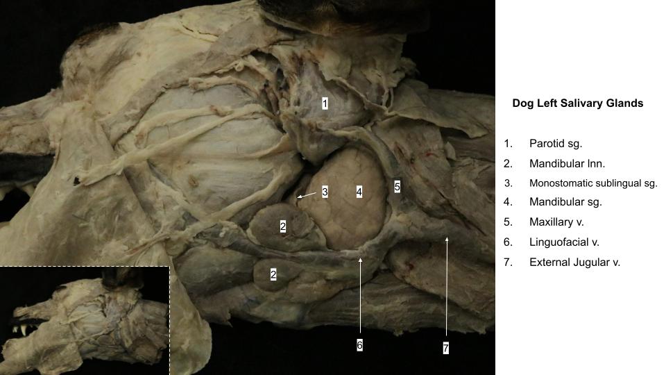
Observe: On the lateral aspects of your cadaver’s head expose the mandibular salivary gland, where it lies between the maxillary and linguofacial veins.
The major sublingual duct is connected caudally with the monostomatic sublingual gland. This is closely associated with the mandibular salivary gland. There are also sublingual gland lobules (the polystomatic sublingual gland) deep to the mucosa of the sublingual fold. These have independent microscopic ducts opening into the oral cavity. The mandibular salivary gland is covered by a thick capsule that also includes the caudal part of the monostomatic sublingual gland.
-
- Salivary glands of the dog. (The right half of the mandible was removed.) 1
-
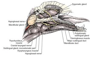
- Salivary glands medial to the right mandible. (The digastric muscles, the mandible, and structures lateral to it have been removed.) 1

-

- Oral cavity of the dog. 8
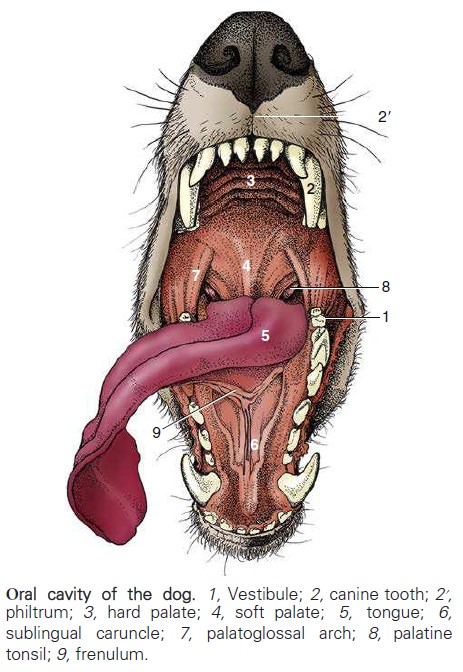
-
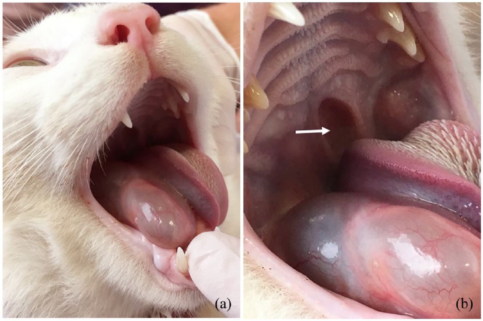
- Muscles and other structures of the neck and head of the cat in lateral view. 5
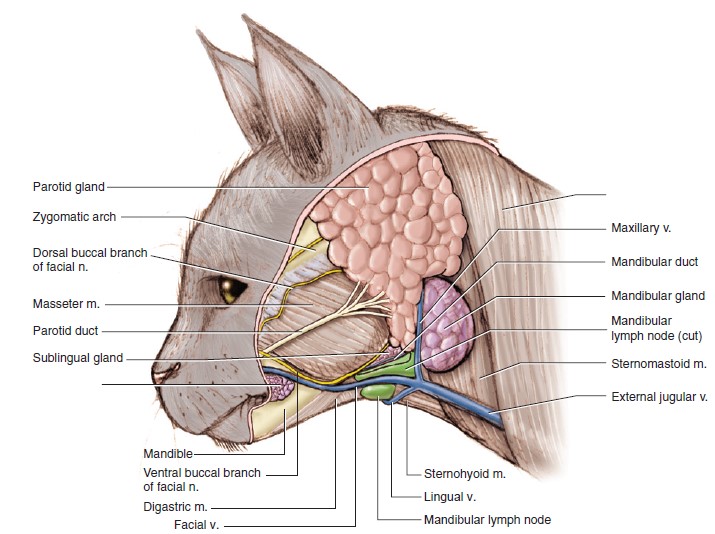
-
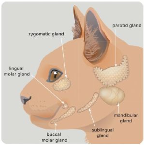
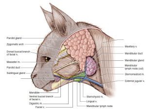
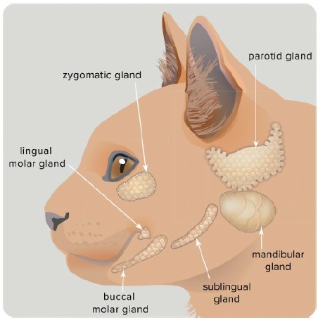
- Cat salivary glands. 35
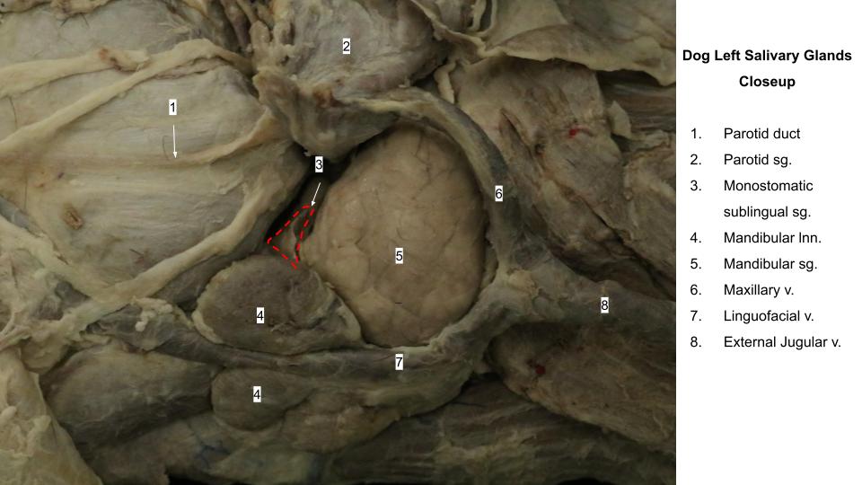
Dissect: If not done so previously, using the superficial dissection, incise, elevate and excise the capsule that surrounds these two glands and identify the fissure between the cranial surface of the mandibular salivary gland and the caudal aspect of the monostomatic sublingual salivary gland. The relationship between the two glands is often visualized as a golf ball (mandibular gland) sitting on a golf tee (monostomatic gland), albeit lying horizontally.
The ducts of both glands leave the rostromedial surface and enter the space between the masseter and the digastricus muscles. These ducts course rostrally beside the frenulum to open in the oral cavity at the sublingual caruncle. Lobules of the sublingual gland continue into the oral cavity with these ducts, where they can be seen beneath the oral mucosa.
The parotid salivary gland lies between the mandibular gland and the ear. It is closely applied to the base of the auricular cartilage of the ear. A small parotid lymph node may be found along the rostral border of this gland. The parotid duct is formed by two or three converging radicles, which unite and leave the rostral border of the gland. It grooves the lateral surface of the masseter muscle as it passes to the cheek. It opens into the vestibule on a small papilla at the level of the caudal margin of the fourth upper premolar.
Observe: Identify the parotid salivary gland on the half of the head used for superficial dissection.
Between the eyeball and the pterygoid muscles is the zygomatic salivary gland. It is hidden laterally by the zygomatic bone. The gland opens into the vestibule by one main and several minor ducts lateral to the last superior molar tooth.
Observe: Observe the zygomatic salivary gland on the deep half of the head. Some extra tissue or the most rostral part of the zygomatic arch may need to be removed in order to spot this gland just ventral to the orbit.
-
- Salivary glands of the dog. (The right half of the mandible was removed.) 1
-
- Salivary glands medial to the right mandible. (The digastric muscles, the mandible, and structures lateral to it have been removed.) 1
-

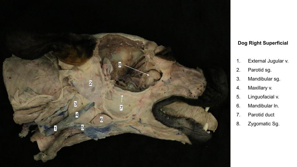
- Dog salivary glands
-
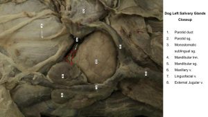
- Dog salivary glands closeup
-

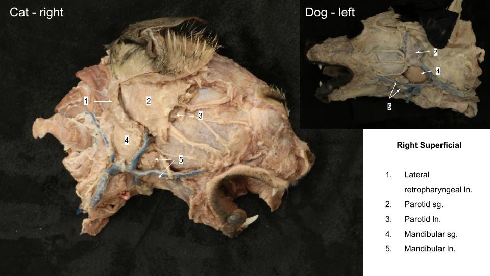
- Cat and dog salivary glands
-

- Zygomatic salivary gland
Clinical Application
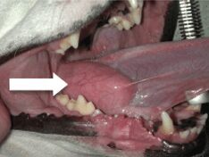
Sublingual Mucocele (Ranula)
Salivary mucoceles are caused by trauma to a salivary gland or duct. Potential inciting causes include bite wounds, chewing on sharp objects, or the use of choke collars. Once damaged, the gland or duct will leak saliva into the surrounding tissues. This saliva causes further damage due to the enzymes present in the secretion, leading to further damage of the gland and duct. Notably, a sublingual mucocele (also known as a ranula [ranula {L} = “frog”, due to its superficial appearance of a frog’s belly]), is related to damage to the mandibular and/or monostomatic sublingual salivary glands (or their shared duct). This type of mucocele can cause difficulty with chewing and swallowing given its presence just ventral to the tongue.
Treatment of a sublingual mucocele invovles marsupialization of the mucocele. Marsupialization involves creating an opening through which the saliva trapped under the tongue can drain directly into the oral cavity (creating a small “pouch”, thus “marsupialization”!).
-
- Dog salivary mucocele Blue Pearl
-
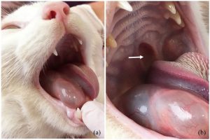
- Cat sublingual sialocoele J. Bassanino
The Muscles of Mastication
The muscles of mastication are a small group of muscles responsible for the action of the mandible during mastication (i.e., chewing). This group includes three muscles responsible for the closing of the mouth (i.e., elevators of the mandible): masseter m., temporalis m., and the pterygoid mm. (Note: the pterygoid mm. are actually two muscles, but we will not bother separating them in this course and will learn them as a single unit). They are innervated by the mandibular nerve, a branch of the trigeminal nerve (CN V). There is then only one muscle responsible for the opening of the mouth (i.e., depressor of the mandible); the digastricus m.
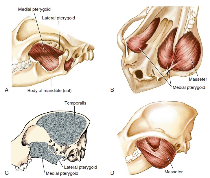
The masseter muscle arises from the zygomatic arch, where its deep portion is intermingled with the fibers of the temporalis muscle. It inserts in the masseteric fossa, the ventrolateral surface of the ramus of the mandible, and the angular process. The muscle is covered by a strong, glistening aponeurosis and contains many tendinous intermuscular strands.
The temporalis muscle arises from the temporal fossa and inserts on the coronoid process of the mandible.
Observe: On the half of the head used for the superficial dissection, observe the masseter m. as the thick mass of muscle covering the lateral surface of the cheek, ventral to the zygomatic arch. The temporalis m. is visible on the lateral surfaces of the parietal and temporal bones and dorsal to the zygomatic arch. It courses deep to the zygomatic arch as it approaches the coronoid process of the mandible.
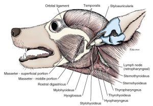
The medial and lateral pterygoid muscles (again, we’ll just refer to them as the pterygoid mm. and need not be distinguished from one another) arise from the pterygopalatine fossa and insert on the medial surface and caudal margin of the ramus of the mandible and angular process, ventral to the insertion of the temporalis muscle. The bulk of the muscle mass is the medial pterygoid.
Observe: On the half of the head used for the deep dissection, observe the lateral surface of the pterygoid muscles, which from this perspective are located ventral to the orbit. Recall that the maxillary nerve and maxillary artery cross over the lateral surface of the pterygoid mm. You do not need to dissect the pterygoid muscles further.
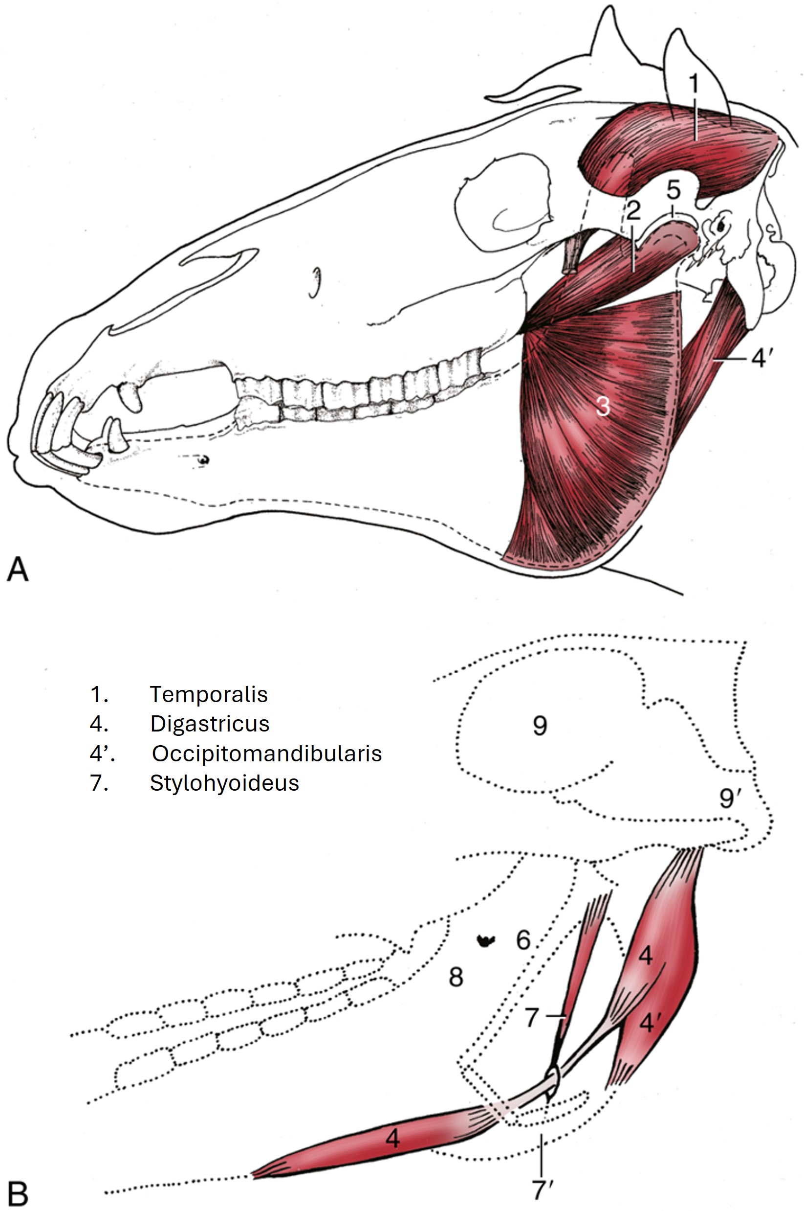
The digastricus m. arises from the paracondylar process of the occipital bone and is inserted on the body of the mandible. A tendinous intersection crosses its belly and divides it into rostral and caudal parts (hence its name digastricus [L] = “two bellies”). It acts to open the mouth. In the carnivores, the two bellies are fused and indistinct. In the ungulates, the rostral and caudal bellies are separated by an intermediate tendon; and then the horse also has an additional part. It forms an obstacle to a lateral surgical approach to the guttural pouch. The rostral portion of the digastricus muscle is innervated by the mandibular nerve, whereas the caudal belly is innervated by the facial nerve (CN VII).
Observe: Observe the digastricus m. on one of the handful of plastinated prosections in which this muscle is clearly defined.
-
- Muscles of mastication, lateral aspect. 1

-
-
Muscles of mastication. A, Medial and lateral pterygoid mm. B, Masseter and
medial pterygoid mm. C, Areas of origin of temporalis m. and pterygoid mm. D, Masseter m. cut to show the deep portion. 1
-
-
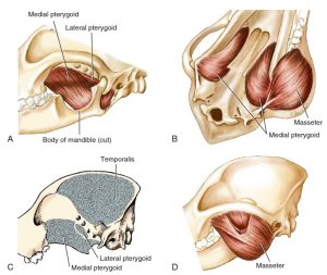
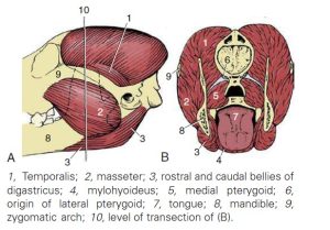
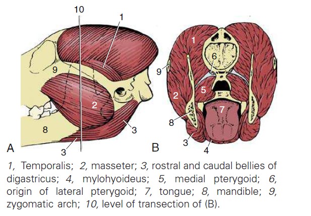
The muscles of mastication of the dog. (A) Left
lateral aspect and (B) in section. 8
-
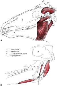
- Equine – (A) the deep masticatory muscles of the left side have been exposed by removal of the left mandibular ramus (stippled). (B) Medial view of the right digastricus and some related structures. 8
The Oral Cavity
The oral cavity, or mouth, is divided into the vestibule and the oral cavity proper. The vestibule is the cavity lying outside the teeth and gums and inside the lips and cheeks. The ducts of the parotid and zygomatic salivary glands open into the dorsocaudal part of the vestibule. The parotid duct opens through the cheek on a small papilla located opposite the caudal end of the upper fourth premolar or shearing tooth. The ducts of the zygomatic gland open into the vestibule lateral to the last upper molar tooth.
The oral cavity proper is bounded dorsally by the hard palate and a small part of the adjacent soft palate, laterally and rostrally by the dental arches, and ventrally by the tongue and adjacent mucosa. Its caudal boundary ventrally is the body of the tongue at the palatoglossal arch.
-
- Oral cavity of the dog. 8
-

- Mouth of the cat. 35
Observe: Pull the tongue away from one mandible and note the fold of tissue that extends from the body of the tongue to the beginning of the soft palate; this is the palatoglossal arch. The oral cavity communicates freely with the vestibule by numerous interdental spaces and is continued caudally by the oropharynx.
The Tongue
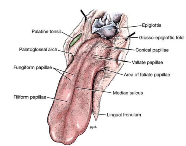
Moving on to the tongue, the tongue is a muscular organ composed of the interwoven bundles of intrinsic and extrinsic muscles. These muscles will be discussed later. It is divided into a root, which composes its caudal third; a body, which is the long, slender, rostral part of the tongue; and a free extremity, the apex. The mucosa covering the dorsum of the tongue is modified to form various types of papillae. A dissecting microscope facilitates examination of these structures. Five types of papillae are recognized by their shape. The filiform papillae are found predominantly on the body and apex of the tongue. They are arranged in rows like shingles, with their multiple pointed tips directed caudally. At the root of the tongue, the filiform papillae are replaced by conical papillae, which have only one pointed tip. The fungiform papillae have a smooth, rounded surface and are fewer in number. They are located among the filiform papillae. A few may be scattered caudally among the conical papillae. The foliate papillae are found on the lateral margins of the root of the tongue, rostral to the palatoglossal arch. They are leaflike but appear as a row of parallel grooves in the fixed specimen. The vallate papillae are located at the junction of the body and root of the tongue. There are four to six in the dog, and they are arranged in the form of a V with the apex directed caudally. They are larger than the others, have a circular surface, and are surrounded by a sulcus. There are taste buds on vallate, foliate, and fungiform papillae.
Observe: Examine the medial cut surface of the apex of the tongue.
On the ventral midline, just under the mucosa, is the lyssa. This fusiform fibrous spicule extends from the apex to the level of the attachment of the frenulum.
Dissect: On the transected surface of the tongue, that is closest to midline or on it, dissect deep to the ventral midline mucosa, separating muscle fibers to isolate a fibrous cord-like structure 3-5 mm diameter and a few centimeters long. If your cadaver’s head was split right on midline, the lyssa may already be visible and partially sectioned (it is typically gray/white and distinguishable from surrounding tongue muscle).
-
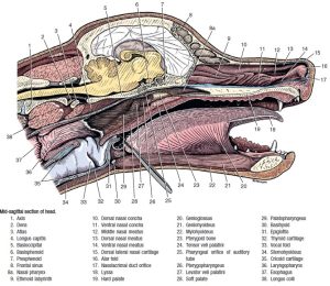
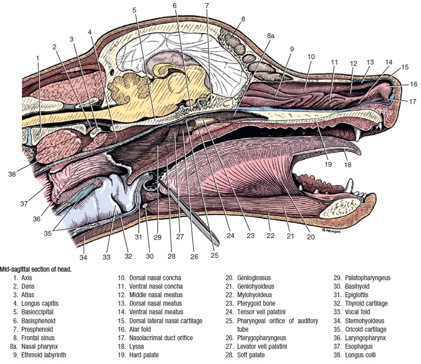
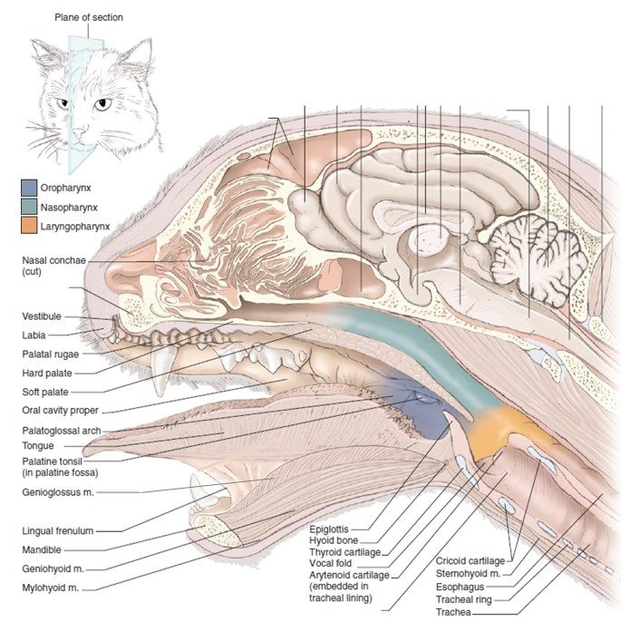
- Mid-sagittal section of head. 1
Muscles of the Tongue
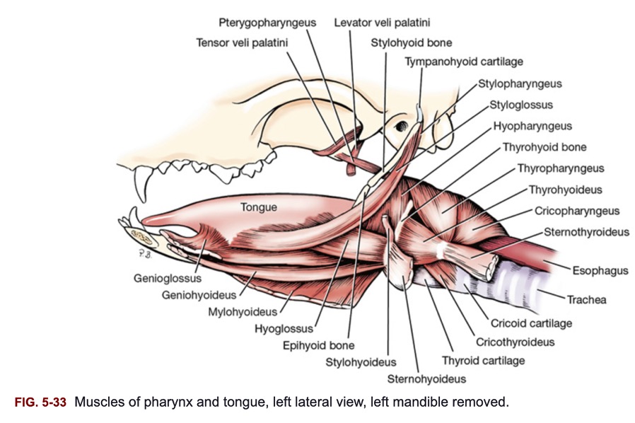
The muscles of the tongue may be divided into extrinsic and intrinsic groups. Three paired extrinsic muscles enter the tongue; the styloglossus and hyoglossus muscles on the lateral aspect of the base of the tongue, and the genioglossus m. on the ventral aspect.
The styloglossus arises from the stylohyoid bone, passes rostroventrally lateral to the palatine tonsil, and inserts in the middle of the tongue. It retracts and elevates the tongue. The hyoglossus arises from the thyrohyoid and the basihyoid and passes into the root of the tongue. It lies medial to the styloglossus and retracts and depresses the tongue. The genioglossus arises from the intermandibular articulation and adjacent surface of the body of the mandible. It joins its fellow at the median plane and is bounded medially by the geniohyoideus and laterally by the hyoglossus. Its caudal fibers protrude the tongue, and its rostral ones retract the apex. It lies partly in the frenulum. These muscles are all innervated by the hypoglossal nerve (CN XII).
Observe: Styloglossus and hyoglossus are best observed in prosection models found around the lab. Genioglossus can be seen on the medial surface of the hemisected heads.
-
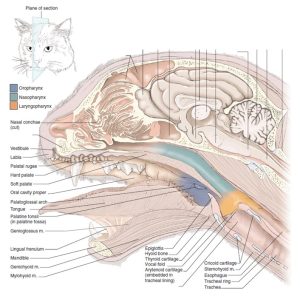
- Sagittal section of the cat head. 5
-

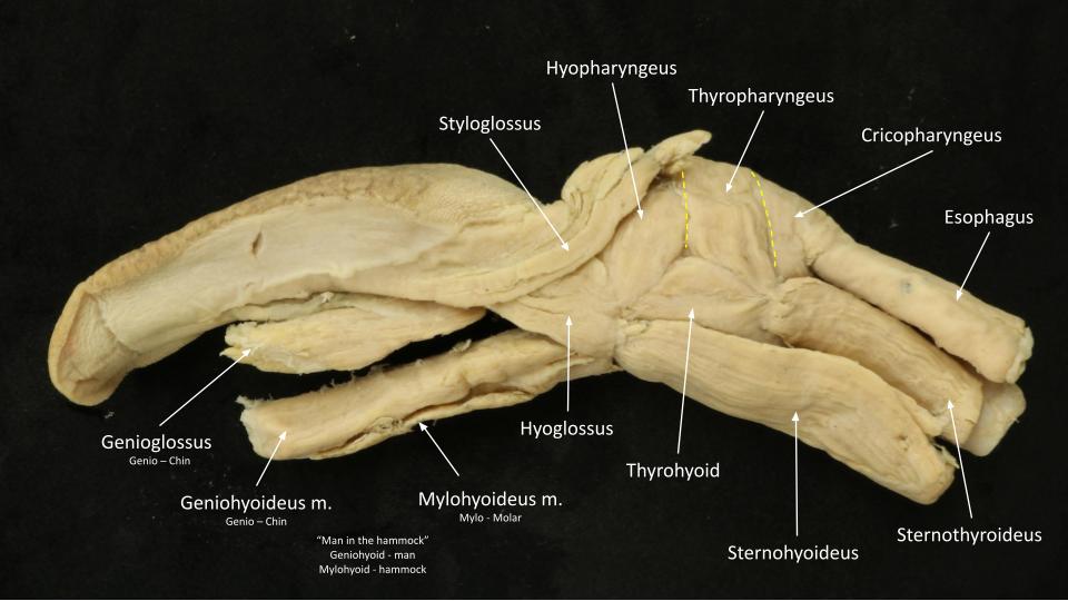
- Tongue and pharynx muscles
Hyoid Muscles
The hyoid muscles are associated with the hyoid apparatus, which suspends the larynx and anchors the tongue. They function in swallowing, lolling, lapping, and retching. All muscles of this group have names with the suffix -hyoideus. The prefixes of the hyoid muscles designate the bone or part from which they arise.
The sternohyoideus m., from its origin on the sternum and first costal cartilage, is fused to the deeper sternothyroideus for the first third of its length. It then separates from this muscle and runs an independent course, adjacent to the ventral midline, to insert on the basihyoid bone.
The thyrohyoideus m. is a short muscle that lies dorsal to the sternohyoideus. It extends from the thyroid cartilage of the larynx to the thyrohyoid bone.
The sternohyoideus and thyrohyoideus muscles are innervated by ventral branches of cervical spinal nerves and the hypoglossal nerve (CN XII).
The mylohyoideus m. spans the intermandibular space. It arises as a thin sheet of transverse fibers from the medial surface of the body of the mandible. It is inserted on its fellow muscle at the midventral raphe. Caudally, it inserts on the basihyoid. It forms a sling that aids in the support of the tongue. It is innervated by the mandibular nerve of CN V.
The geniohyoideus m. lies deep to the mylohyoideus. It is a muscular strap that arises on and adjacent to the intermandibular articulation. It parallels its fellow along the median plane and attaches to the basihyoid. Contraction of the geniohyoideus draws the hyoid apparatus and larynx rostrally. It is innervated by the hypoglossal nerve.
Observe: Observe the hyoid mm. on plastinated specimens and wet prosections made available to you in the lab.
-
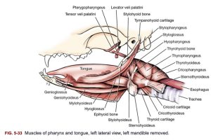
- Muscles of tongue, pharynx, and hyoid of the dog, mandible removed. 1

-
- Tongue and pharynx muscles
The Pharynges
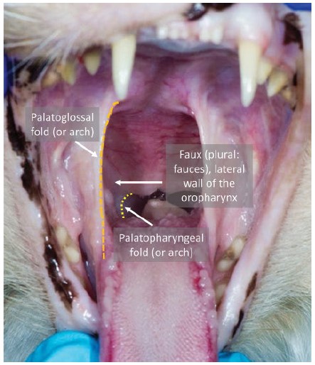
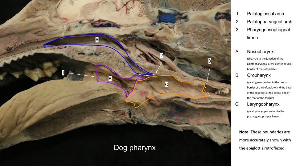
The pharynx is a passage that is common, in part, to both the respiratory and digestive systems. It is located between the oral cavity and esophagus and is divided into oral, nasal, and laryngeal parts. The oropharynx extends from the level of the palatoglossal arches to the caudal border of the soft palate and the base of the epiglottis at the caudal end of the root of the tongue. The dorsal and ventral boundaries of the oropharynx are the soft palate and the root of the tongue. The lateral wall of the oropharynx contains the palatine tonsil in the tonsillar fossa.
The palatine tonsil is elongated and is located caudal to the palatoglossal arch. The medial wall of the fossa, which partially covers the tonsil, is the semilunar fold. The tonsil is attached laterally throughout its entire length.
-

- Tongue, dorsal aspect. 1
-

- Palatine tonsil
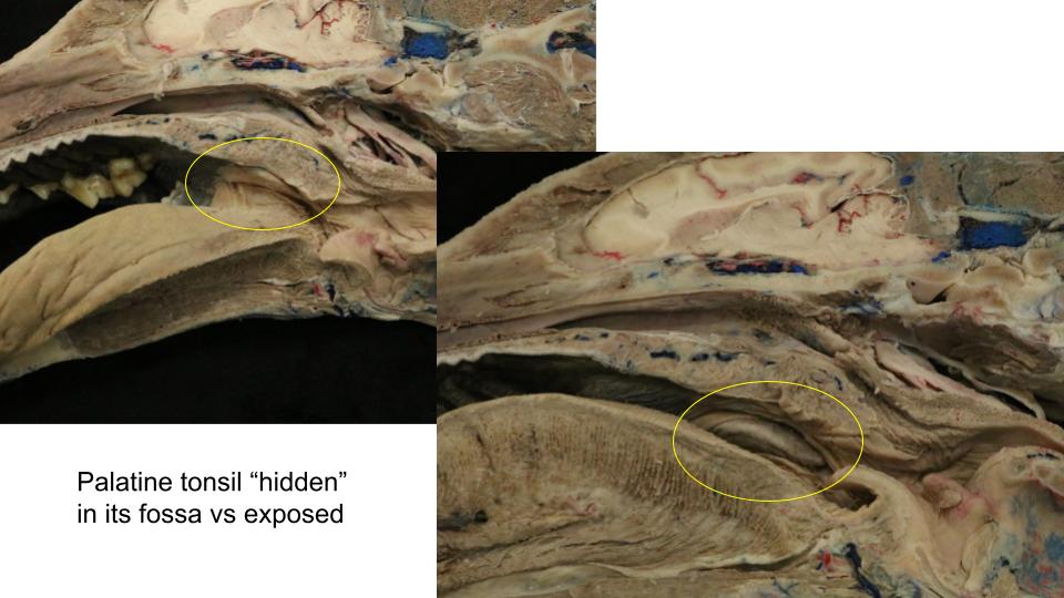
Observe: Reflect the palatine tonsil from the tonsillar fossa on the medial surface of the hemisected head.
The nasopharynx extends from the choanae to the junction of the palatopharyngeal arches at the caudal border of the soft palate. A palatopharyngeal arch extends caudally on each side from the caudal border of the soft palate to the dorsolateral wall of the nasopharynx. It is a fold of mucosa that covers the palatopharyngeus muscle. On the lateral wall of the nasopharynx, dorsal to the middle of the soft palate, is an oblique, slitlike opening, the pharyngeal opening of the auditory tube.
The laryngopharynx is dorsal to the larynx. It extends from the palatopharyngeal arches to the beginning of the esophagus. The esophagus begins at an annular constriction at the level of the cricoid cartilage, the pharyngoesophageal limen.
-
- Sagittal section of the cat head. 5
-

- Dog pharynx
-
- Dog pharynx boundaries

Observe: Identify the borders of the subdivisions of the pharynx, and be able to identify the oropharyx, nasopharynx, and laryngopharynx as discrete areas.
The pharyngeal muscles aid directly in swallowing. The cricopharyngeus m. arises from the lateral surface of the cricoid cartilage. Its fibers are inserted on the median dorsal raphe of the laryngopharynx. Caudally, its fibers blend with the esophagus.
The thyropharyngeus m. arises from the lateral side of the thyroid lamina and is inserted on the median dorsal raphe of the pharynx. This muscle is rostral to the cricopharyngeus m. and caudal to the hyopharyngeus m.
The hyopharyngeus m. is in two parts as it arises from the lateral surface of the thyrohyoid bone and the ceratohyoid bone. This origin was previously transected. The fibers of both parts form a muscle plate that passes dorsally over the larynx and pharynx to be inserted on the median dorsal raphe of the pharynx with its fellow from the opposite side. These pharyngeal muscles are all innervated by pharyngeal branches of the glossopharyngeal and vagus nerves. The remaining pharyngeal muscles and muscles of the palate listed here need not be dissected.
-

- Muscles of tongue, pharynx, and hyoid of the dog, mandible removed. 1
-
- Tongue and pharynx muscles
Observe: Observe the pharyngeal muscles on wet and plastinated prosections. Come back to your cadaver’s head and take a shot at identifying these muscles on the cadaver as well. Recognize that the ability to do so is variable and dependent on the nature of the bisecting cut.
The Esophagus
The esophagus is the connecting tube between the laryngeal part of the pharynx and the stomach. In medium-sized dogs it is approximately 30 cm long and 2 cm in diameter when it is collapsed. Because this passage traverses most of the neck and all of the thorax, and ends on entering the abdomen, it is divided into cervical, thoracic, and abdominal portions.
The cervical portion (pars cervicalis) of the esophagus is related mainly to the left longus colli and longus capitis muscles dorsally, and to the trachea ventrally and to the right. At its origin it starts to incline to the left, so that at the thoracic inlet it usually lies left lateral to the trachea. Its position here varies; in some specimens it is left dorsal and in others it is left ventral to the trachea. On the left side, the left common carotid artery, vagosympathetic nerve trunk, internal jugular vein, and tracheal trunk (duct) run in the angle between the esophagus and the longus capitis muscle. The corresponding structures on the right side are located lateral to the trachea.
The thoracic portion of the esophagus extends from the thoracic inlet to the esophageal hiatus of the diaphragm. At first, it usually lies to the left of the trachea between the widely separated leaves of the dorsal part of the cranial mediastinum. It obliquely crosses the left face of the trachea to gain its dorsal surface as the trachea bifurcates into the principal bronchi ventral to the fifth and sixth thoracic vertebrae. In reaching this level it crosses the right face of the aortic arch and lies ventral to the right and left longus colli muscles. Caudal to a transverse plane through the termination of the trachea the esophagus lies nearly in the median plane as it passes in the mediastinum between the two pleural sacs. The aorta obliquely crosses the left side of the esophagus between the fifth and ninth thoracic vertebrae, and thereafter diverges from it in a progressive manner caudally so that at the diaphragm the two structures are separated by approximately 3 cm.
The abdominal portion (pars abdominalis) of the esophagus is its wedge-shaped terminal part. Dorsally, the esophagus immediately joins the stomach. Ventrally, it notches the thin, dorsal border of the caudate lobe of the liver.
Observe: Identify the cervical and thoracic portions of the esophagus in your dog or cat cadaver.
Salivary Glands of the Ungulates
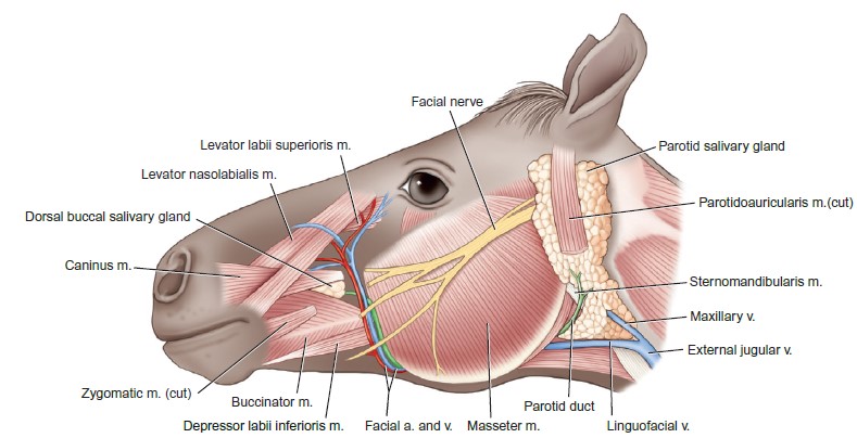
Observe: Identify the parotid salivary gland in the ungulate cadavers. It is found in a similar position to that in the carnivores; ventral to the ear and caudal to the masseter m.
The parotid salivary gland is relatively large compared to carnivores, commensurate with the high-fiber diet of ungulates, and is particularly large in the pig. The parotid duct has a species-varying path. In the carnivores and sheep, the parotid duct arises from the rostral border of the sg. and crosses the masseter muscle, whereas in the horse, ox, goat, and pig, it arises from the ventrorostral aspect of the sg. and follows a curving course around the ventral part of the masseter. The duct terminates in the familiar location, on the parotid papilla, found in the mucosa approximately dorsal to the upper 4th PM in most species, in the vestibule of the oral cavity.
The mandibular salivary gland is medial to the tendon of the sternocephalicus m. (mandibularis) in the horse and it is an elongated, larger structure compared to the smaller ovoid gland of the pig, dog, and cat. The mandibular sg. duct (no need to identify it) passes rostrally in a submucosal position ventrolateral to the tongue on the floor of the oral cavity. The duct discharges at the sublingual caruncle. Extensive sublingual and buccal salivary glands are present in the ungulates – we will not dissect these.
Observe: Locate the mandibular salivary gland lying partly deep and ventral to the parotid salivary gland. Reflect the ventral aspect of the parotid sg. to find it.
-

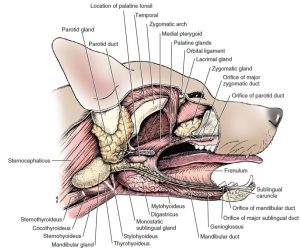
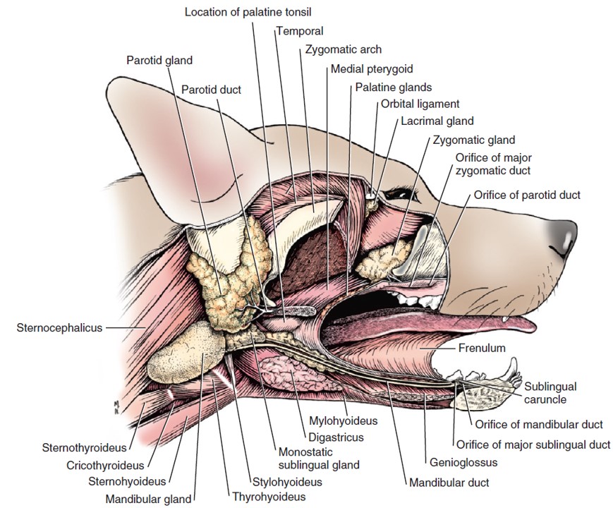
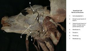
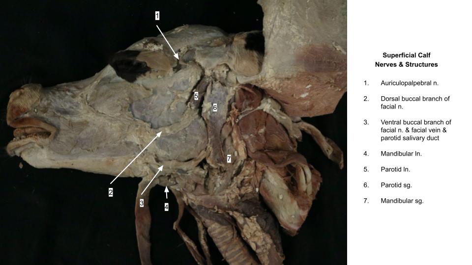
- Superficial view of the lateral head of the horse, focusing on the salivary glands and related structures. 9
-
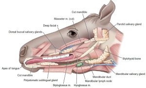
- Deep view of the lateral head of the horse, focusing on the salivary glands and related structures. 9

-
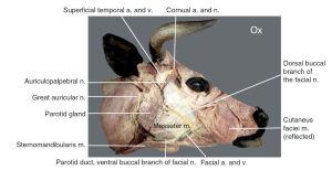
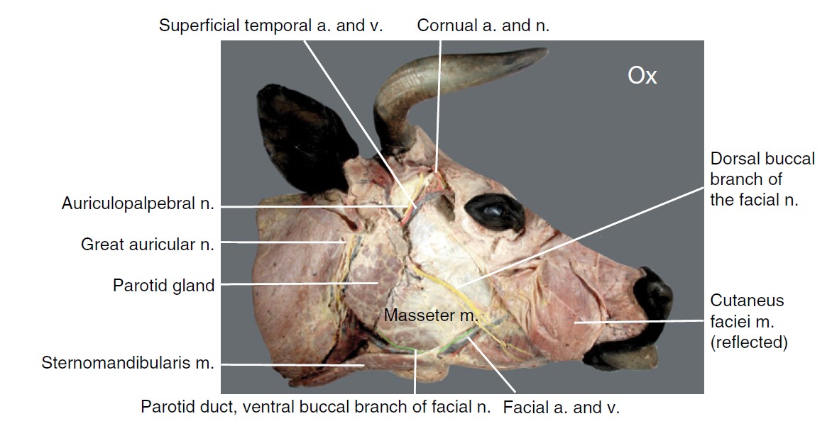
- Dissection of superficial structures of the bovine head: lateral view. 12
-
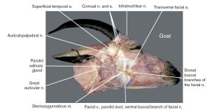
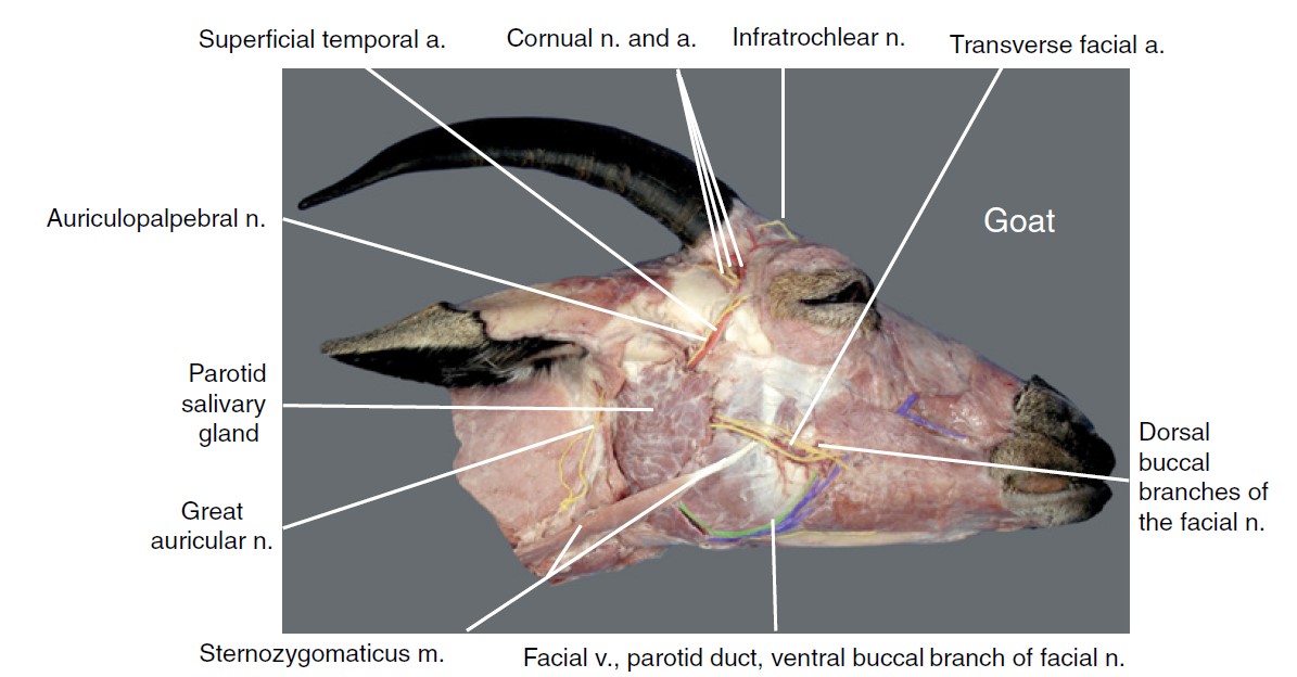
- Superficial structures of goat head: lateral view. 12
-

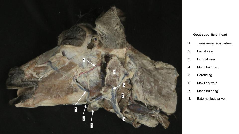
- Goat salivary glands
-
- Calf salivary glands
-
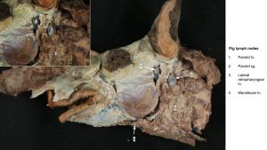
- Pig salivary glands

The Oral Cavity and Pharynges of the Ungulates
In gross structure, the oral cavity and pharynx of the ungulate is very similar to that of the carnivore.
Observe: Identify the following structures of the oral cavity in the ungulates. Note their similarities in position and structure to
- Vestibule
- Hard palate
- Tongue (apex, body, root)
- Oropharynx, nasopharynx, laryngopharynx
- Palatoglossal arches
- Palatine tonsil
- Palatopharyngeal arches
- Soft palate
- Nasopharyngeal openings of auditory tubes (in the horse, these are located in the large slits present in the caudal end of the nasopharynx)
However, there are notable differences in the ungulate species. In the horse, identify a small blind pocket in the caudodorsal aspect of the nasopharynx, dorsal to the slit containing the nasopharyngeal opening of the auditory tube. This blind pocket is known as the (dorsal) pharyngeal recess.
The nasopharynx ends caudally at the palatopharyngeal arches and caudal border of the soft palate. The arches and the soft palate form a complete ring of soft tissue, creating an opening between the space of the nasopharynx and the space of the laryngopharynx. This opening is called the intrapharyngeal ostium, and the rostral aspect of the larynx projects through this opening into the nasopharynx space, in the normal horse.
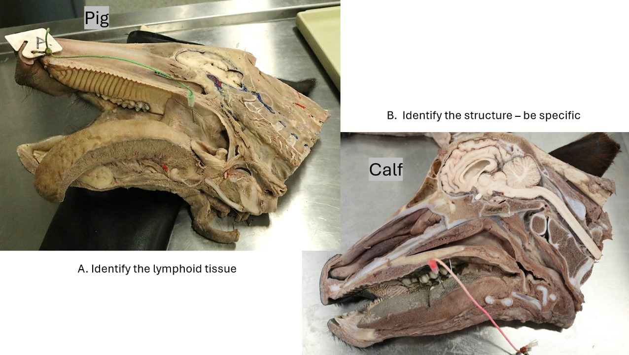
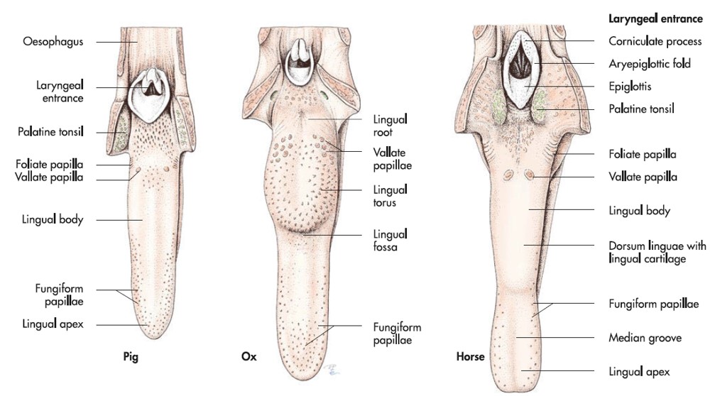
Observe: On the surface of the base of tongue, notice an aggregate of lymphoid tissue. This is the lingual tonsil.
In the ruminants, notice the lack of upper incisors. In their place, we see a flat dental pad. The tongue of the ruminant also has a large eminence in the middle of the body of the tongue, known as the torus linguae. Just rostral to the torus linguae is a groove, known as the fossa linguae. The fossa linguae is a common site of entrapment for foreign bodies in the oral cavity of the ruminant.
Observe: Identify the torus linguae and fossa linguae in a ruminant head.
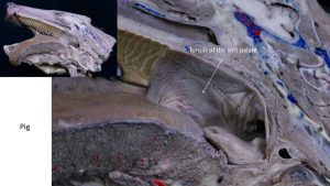
Observe: Finally in the oropharynx of the pig, look on the ventral surface of the soft palate to find a series of small pin-holes, which represent the tonsils of the soft palate (colloquially referred to as the “tonsils” in the clinic).
Moving caudally to the laryngopharynx of the pig, identify a small pouch dorsal to the laryngopharynx. This is the pharyngeal diverticulum.
-

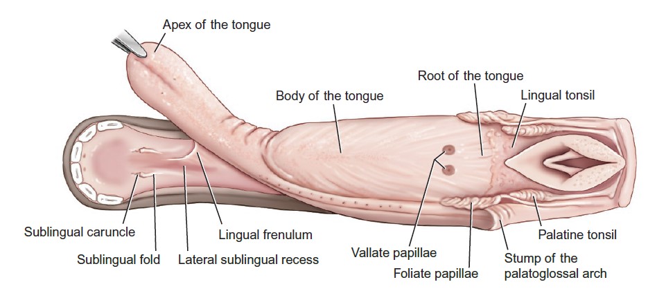
- Dorsal view of the tongue and additional structures in the oral cavity proper of the horse. 9
-
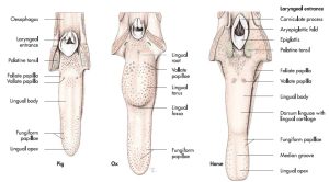
- Tongue, pharynx and esophagus of pig, ox and horse. 7
-
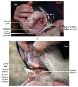
- Dental pad of the ox and goat. 12

-

- Sublingual caruncle of the cow. 8
-
- Tonsils of the soft palate

Clinical application:
Review videos
Dog pharynx & pharyngeal muscles – 10 min, watch until 11:30
Ungulate pharynx – 11 min
Dog salivary glands – 5 min
TERMS
| Term | Species/Notes |
| Parotid sg. | All |
| Parotid duct | All |
| Mandibular sg. | All |
| Monostomatic sublingual sg. | Carnivore |
| Sublingual caruncle | All |
| Sublingual mucocele (ranula) | Caused by damage to mandibular and/or monostomatic sublingual sgg.; treatment includes marsupialization |
| Zygomatic sg. | Carnivore; there may not be much of this gland left to identify in many cadavers |
| Oral cavity | All |
| Vestibule | All |
| Hard palate | All |
| Soft palate | All |
| Palatoglossal arches | All |
| Palatopharyngeal arches | All |
| Tongue | All |
| Apex; body; root | All |
| Lyssa | Carnivore |
| Lingual tonsil | Horse; ruminant |
| Torus linguae | Ruminant |
| Fossa linguae | Ruminant |
| Muscles of mastication | Innervated by mandibular n. (CN V3) |
| Masseter m. | All |
| Temporalis m. | All |
| Pterygoid mm. | Carnivore |
| Digastricus m. | All; also innervated by facial n. (CN VII) |
| Occipitomandibularis m. | Horse; part of digastricus m. in horse |
| Lingual muscles | Innervated by hypoglossal n. (CN XII) |
| Styloglossus m. | Plastinated models |
| Hyoglossus m. | Plastinated models |
| Genioglossus m. | Plastinated models |
| Muscles of hyoid appartus | |
| Sternohyoideus m. | Plastinated models |
| Thyrohyoideus m. | Plastinated models |
| Geniohyoideus m. | Carnivore cadaver heads and Plastinated models |
| Mylohyoideus m. | Carnivore cadaver heads and Plastinated models |
| Oropharynx | All |
| Palatine tonsils | All |
| Tonsils of the soft palate | Pig |
| Nasopharynx | All |
| Nasopharyngeal opening of auditory tube | Carnivore; Horse; Ruminant |
| Intrapharyngeal ostium | Horse |
| (Dorsal) Pharyngeal recess | Horse |
| Laryngopharynx | All |
| Piriform recess | All |
| Pharyngoesophageal limen | All |
| Pharyngeal diverticulum | Pig |
| Pharyngeal muscles | Innervated by glossopharyngeal n. and vagus n. (CNs IX and X) |
| Hyopharyngeus m. | Plastinated models |
| Thyropharyngeus m. | Plastinated models |
| Cricopharyngeus m. | Plastinated models |
| Esophagus | All |