LAB 1: The Kidneys, Ureters, and Adrenal Glands
lecobb
LAB 1: The Kidneys, Ureters, and Adrenal Glands
Learning Outcomes
- Identify the adrenal glands in the domestic species.
- Identify the adrenal cortex and medulla in the domestic species and recall what hormones are produced in each part.
- Define Cushing disease and Addison disease and state what part of the adrenal gland is affected in each condition (cortex or medulla?). Describe and differentiate between probable imaging findings in Cushing disease (PDH vs. ADH).
- Describe the position of the kidneys within the abdominal cavity of the domestic species.
- Identify the structures of the kidney and ureters in the domestic species.
- Define ectopic ureters and describe clinical and diagnostic findings thereof.
- Identify the structures of the kidney and ureter on ultrasonographic images of the canine kidney and differentiate three different ultrasonographic views from each other. Differentiate between a normal and abnormal ultrasonographic image of the carnivore kidney.
- Relate structures of the nephron to gross anatomical structures.
- Define pyelonephtritis and describe clinical and diagnostic findings thereof.
Carnivore Adrenal Glands
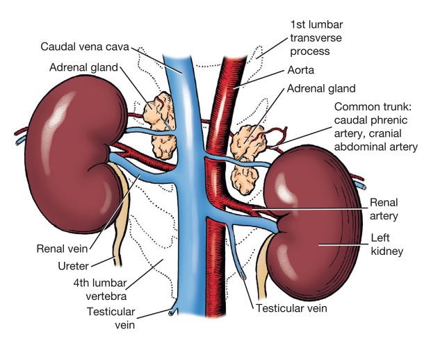
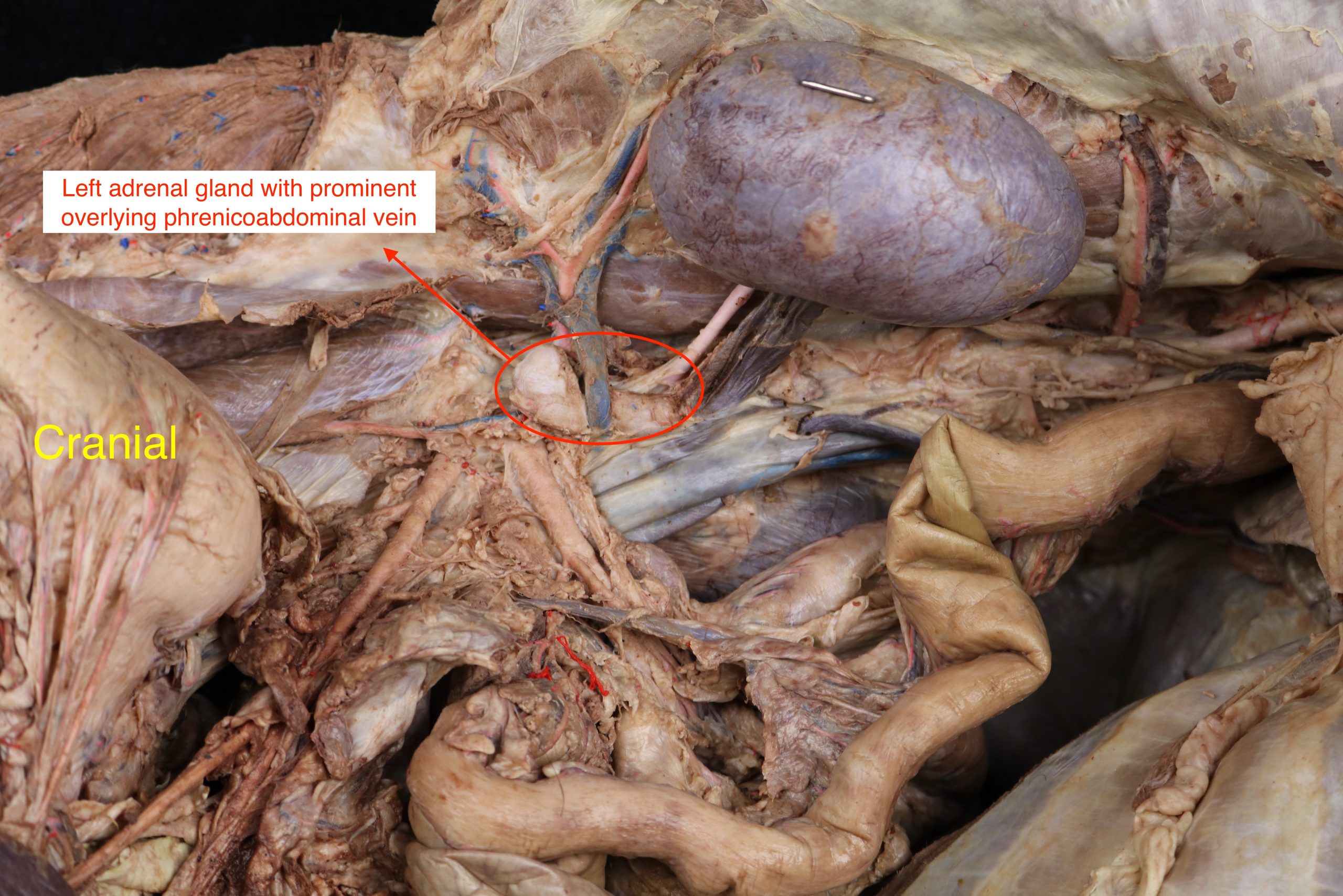
The adrenal glands are light-colored and are located at the cranial aspect of each kidney. Each gland is crossed ventrally by the common trunk of the caudal phrenic and cranial abdominal veins, called the phrenicoabdominal vein, which leaves a deep groove on its ventral surface. The right adrenal gland lies between the caudal vena cava and the caudate lobe of the liver ventrally and the sublumbar muscles dorsally.
The adrenal glands possess an outer layer, the adrenal cortex, and an inner layer, the adrenal medulla.
Recall from physiology that the adrenal cortex consists of three different zones which produce the following hormones:
Zona glomerulosa > produces mineralocorticoids (e.g., aldosterone) – hormones responsible for salt regulation.
Zona fasciculata > produces glucocorticoids (e.g. cortisol) – hormones closely related to sugar control.
Zona reticularis > produces androgens, or sex hormones.
The adrenal medulla hormones (catecholamines) include norepinephrine and epinephrine.
Dissect:
- Expose the left adrenal gland by dissection between the aorta and the kidney, cranial to the renal artery and vein. It is readily located by finding the phrenicoabdominal vein, which courses over it at this location. Dissect carefully, sparing the neural structures which cover it, to whatever extent possible.
- Transect the left adrenal and note the lighter-colored cortex and darker medulla. If a distinct cortex and medulla cannot be observed, it may be that a renal lymph node has been transected rather than the adrenal gland. Recall which hormones are produced in which sections of the cortex and in the medulla.
- The right adrenal gland need not be dissected.
-
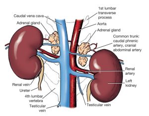
- Kidneys and adrenal glands, ventral view. 1
-
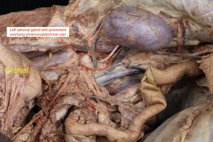
- Phrenicoabdominal vein
-

- Canine left kidney and adrenal gland
Clinical Application: Cushing Disease (Hyperadrenocorticism)
Cushing syndrome is any disorder characterized by elevated cortisol concentrations. The main types are pituitary-dependent hyperadrenocorticism and adrenal-dependent hyperadrenocorticism, in which a unilateral or bilateral adrenal tumor secretes cortisol. Clinical signs include polyuria/polydipsia, polyphagia, a pendulous abdomen, panting, muscle wasting, and dermatological changes. History, physical examination, clinicopathological testing, endocrine testing, and diagnostic imaging are all part of diagnosis. Surgery and adrenocortical suppressants are used for treatment.
-
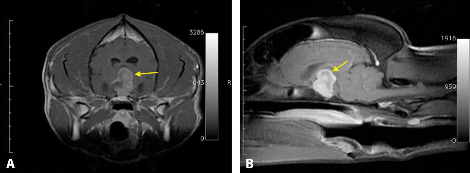
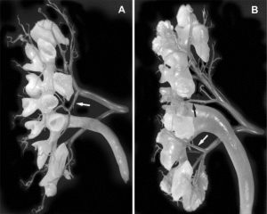
Pituitary-dependent hyperadrenocorticism (PDH) is due to an adrenocorticotropic hormone (ACTH) -secreting tumor of the pituitary gland (pituitary adenoma). It is the most common form of hyperadrenocorticism in dogs and cats, accounting for 80%–85% of reported cases. The tumor produces excess ACTH, which causes the adrenal glands to enlarge and produce too much cortisol. In animals with PDH, the adrenal glands are usually enlarged symmetrically; they may not be identical in size. MRI/CT of the brain may reveal a pituitary tumor.
-
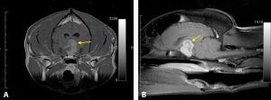
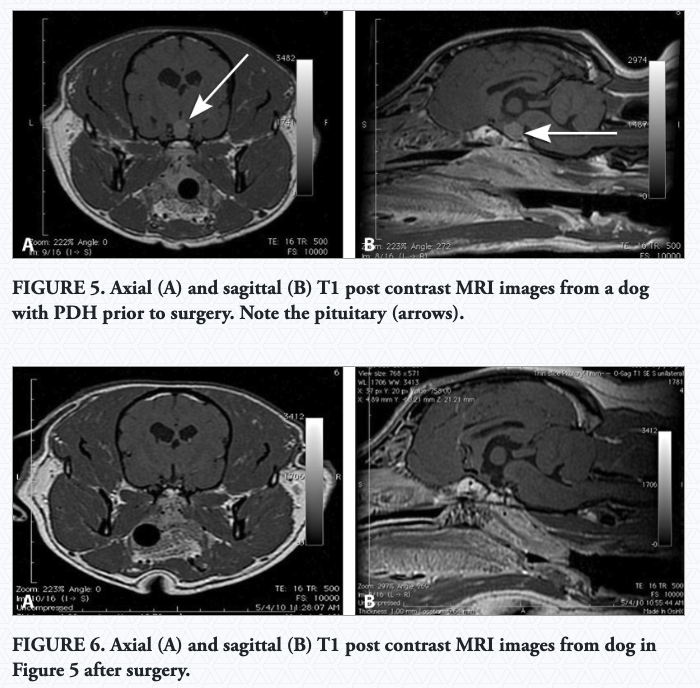
- Axial (A) and sagittal (B) T1 post contrast MRI images from a dog with PDH and large pituitary macroadenoma.
-
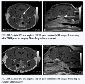
- Pituitary-dependent hyperadrenocorticism (PDH)
-
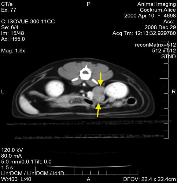
Adrenal-dependent hyperadrenocorticism (ADH) results from the autonomous secretion of glucocorticoids from an adrenal cortical tumor. It accounts for 10%–15% of cases. In dogs with an adrenocortical tumor, one adrenal gland is often enlarged and irregularly shaped.
-
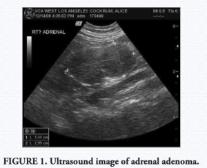
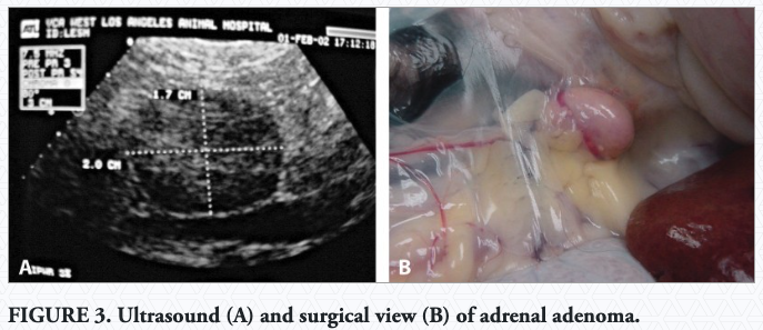
- Adrenal adenoma

-
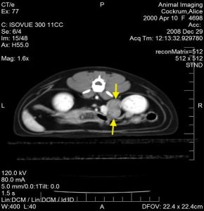
- CT image of large right adrenal adenoma between the right kidney and caudal vena cava
-

- Adrenal adenoma
Read More: Cushing’s Disease in Dogs Part 2: Diagnostic Approach
(Images sourced from link above.)
Clinical Application: Addison Disease (Hypoadrenocorticism)
Addison disease (hypoadrenocorticism) results from a lack of glucocorticoids, mineralocorticoids, or both. Isolated aldosterone insufficiency appears to be very rare, whereas isolated glucocorticoid insufficiency is likely underdiagnosed because of the lack of electrolyte abnormalities. The clinical signs of hypoadrenocorticism are often vague, can wax and wane over time, are rarely pathognomonic, and can be present for days, weeks, or months before diagnosis. Diagnosis depends on an accurate history, physical examination, and screening laboratory tests (CBC, chemistry panel, urinalysis). Confirmation requires adrenal function testing using the ACTH stimulation test. Treatment is highly successful but often lifelong.
Carnivore Kidneys and Ureters
The canine kidneys are dark brown. They are partly surrounded by fat and are covered only on their ventral surface by peritoneum. For this reason, they are considered to be retroperitoneal organs. The lateral border is strongly convex, and the medial, nearly straight. At the middle of the medial border is an indentation, the hilus of the kidney, where the renal vessels and nerves and the ureter communicate with the organ.
The right kidney lies opposite the first three lumbar vertebrae. It is farther cranial than the left kidney by the length of half a kidney. The right kidney is more extensively related to the liver than to any other organ. Its cranial third is covered by the caudate process of the caudate lobe of the liver. The remaining ventral surface is related to the descending duodenum, the right lobe of the pancreas, the cecum, and the ascending colon. The caudal vena cava is on the medial border of the right kidney.
The left kidney lies opposite the second, third, and fourth lumbar vertebrae. It is related ventrally to the descending colon and the small intestine. The spleen is related to the cranial extremity of the kidney. The medial border is close to the aorta.
Feline kidneys are situated relatively farther caudally in the body cavity than in dogs. The right kidney typically extends from the first to the fourth lumbar vertebrae, while the left kidney typically extends from the second to the fifth lumbar vertebrae. The kidneys are covered by peritoneum only on their ventral surfaces. Thus, renal hemorrhage or rupture of the ureter may result in localized collection of fluid near the dorsal body wall rather than diffusion of the fluid among the abdominal organs. The attachments of the feline kidneys to the body wall are relatively loose, rendering the kidneys somewhat mobile and therefore relatively easy to palpate.
Observe: Be able to state where the feline and carnivore kidneys are located.
-
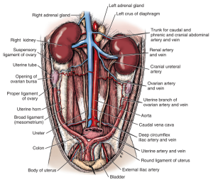
- Female urogenital system, ventral aspect. 1

The ureters are muscular tubes conducting urine from the kidney to the urinary bladder via peristaltic contractions. Each ureter leaves the kidney at the hilus and courses retroperitoneally along the dorsal body wall toward the urinary bladder. On nearing the urinary bladder, each ureter inclines slightly ventrally to access the rounded upper part of the bladder.
The ureters enter the dorsal surface of the bladder at an oblique angle, tunneling through the muscular bladder wall a short distance before entering the lumen. This slanted entrance into the bladder results in pressure being put along the length of the ureter within the bladder wall as the filling bladder stretches, assisting in prevention of reflux of urine from the bladder into the ureter and potentially the kidney. The two ureteral openings, along with the internal urethral orifice, define the trigone of the bladder, an important clinical anatomic landmark. While the urinary bladder, the ureteral openings, and internal urethral orifice (and trigone) will be observed during the urogenital unit, it is important to understand now where the ureters drain.
The expanded part of the proximal ureter within the kidney is called the renal pelvis, extensions of which are called the pelvic recesses.
Observe: Follow the course of the left ureter as far caudally as possible. In the female cadavers, do so without disrupting the broad ligament of the uterus. In the female, the ureter passes deep to the broad ligament as it runs caudally. The broad ligament is the connecting peritoneum of the female reproductive tract which connects the tract to the abdominal wall.
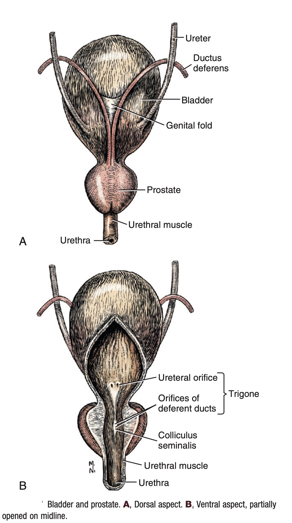
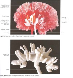
There is at least one cast of the proximal ureter/renal pelvis in the lab on which the pelvic recesses of the renal pelvis can be observed. If one cannot be located, use the photographs below to understand the anatomy of the renal pelvis and pelvic recesses in the carnivore (and small ruminant).
-
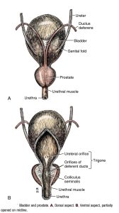
- Canine urinary bladder 1
-
- Renal pelvis corrosion cast.7

-
- Dog renal cast

Clinical Application: Ectopic Ureters
-
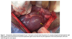
- Hydronephrosis and hydroureter

-
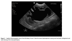
- Hydronephrosis secondary to ectopic ureters (ultrasound)
-
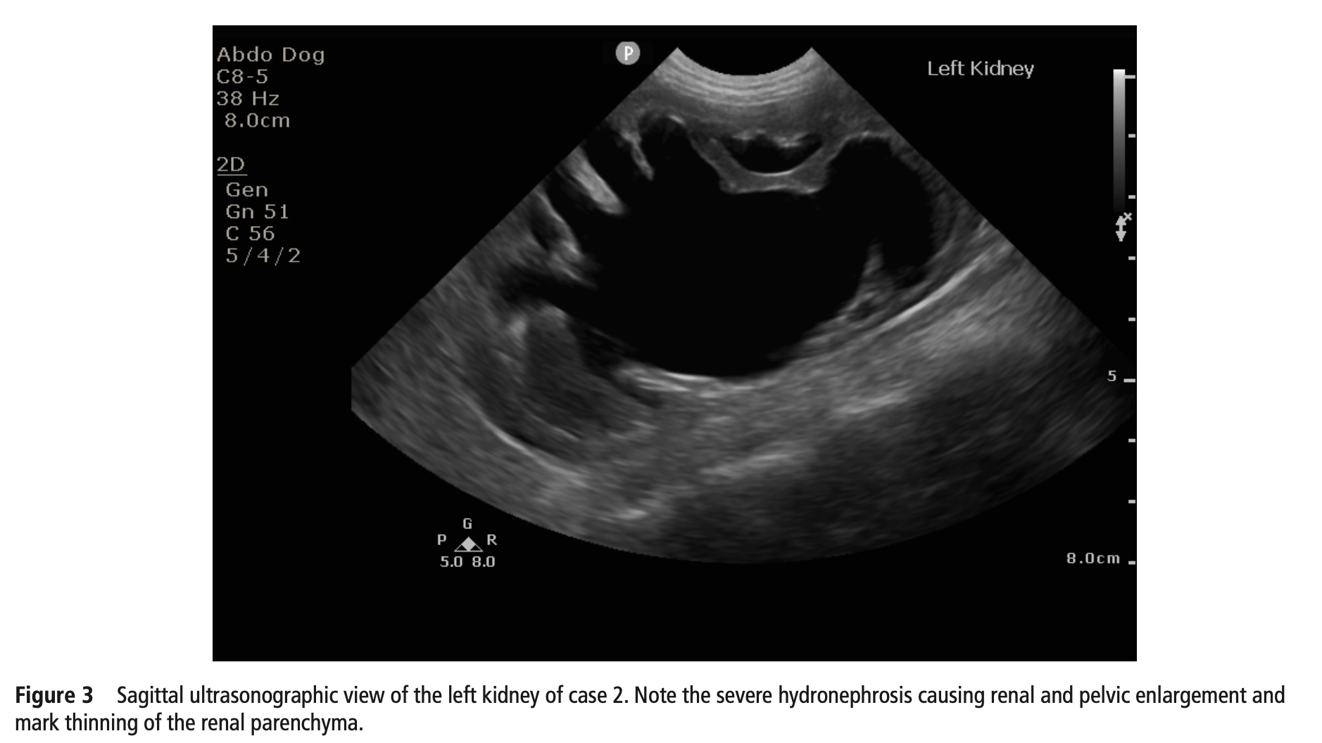
- Hydronephrosis secondary to ectopic ureters (radiograph)

Read More: Ureteral Anomalies in Animals
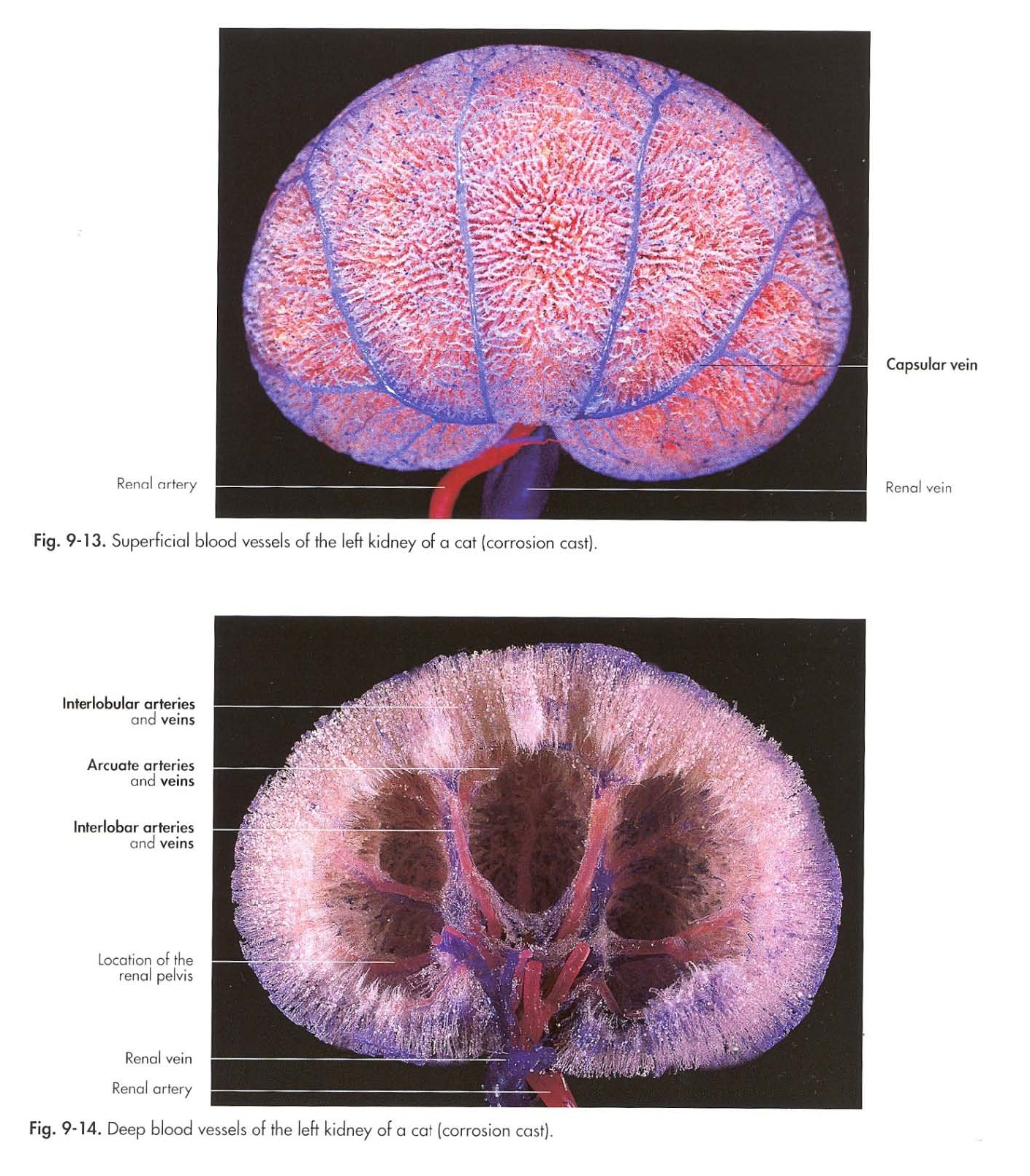
A thin fibrous renal capsule covers the surface of the kidney. The capsule follows the hilus to line the walls of the sinus and to form the adventitia of the renal pelvis. It also invests the renal vessels and nerves before they pass into the sinus. The fibrous capsule of normal kidneys is easily removable, except in the renal sinus, where it is adherent to blood vessels and to the renal pelvis. Note that in the cat, there are multiple large capsular veins on the cortical surface of the kidneys, just deep to the capsule. These veins converge to the renal vein at the hilus. They are present but much less pronounced in the canine kidney and should not be interpreted as abnormal in the cat.
Recall that the term parenchyma refers to the functional part of an organ. In the kidney, the parenchyma is made up of the outer renal cortex and the inner medulla. Note the granular appearance of the peripheral portion of the renal parenchyma. This is the renal cortex, which contains primarily the renal corpuscles and convoluted portions of the tubules. The more centrally positioned parenchyma is the medulla. It has a striated appearance owing to numerous collecting ducts.
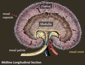
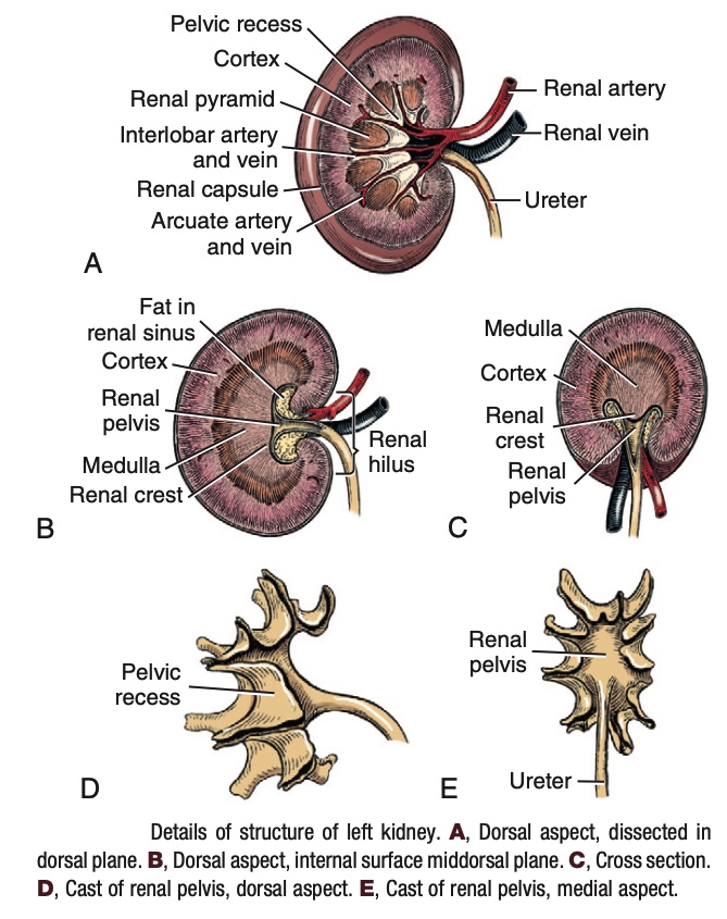
Recall the expanded part of the ureter within the kidney is called the renal pelvis – note that the renal pelvis and ureter are not part of the renal parenchyma. A median plane longitudinal section of the kidney shows the medulla as a continuous striated structure with its free edge facing the renal pelvis. This is the renal crest, through which collecting tubules of the kidney excrete urine into the renal pelvis.
In some specimens, it may be possible to differentiate between adjacent renal pyramids, which are the sections of medulla separated by each pelvic recess. The apex of each renal pyramid is call the renal papilla. In animals with unilobar kidneys (fused cortex and medulla), the apices of each renal pyramid combine to form the renal crest.
A variable number of papillary foramina open on the border of the renal crest that faces the renal pelvis. These are the openings of the papillary ducts that pass urine into the renal pelvis (expanded proximal ureter), which leads to the distal ureter.
The vessels that are apparent at the corticomedullary junction are the arcuate branches of the interlobar renal vessels.
Dissect: Free the left kidney from its overlying peritoneum and fascia. Do not cut its vascular attachment. Make a midline dorsal plane longitudinal section of the left kidney from its lateral border to the hilus, dividing it into dorsal and ventral halves (see photographs below).
Identify the following structures: renal capsule, renal cortex and medulla, interlobar a/v and arcuate branches, renal pelvis, ureter, and renal crest. Recognize that the papillary foramina (the openings of the papillary ducts) exist along the border of the renal crest. Urine drips from the renal crest into the renal pelvis and then drains into the ureter.
Observe the renal pyramids, as defined by each section of medulla located within one pelvic recess. The apex of each of the renal pyramids is the renal papilla. The renal crest is formed by the convergence of the papilla of the renal pyramids in kidneys with a fused medulla.
-
- Internal structures of unilobar kidney
-
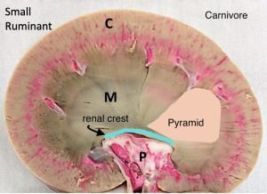
- Internal structures of carnivore and small ruminant kidney
Para-median sagittal sections of the kidney, i.e. a longitudinal section parallel to the midline cut, allows for visualization of the exterior portion of the medulla separated into cone-shaped renal pyramids with interlobar vessels between them. The base of the renal pyramids are at the level of the renal cortex. These pyramids extend from the cortex on the dorsal and ventral surfaces of the kidney into the center where they fuse into the renal crest. On this view, the pelvic recesses have been transected and can be observed surrounding the exterior portion of the renal pyramids.
Dissect: Make a second longitudinal section parallel to the first and note the exterior portion of the renal pyramids surrounded by the transected pelvic recesses. *Some resources identify the apices of the renal pyramids as seen in this view as the renal papilla. However, what can be observed from this section is the exterior portion of the renal pyramid (the internal portion has been cut). Recall that the renal papilla converge to form the renal crest in these species.
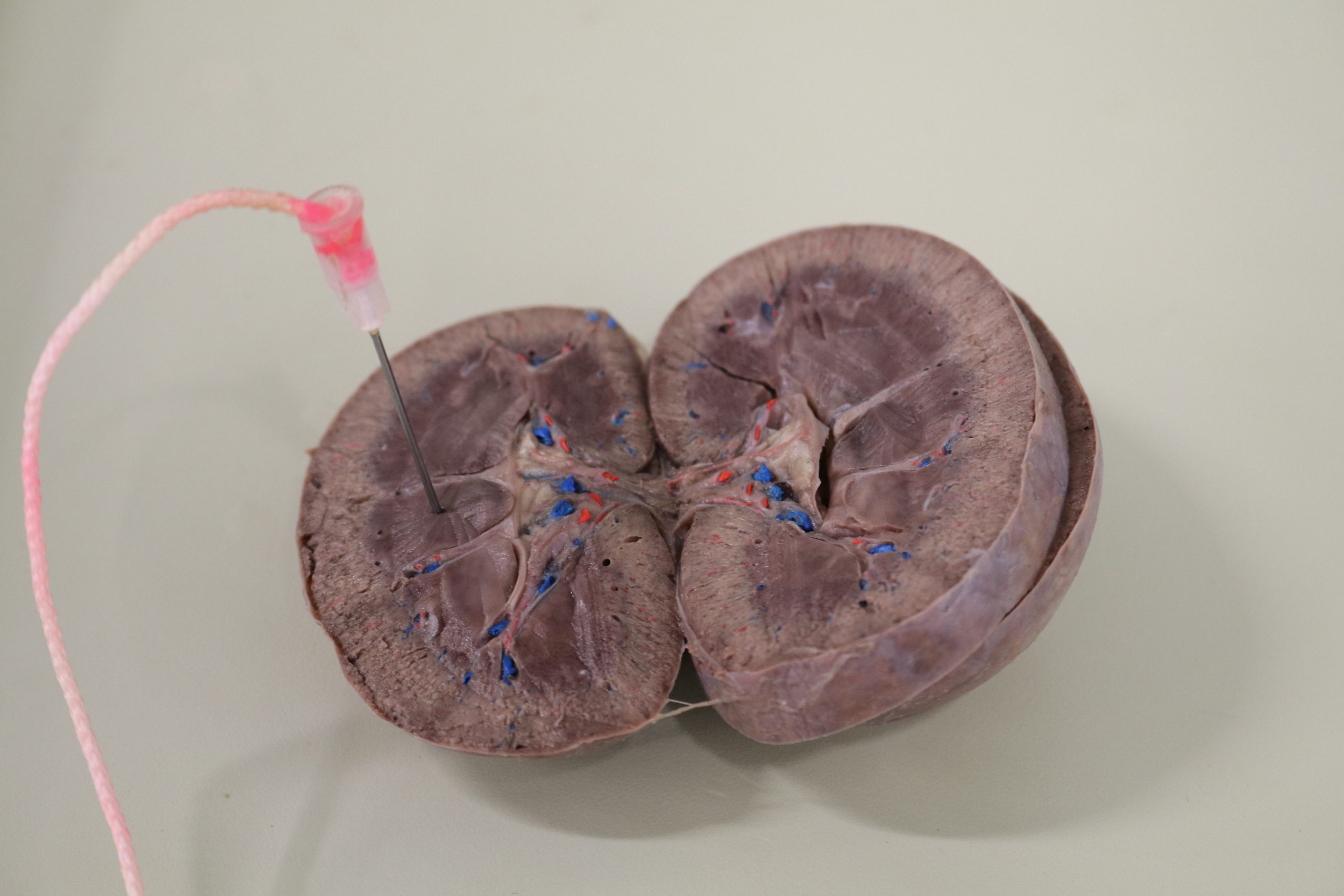
Dissect: Now free the right kidney from its overlying peritoneum and fascia keeping its vascular attachment intact. Transect the kidney (“hamburger style”) at the level of the renal hilus and observe the renal cortex, medulla, crest, and pelvis on the cut surfaces.
-
- Left kidney in cross section. 1
-

- Details of structure of left kidney. 1
-
- Renal pelvis corrosion cast.7
-
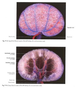
- Cat kidney corrosion cast7
-
- Capsular veins on a feline kidney

-
- Dog renal cast
The Nephron
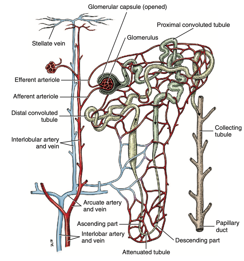
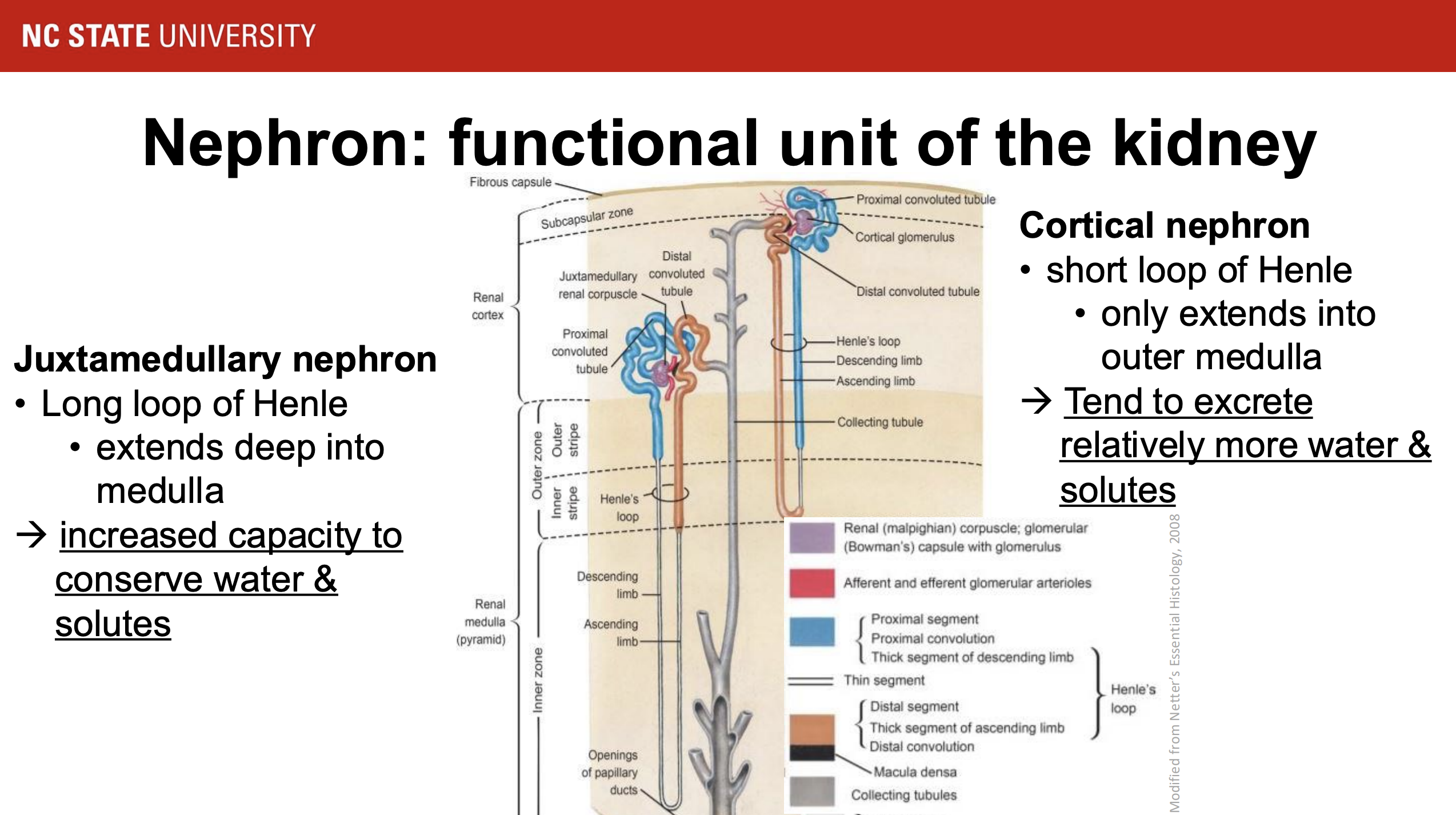
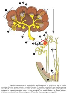
The nephron is a continuous contorted tube that serves for urine production and for the regulation of the volume and composition of the extracellular fluid. There are approximately 500,000 nephrons in the dog kidney. Each nephron begins at the double-layered glomerular capsule, which is invaginated by a spherical rete of blood capillaries, the glomerulus. The glomerulus and capsule together form the renal corpuscle. Renal corpuscles are present in the renal cortex, but not in the medulla. Recall from the physiology portion of this unit where juxtamedullary and cortical nephrons are located and where each part of these nephrons are located.
-
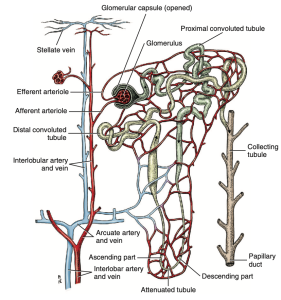
- Vessels around the nephron or renal tubule. 1
-

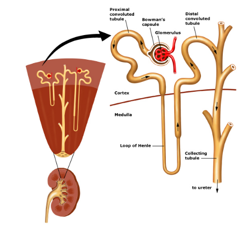
- Structures of the cortex and medulla. safarivet.com
-

- Nephron
Clinical Application: Renal Ultrasound
- Chronic kidney disease (CKD) is prevalent among older cats and dogs.
- Early recognition and staging of CKD can prompt the initiation of supportive care.
- Ultrasonography may help detect and stage CKD before abnormalities are detected with biochemical testing.
- Ultrasonography is valuable for excluding CKD sequelae or other renal diseases, which may warrant more aggressive therapy, require other diagnostics, or alter prognosis.
- CKD is progressive, and serial ultrasonographic evaluations are valuable for monitoring longitudinal changes.
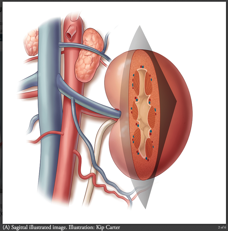
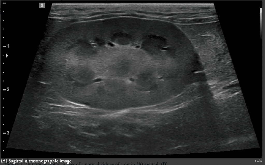
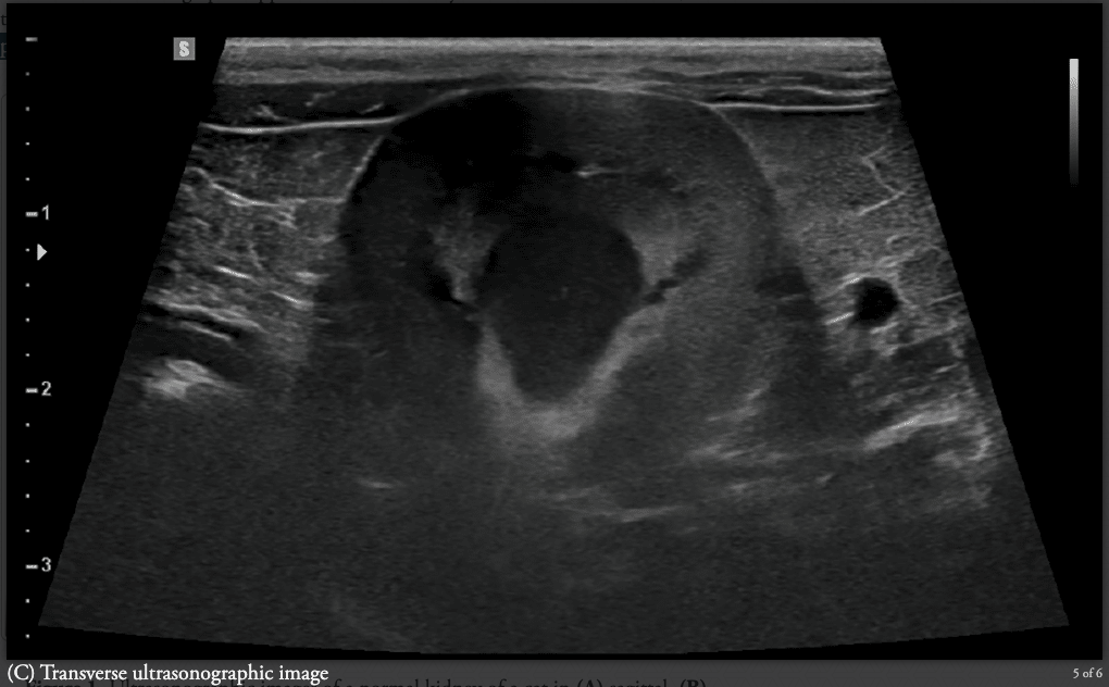
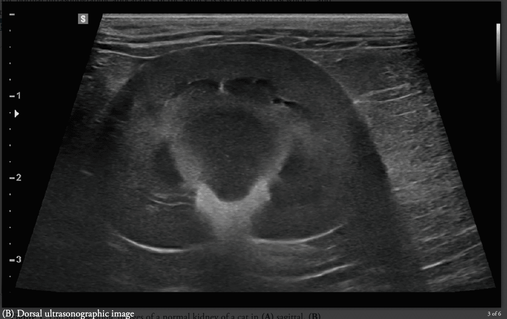
To assess renal morphology via ultrasonography, the kidneys should be imaged in three planes: sagittal, dorsal, and transverse. Be able to differentiate between images of each plane. Use the images below and review the renal lecture to identify the following structures on each of the three ultrasonographic images below: cortex, medulla, renal pelvis, and renal crest. On the sagittal section only, also identify the renal pyramids and pelvic recesses. Be able to distinguish between a normal and abnormal ultrasonographic image of the carnivore kidney (see clinical application exercises below for examples of abnormal images).
-

- Illustration of sagittal image of canine kidney.
-

- Sagittal ultrasonographic image of normal canine kidney.
-
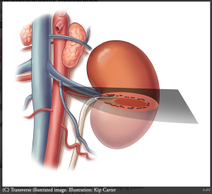
- Illustration of transverse ultrasonographic image of canine kidney.

-

- Transverse ultrasonographic image of canine kidney.
-
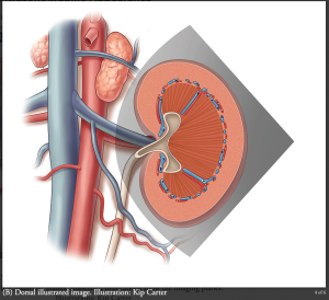
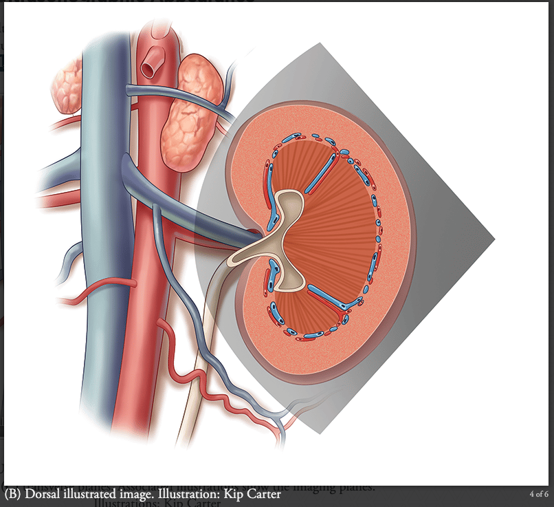
- Illustration of dorsal image of canine kidney.
-
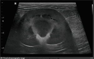
- Dorsal ultrasonographic image of normal canine kidney.
Read More: Ultrasonography for Diagnosing Chronic Kidney Disease in Dogs and Cats
(Images sourced from link above.)
Clinical Application: Gross Anatomic Changes in Acute and Chronic Kidney Disease
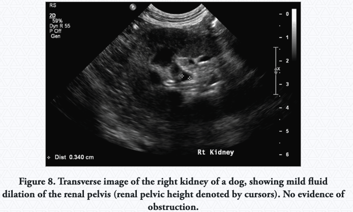
Pyelectasia: the collection and subsequent dilation of the renal pelvis with fluid secondary to a nonobstructive process.
-
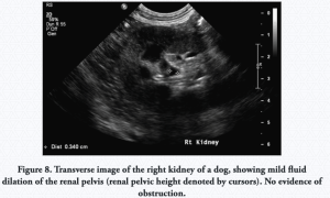
- Pyelectasia
Reduced Kidney Size: Decreased kidney size is expected with fibrosis; however, size may not directly correlate with the degree of fibrosis in the presence of concurrent compensatory hyperplasia, renal cysts, and/or pyelectasia.
-
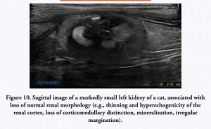
- Small left kidney (feline)

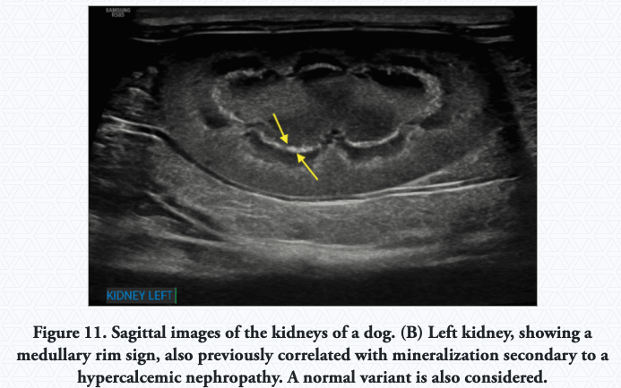
Mineralization (Nephrocalcinosis): Dystrophic mineral deposition within the nephrons and interstitium can occur secondary to renal damage and can be difficult to differentiate from fibrosis.
-
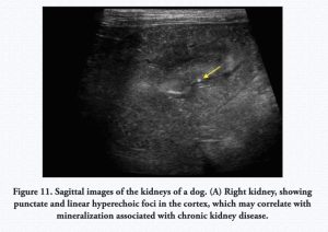
- Nephrocalcinosis
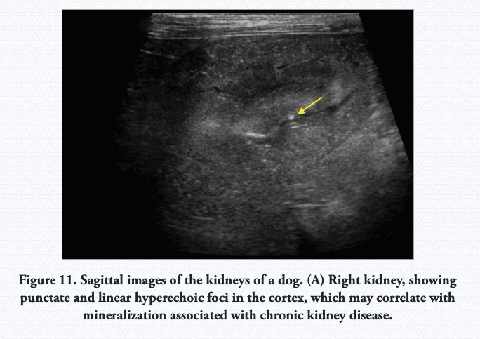
-

- Nephrocalcinosis
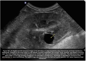
Irregular Renal Contour: Irregular renal margins may be associated with one or more, often a combination, of fibrosis, sclerosis, atrophy, cyst formation, and infarction.
-
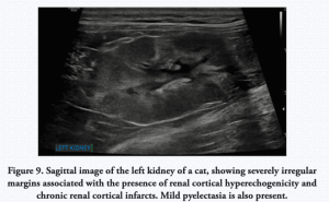
- Irregular Renal Contour (feline)

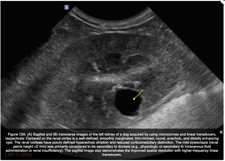
Cysts: Renal cysts occur more commonly in the cortex and can be acquired or congenital; therefore, without prior ultrasonographic examination for comparison, they are not a sensitive indicator of CKD in dogs. Renal cysts are common in dogs with CKD and most often do not affect renal function. They can distort the renal margins and may be a contributing cause of renal margin irregularity.
-
- Renal cortical cyst (dog) sagittal view
-

- Renal cortical cyst (dog) transverse view

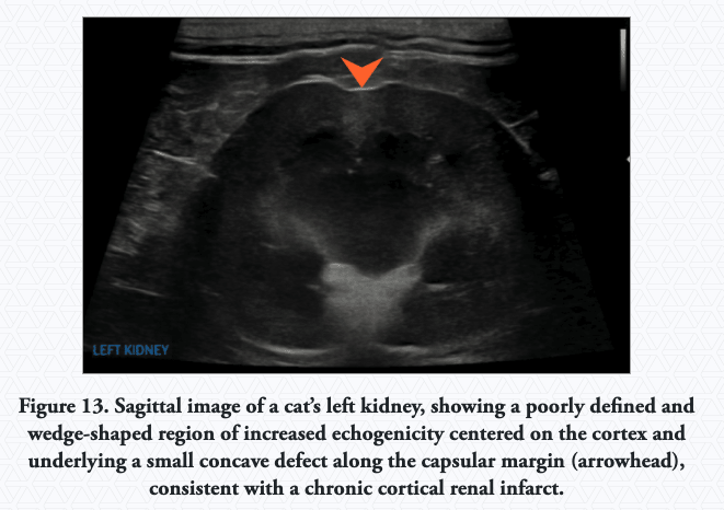
Infarcts: Renal infarcts most commonly affect the renal cortex and can vary in number and size. They are associated not only with CKD but with other causes, including underlying coagulopathy, sepsis, or neoplasia.
-
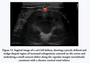
- Chronic cortical infarct (feline)
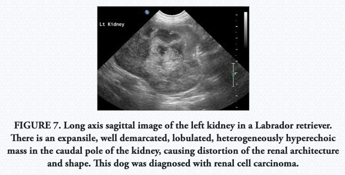
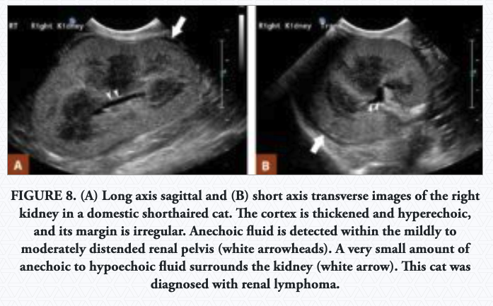
Renal Neoplasia: The most common renal tumor in the dog is renal cell carcinoma. The most common renal tumor in the cat is renal lymphoma, where diffuse changes can be seen. The kidneys are usually enlarged, irregular, hyperechoic, with hypoechoic subcapsular thickening.
-
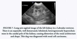
- Renal cell carcinoma (canine)
-
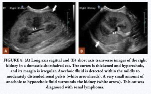
- Renal lymphoma (feline)
Renal Abscess: A renal abscess can occur in conjunction with fever and leukocytosis. Abscesses appear as poorly demarcated cavitary lesions that are irregular in contour with internal echoes and sedimentation, and they have variable degrees of distal acoustic enhancement. Typically, the renal pelvis is enlarged.
-
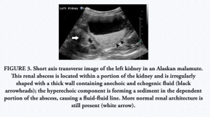
- Renal abscess (Alaskan malamute)

Autosomal Dominant Polycystic Kidney Disease: This is an inherited disease reported mostly in Persian or Persian-cross cats, cairn terriers, and bull terriers.
-
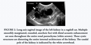
- Polycystic Kidney Disease (Ragdoll cat)

Hydronephrosis: Accumulation of fluid in the renal pelvis secondary to post-renal obstruction, such that occurs secondary to urolithiasis (stones in the urinary system).
-
- Hydronephrosis and hydroureter
-
- Hydronephrosis secondary to ectopic ureters (ultrasound)
-
- Hydronephrosis secondary to ectopic ureters (radiograph)
Read More: Ultrasonography for Diagnosing Chronic Kidney Disease in Dogs and Cats
Read More: Ultrasonography of the Urinary Tract: Kidneys and Ureter
(Images sourced from links above.)
Ungulate Adrenal Glands and Kidneys
Kidneys
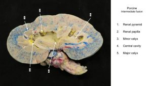
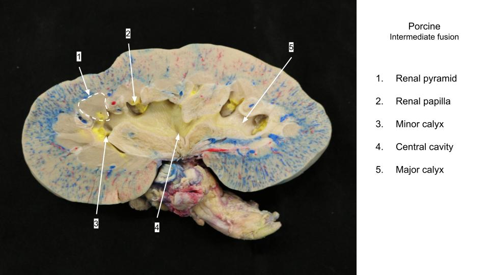
The kidneys of domestic mammals vary in appearance on the surface. Embryologically, kidneys undergo some fusion, and basically, the difference in the degree of fusion accounts for the variation in their shape and structure. The kidney of the ox has the least amount of fusion, the kidney of the pig is intermediate in degree of fusion, and the kidney of the horse, and small ruminants have the most complete fusion.
Ox and Pig
In the ox, the cortex remains unfused while in the pig, the cortex IS fused. Therefore the outer surface of the ox kidney is lobulated while that of the pig is smooth.
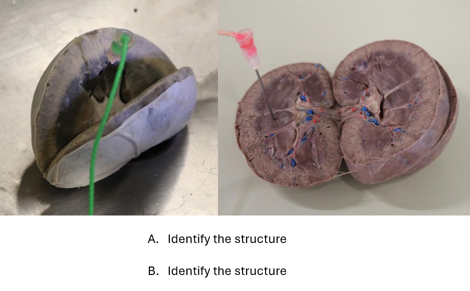
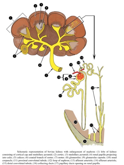
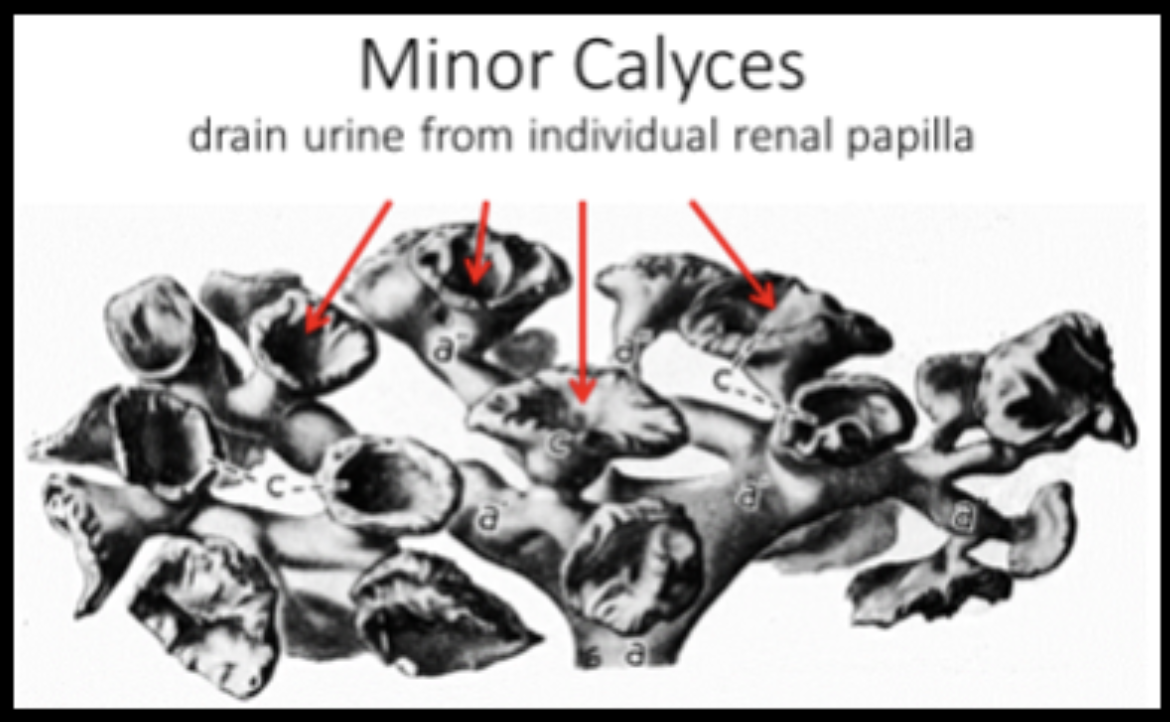
In the kidneys of both the ox and the pig, the medulla remains unfused. The apex of each renal pyramid is the renal papilla; it is here that the papillary ducts (large urine-collecting ducts), open. There is no fusion of renal papillae to form a ridge (as in the carnivore kidney); therefore, no renal crest is present. Each renal papilla projects into a funnel-shaped collecting chamber of the proximal ureter, called a minor calyx.
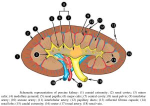
In the ox, the minor calyces drain to the cranial and caudal branches of the ureter. In the pig, the minor calyces drain to one of two larger chambers known as the major calyces, then into the central cavity.
Note that in the newborn calf, the left kidney is closely associated with the sublumbar muscles and, as expected, is located to the left of the median plane. Because of the tremendous growth of the rumen, however, the left kidney of adult ruminants is displaced toward the right side of the abdominal cavity. This displacement is accompanied by a longitudinal, pendulum-like rotation of the kidney so that the hilus is directed dorsally.
The location of the pendulous left kidney varies with the fullness of the rumen, and when the dorsal sac is filled, the left kidney lies to the right of the median plane, caudoventral to the right kidney. The surface of the left kidney that is in contact with the rumen becomes flattened, so that the fixed (hardened) left and right kidneys of the adult ox can usually be differentiated after removal from the animal.
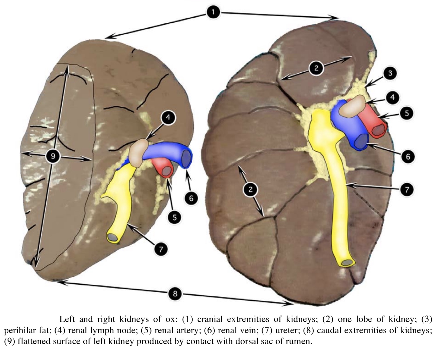
Identify: Ox kidney external shape, cortex, medulla, ureter, renal a./v., renal lobe, renal pyramid, renal papilla, minor calyx (-ices), branches of ureter, interlobar aa. Note the location of the left kidney in adult ruminants.
Pig external shape, cortex, medulla, ureter, renal a./v., interlobar a., renal pyramid, renal papilla, minor calyx (-ices), major calyx (- ices), central cavity of ureter (the latter two structures form the renal pelvis).
-
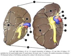
- Ox kidney 2
-
- Ox kidney 2
-
- Pig kidney 2

-
- Pig kidney
-
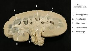
- Pig kidney

-
- Cast of porcine ureter and renal pelvis

-
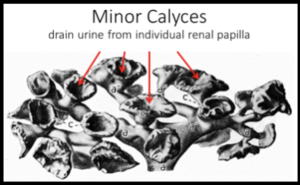
- Cast of bovine ureter, branches of the ureter, and minor calyces
Goat and Sheep
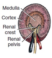
The kidneys of the goat and sheep are almost identical to those of the dog. Embryonic fusion has resulted in a smooth surface, with no external evidence of lobation. The renal papillae have aligned themselves into a row and have fused to form the renal crest, on which the papillary ducts open. The crest is surrounded by the dilated proximal ureter, i.e. the renal pelvis. Interlobar diverticula, known as the pelvic recesses are extensions of the renal pelvis that are closely associated with the interlobar blood vessels, branches of the renal a/v.
Identify: Goat/sheep external shape, cortex, medulla, ureter, renal a./v., renal crest, renal pelvis, pelvic recesses.
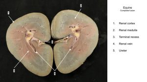
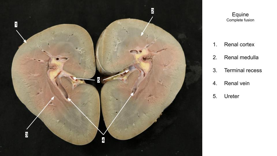
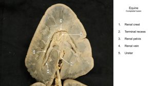
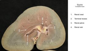
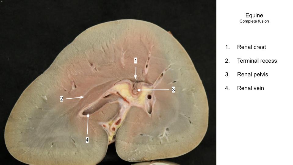
Equine
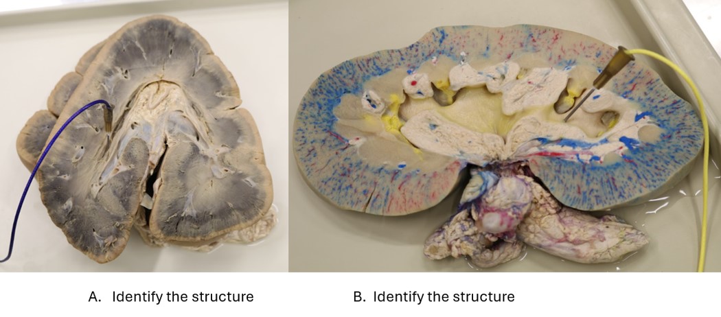
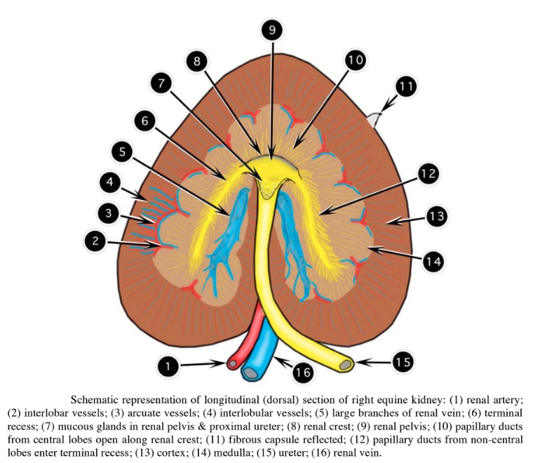
The kidney of the horse is not kidney-shaped but shaped more like the heart on a playing card. The right kidney is compressed in a craniocaudal direction so that its craniocaudal dimension is equal to or less than its mediolateral dimension. Embryonic fusion has resulted in a smooth surface. Internally, there is a hint of lobation as seen by the branches of
the renal a. There is a small renal crest surrounded by a small centrally located renal pelvis. The renal crest is short and represents the fusion of the more centrally located renal papillae only. The papillary ducts from those lobes near the extremities of the kidney, unite to form two large collecting ducts known as the terminal recesses. These ducts conduct the urine from the cranial and caudal extremities to the renal pelvis. The terminal recesses are best demonstrated by corrosion casts of the urine collection system. Do not confuse the subtle terminal recesses with the prominent branches of the renal vein. In the horse, the renal pelvis and proximal part of the ureter contain mucous glands, the secretion of which accounts for the thick mucus-like nature of equine urine.
Identify: Horse external shape, cortex, medulla, ureter, renal a./v., interlobar a., renal pelvis, renal crest, terminal recesses. Note that mucous glands are present in the proximal ureter of the horse.
-

- Equine kidney longitudinal section 2
-

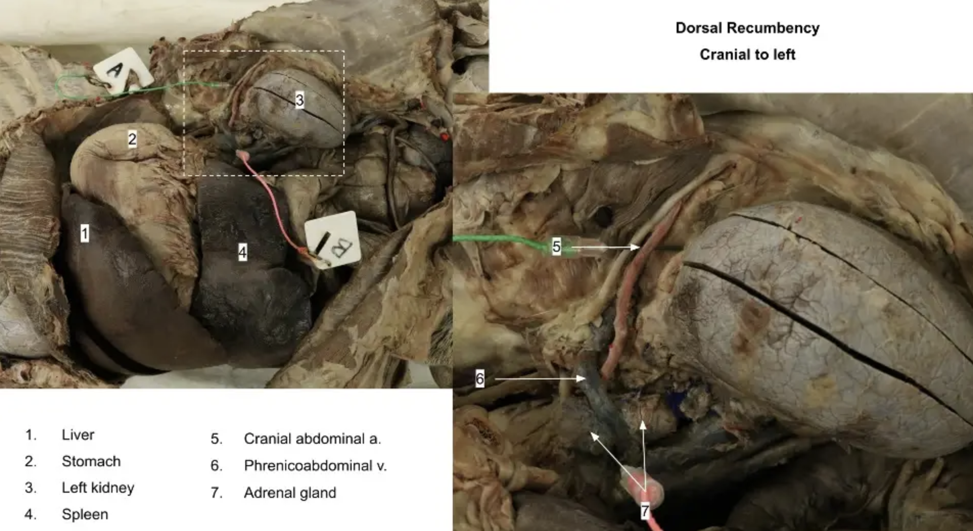
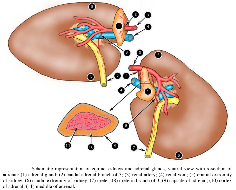
- Equine kidney and adrenal gland 2
-
- Horse kidney
-
- Horse kidney

-
- Horse kidney
Clinical Application: Pyelonephritis
Cystitis is an inflammation of the urinary bladder that may ascend the ureters to cause infection of the kidneys (pyelonephritis). The most common causative bacteria are ubiquitous in the environment and are common inhabitants of the vagina and prepuce. Pyelonephritis develops from an ascending infection from the bladder and may occur in any of the domestic species. Cystitis may be present without involving the ureters or ascending to the kidney until some event occurs that compromises the defense mechanism of the ureteral mucosa. The organisms attack or colonize the mucosal lining of the bladder and ureters, usually after some traumatic insult (such as parturition or abnormal deformity of the vaginal tract). In cattle in particular, the stresses of parturition, peak lactation, and a high-protein diet (which increases the pH of the urine and is therefore conducive to colonization of the urinary tract by Corynebacterium spp) are all contributing factors.
The first clinical sign observed in a case of bovine cystitis or pyelonephritis may be the passage of blood-stained urine in an otherwise healthy cow. As the infection proceeds up the ureters, causing inflammation and subsequent involvement of the kidney, the animal exhibits discomfort manifest by frequent attempts to urinate, anorexia, a slight fever, loss of production, colic with restlessness, tail switching, polyuria, hematuria, or pyuria. In chronic cases, the animal may show colic, diarrhea, polyuria, polydipsia, stranguria, and anemia. As the disease progresses, the bladder becomes thickened and inflamed. The ureters become thickened and dilated with a purulent exudate. The involved kidneys develop multiple small abscesses on the surface that may extend into the cortex and medulla.
Read More: Bovine Cystitis and Pyelonephritis

Adrenal Glands
Identify: Adrenal glands of each species. Observe the cut surface of transected adrenal glands, and distinguish between an adrenal gland and a renal lymph node by the presence of a distinct cortex and medulla. Note that even in ruminants, in which the left kidney is displaced to the right by the rumen, the left adrenal gland remains on the left side of the abdomen, as usual.
Review videos
Terms
| Term | Species/Notes |
| Kidney (Right and Left) | |
| Hilus | |
| Renal capsule | |
| Renal cortex | |
| Renal medulla | |
| Renal crest | All except pig and bovine |
| Renal pyramids | |
| Renal papilla | Not in carnivores or small ruminants. |
| Papillary ducts | Conceptual only – know where located |
| Papillary foramina | Conceptual only – know where located |
| Ureter | |
| Renal pelvis | All except bovine |
| Pelvic recesses | Carnivores and small ruminants |
| Terminal recesses | Equine only |
| Minor calyx | Cow and pig |
| Major calyx | Pig |
| Central cavity | Pig |
| Mucous glands | Conceptual; Equine only |
| Renal a. | |
| Interlobar aa. | |
| Arcuate aa. | |
| Renal v. | |
| Nephron (Juxtamedullary vs. cortical) | Know where each part is located in the kidney |
| Renal corpuscle | |
| Glomerulus | |
| Glomerular capsule | |
| Loop of Henle | |
| Proximal and distal convoluted tubules | |
| Collecting tubules | |
| Adrenal gland | |
| Adrenal cortex | |
| Zona glomerulosa | produces mineralocorticoids (e.g., aldosterone) – hormones responsible for salt regulation. |
| Zona fasciculata | produces glucocorticoids (e.g. cortisol) – hormones closely related to sugar control.
|
| Zona reticularis | produces androgens, or sex hormones. |
| Adrenal medulla | produces catecholamines, including norepinephrine and epinephrine. |
| Phrenicoabdominal v. | ID in carnivore only |
| Urinary bladder | Conceptual |
| Ureteral openings | Conceptual |
| Internal urethral orifice | Conceptual |
| Trigone | Conceptual |