
Example Practical Exam Answers
Lab 1 Tongue Muscles
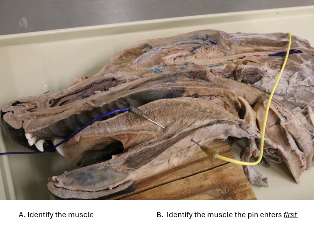
A. Genioglossus m.
B. Mylohyoideus m.
Lab 1 Tongue Structures
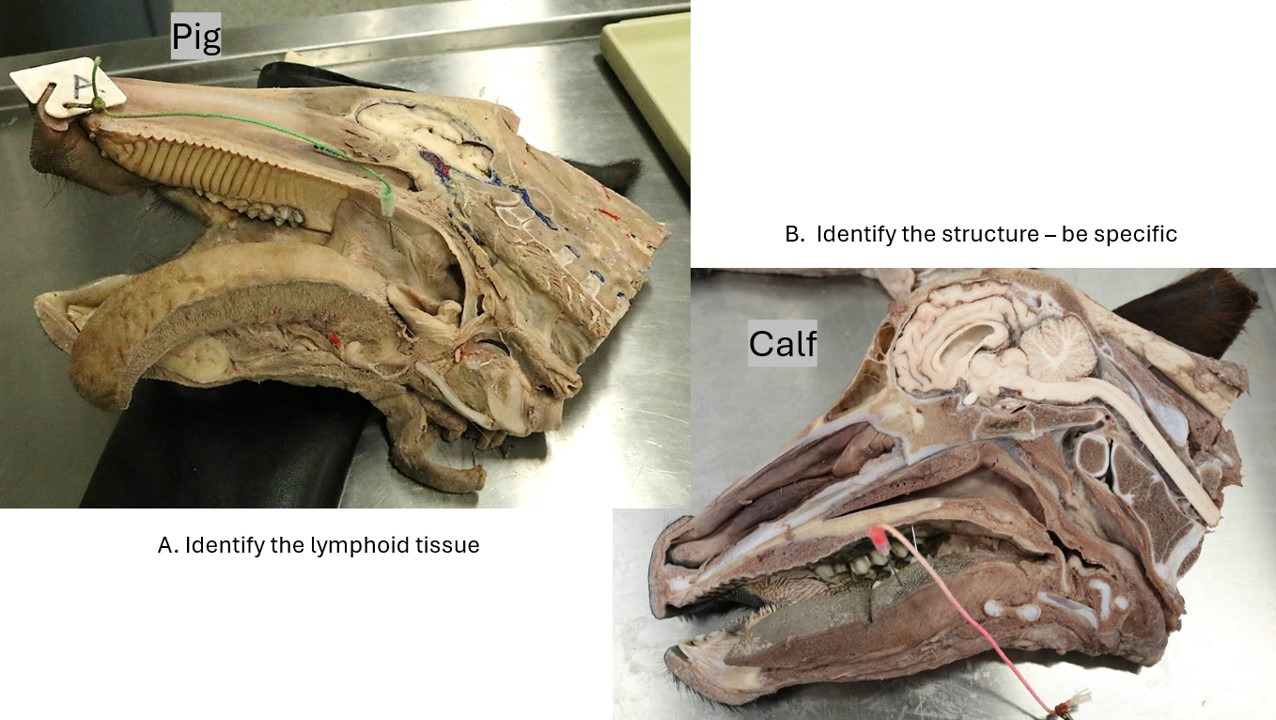
A. Tonsils of the soft palate
B. Torus linguae
Lab 2 teeth
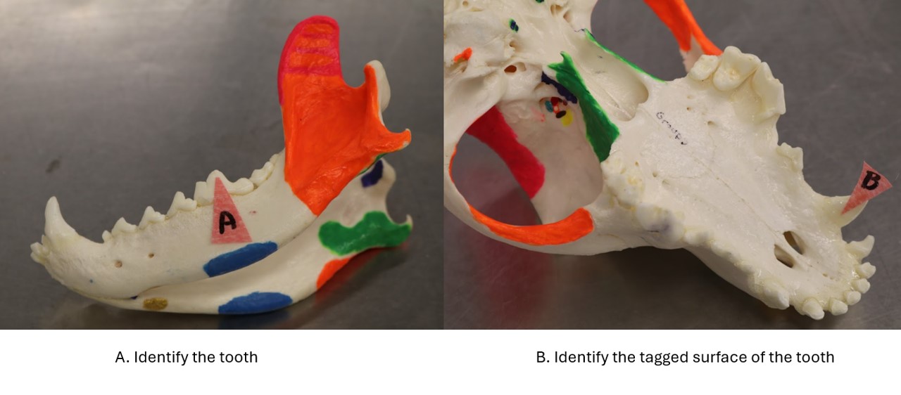
A. Mandibular 1st molar (309)
B. Palatine surface
Lab 2 horse teeth
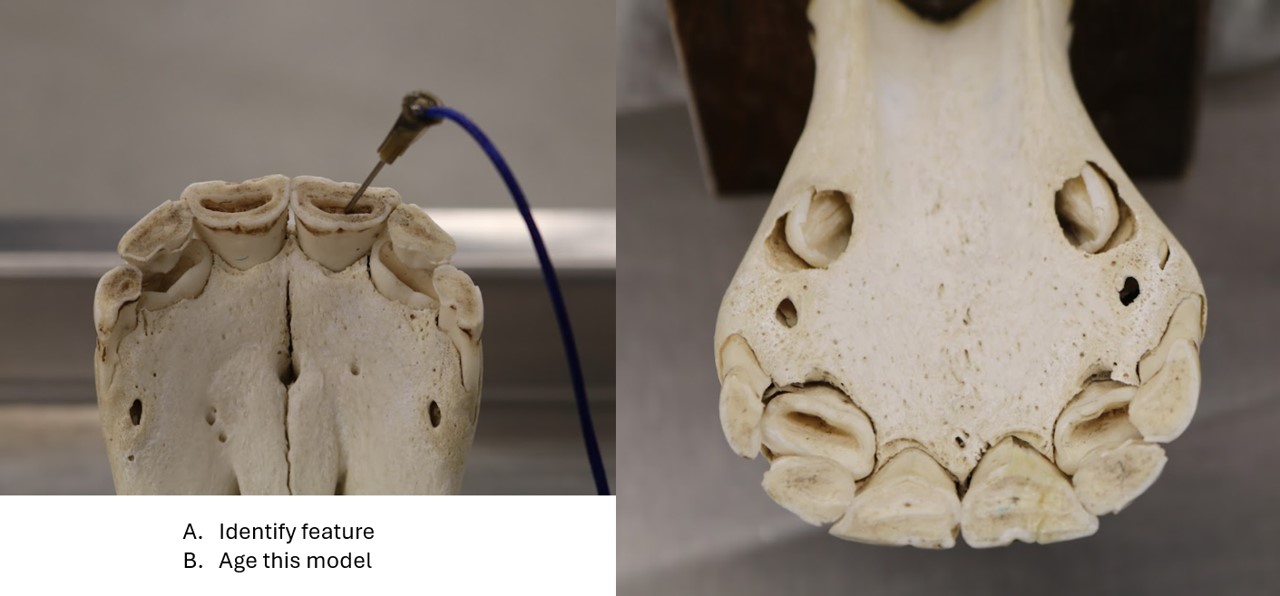
A. Infundibulum (dental cup)
B. 3.5 years (will accept 3-4 years)
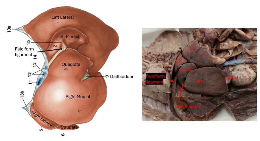
Lab 3 Cat liver
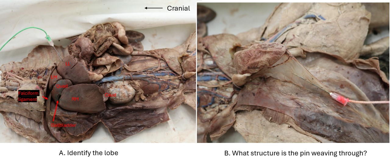
A. Left lateral lobe
This isn’t an easy question to answer from a photo. It’s showing the diaphragmatic view of the liver. There are several landmarks to make this more straightforward: the falciform ligament’s free edge is the round ligament of the liver (which developed from the embryologic umbilical vein) always denotes the division between the left and quadrate lobes; the gallbladder lies between the quadrate lobe and right medial lobe; and the caudate process of the caudate lobe has a renal impression from the right kidney. Hopefully, comparing the photo to the diagram below will help make a “challenging in 2D” question more understandable.
B. Median ligament of the bladder
Again, possibly a “challenging in 2D” question (hence the “bladder” hint). Note the strip of abdominal muscles reflected caudally, which shows this structure is connecting on midline. The lateral ligaments of the bladder (not learned yet) in this photo have blobs of embedded fat and are located to the top and bottom (in this photo’s orientation) of the bladder.
Lab 3 Omentum
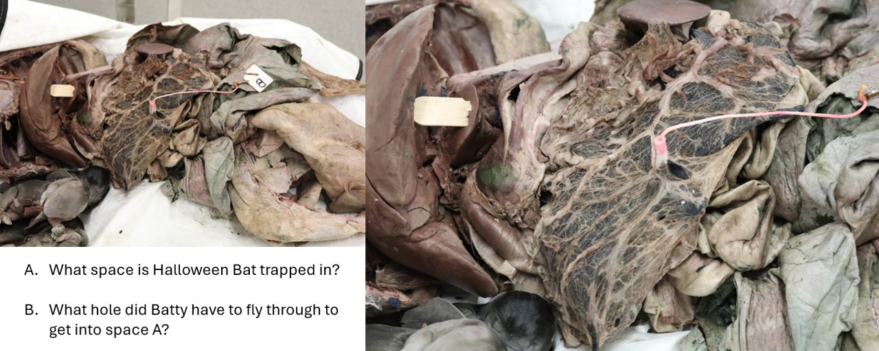
A. The omental bursa
B. The epiploic foramen (don’t ask how, he’s clearly *magical*, and totally doesn’t have rabies…).
Lab 6 Cat GI
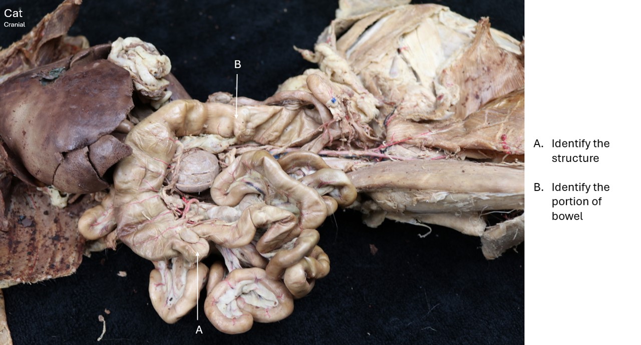
A. Cecum (FYI – with an adjacent cecal lymph node)
B. Descending colon
Lab 6 Dog GI
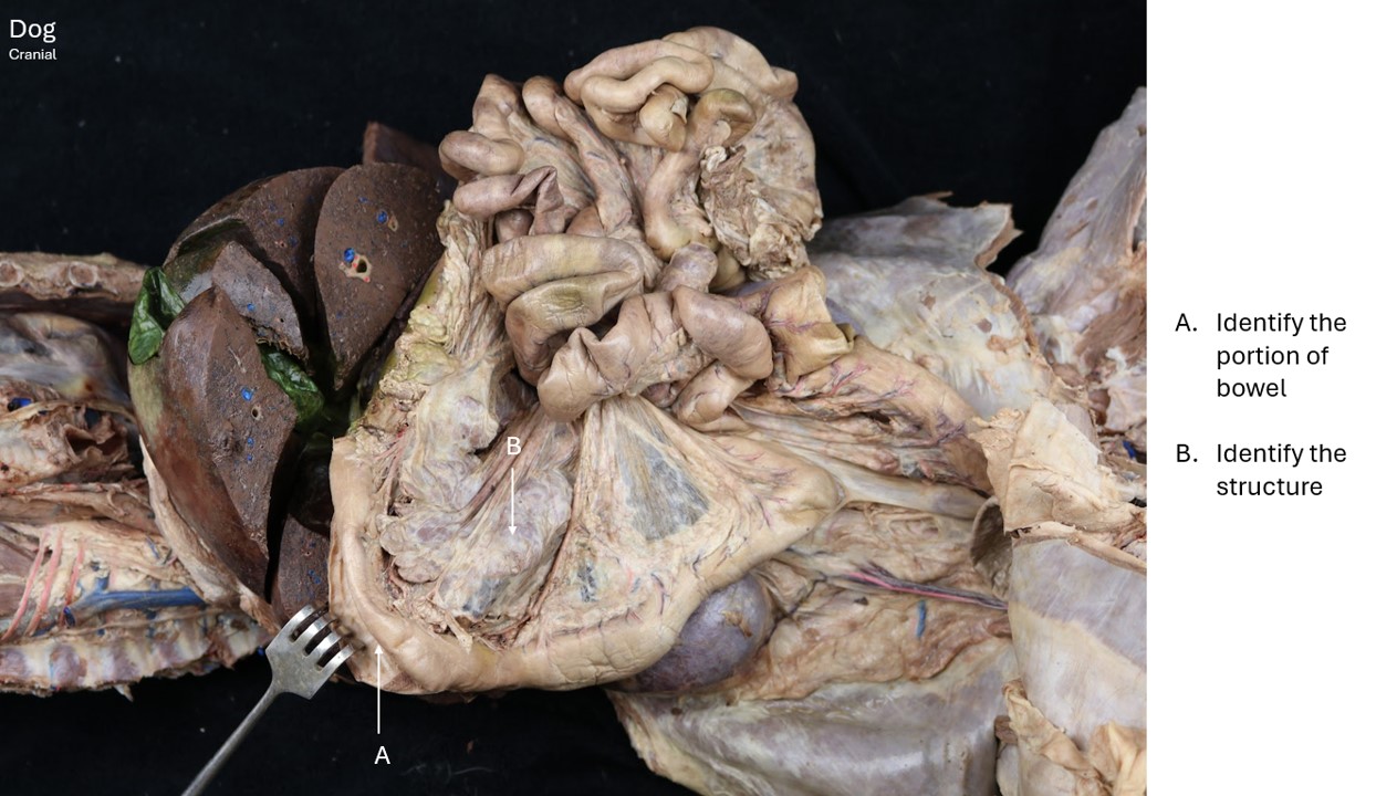
A. Descending duodenum
B. Right lobe of pancreas
Lab 7 Vessels
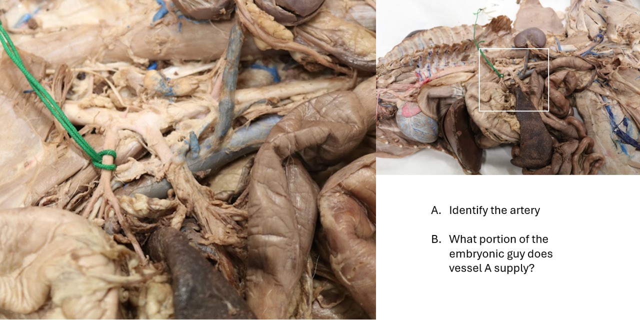
A. Celiac artery
B. Foregut
Lab 7 veins
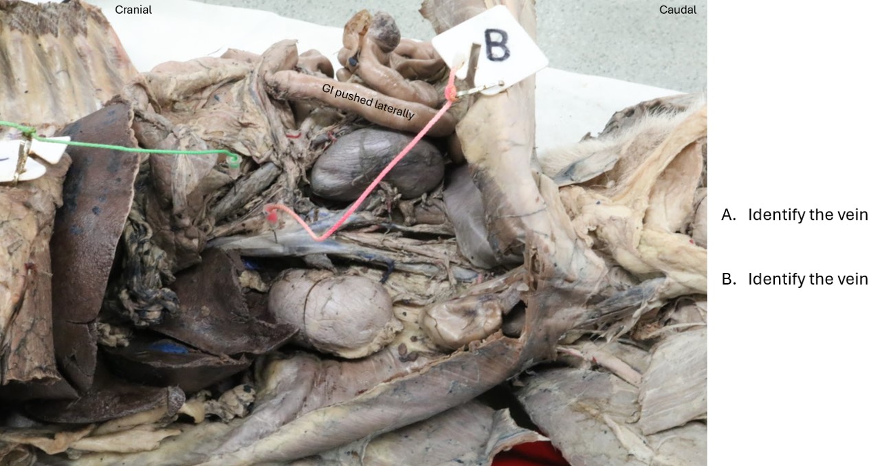
A. Hepatic portal vein
B. Vena cava