Lab 3A: Male Carnivore Reproductive Anatomy
Learning Objectives
- Identify the structures of the male reproductive tract, and related anatomy, in the carnivores.
- Identify the structures passing through the inguinal canal of the male and female of the carnivorous species.
- Identify the structures of the carnivore penis and prepuce.
- Identify the vaginal tunics, mesorchium, and mesoductus deferens.
- Identify the anatomic spermatic cord and the structures that comprise it.
- Identify the testis and associated structures.
- Identify the tissue layers surrounding the descending testicle and match them with the abdominal structures from which they were derived.
- Differentiate between an open and closed castration technique on cadaveric specimens.
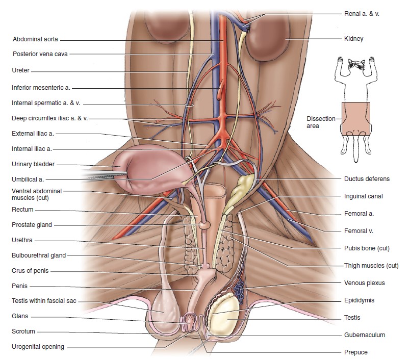
Male reproductive tract – carnivore
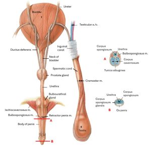
Accessory Sex Glands
Prostate
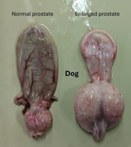
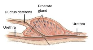
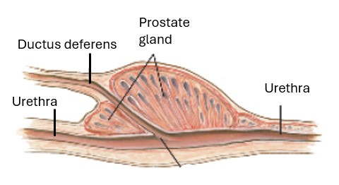
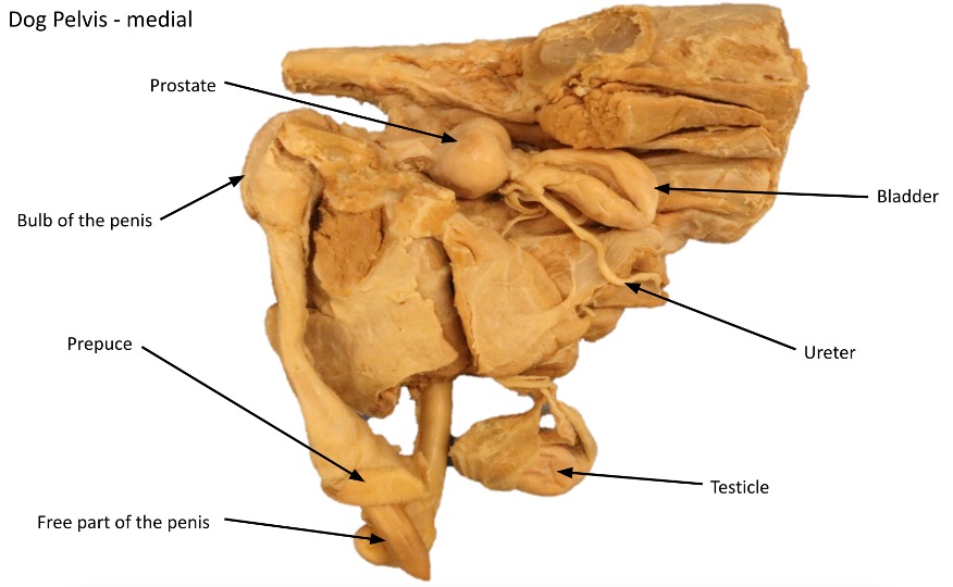
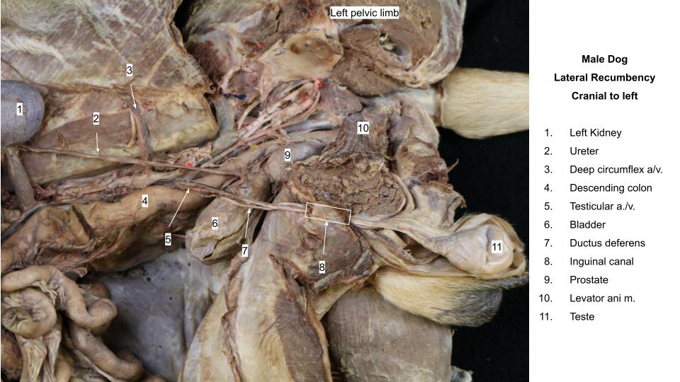
The prostate gland completely surrounds the neck of the bladder and the beginning of the pelvic urethra in the dog.
Note that the prostate of the male cat does not completely surround the pelvic urethra and does not enlarge very much with age.
The normal size and weight of the prostate vary greatly. The organ generally lies at the pelvic inlet. It is larger and extends farther into the abdomen in older dogs. The prostate is flattened dorsally and rounded ventrally and on the sides. It is heavily encapsulated. Muscle fibers from the bladder run caudally on its dorsal surface. A longitudinal septum leaves the ventral part of the capsule and extends dorsally to reach the prostatic part of the pelvic urethra, thus partially dividing the gland ventrally into right and left lobes. This is indicated on the ventral surface by a shallow but distinct longitudinal furrow.
Observe: Examine the surface, form, size, and location of the prostate on several specimens.
Again notice that the pelvic (prostatic) urethra runs through the center of the gland. The prostatic urethra is simply a subpart of the pelvic urethra surrounded by the prostate gland.
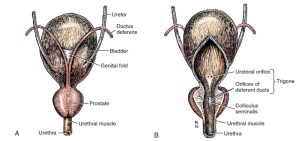
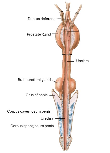
On the dorsal surface of the bladder, locate the two ductus deferentes (singular: ductus deferens) where they are joined by the genital fold. The genital fold is where the left and right mesoductus deferens combine to connect the left and right ductus deferens. The mesoductus deferens is the connecting periotoneum of the ductus deferens. It covers the ductus deferens itself and connects it to the abdominal wall and to the other ductus deferens by the genital fold.
-
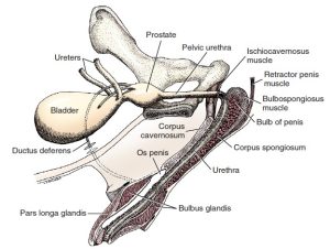
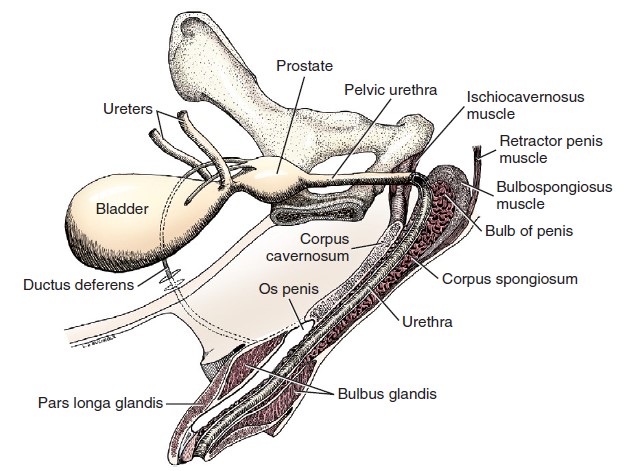
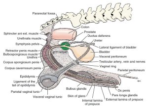
- Schematic left lateral aspect of pelvic structures and a median section of the penis. 1
-
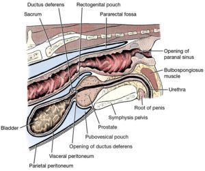
- Median section through male pelvic region. 1

-
- Bladder and prostate. A, Dorsal aspect. B, Ventral aspect, partially opened on midline. 1

-
- Dog bladder and prostate glands

-
-
Pelvic cavity of the male cat with many of the viscera removed and urinary bladder
reflected to the right to show vessels and urogenital structures, in ventral view. 5
Clinical Application: Benign Prostatic Hyperplasia
Benign prostatic hyperplasia (BPH) is the most common prostatic disorder in intact male dogs and results from androgenic stimulation or an altered androgen:estrogen ratio. It is not known why some males are affected and others are not. In some dogs, hyperplasia may begin as early as 2.5 years of age and, after 4 years of age, cystic hyperplasia tends to develop. Clinical signs may be absent, but persistent or intermittent hematuria, hemospermia, and/or hemorrhagic preputial discharge are most commonly reported.
Castration is the treatment of choice in male dogs not intended for breeding and/or showing. With castration, prostatic involution is usually evident within a few weeks and is often complete in several months.
Bulbourethral Glands
Tomcats only: The bulbourethral glands, located dorsolateral to the pelvic urethra at the ischiatic arch should be identified.
-
- Male genital system of the cat with some muscles and prepuce removed, dorsal view. 4

-

- Structures common to male cat genital and urinary systems with urethra opened. 4
-
- Junction of male genital and urinary systems, median section. 4
Prepuce
The prepuce is a tubular sheath or fold of integument that is continuous with the skin of the ventral abdominal wall and is reflected over the glans. It has a smooth internal lamina and a haired external lamina, which meet at the preputial orifice. At its deepest recess, the fornix of the prepuce, the internal layer is reflected onto the glans as the skin of the glans. In the erect state the fornix is eliminated, because the external layer of the prepuce is closely applied to the body of the penis.
Dissect: Open the prepuce by a mid ventral incision from the orifice to the fornix (where the prepuce folds back onto the free end of the penis). Continue the midventral skin incision to the anus so as to expose the entire length of the penis.
Observe: Everyone is responsible for observing the tomcat penis after the prepuce has been incised. Note the spines of the penis and the external anatomy of the penis, for which you are responsible. Further dissection of the penis itself is not mandatory for teams dissecting tomcats. If you would like to give it a go, you are encouraged to do so. The anatomy is the same, just much smaller. You are responsible for learning the internal structures on the male dogs.
-

- Male genitalia, ventral view. 1
-

- Dog prepuce and penis
Penis
The penis is composed of a root, a body, and a glans. The dorsal surface of the penis faces the pelvic symphysis and the abdominal wall. In the nonerect state the glans is entirely withdrawn into the prepuce.
Root of the Penis
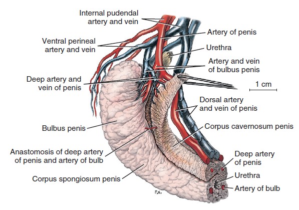
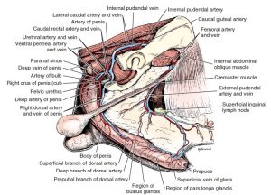
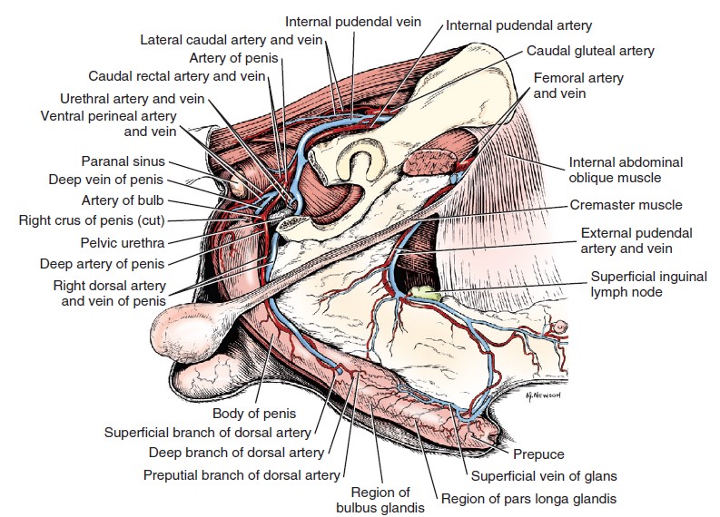
The root of the penis is formed by right and left crura and the bulb of the penis. The crura originate on the ischiatic tuberosity of each side. The root ends where the crura blend with each other on the midline to form the body. Each crus is composed of vascular cavernous tissue, corpus cavernosum (penis), supplied by the deep artery of the penis and surrounded by a thick fibrous tunic, the tunica albuginea penis.
At the root of the penis between the crura is the bulb of the penis covered by the bulbospongiosus muscle. This bilobed dorsal expansion of the corpus spongiosum (penis) surrounds the urethra and is located at the ischial arch. It is supplied by the artery of the bulb. A thin layer of corpus spongiosum penis surrounds the entire length of the urethra in the penis. FYI: Corpus spongiosum also surrounds the pelvic urethra, but for our purposes, we will focus on the corpus spongiosum as part of the penis.
The artery of the bulb of the penis arborizes in the bulb and continues to supply the corpus spongiosum penis and penile urethra. While we will not be dissecting the origin of this vessel, knowing what part of the penis it vascularizes is required.
The deep artery of the penis arises close to the artery of the bulb and enters the corpus cavernosum penis at the crus. This is at the level of the ischial arch lateral to the penile bulb. Like the artery of the bulb, we will not be dissecting the origin of this vessel. However, naming the part of the penis it vascularizes is required.
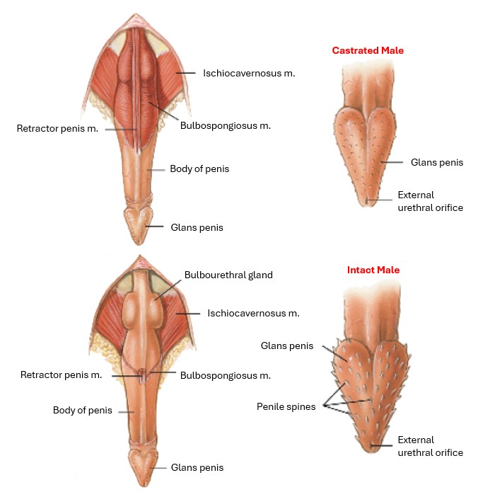
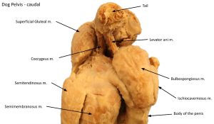
The retractor penis muscle is an elongated slip of mixed smooth and striated muscle fibers. It originates from the ventral surface of the sacrum, or first two caudal vertebrae, blends with the external anal sphincter, and extends distally on the ventral surface of the penis to the level of the glans, where it inserts. In the region of the anal sphincter, there is muscle fiber exchange between the retractor penis muscle and the external anal sphincter.
The bulbospongiosus muscle covers the bulb of the penis and thus bulges between the ischiocavernosus muscles (which cover the crura), ventral to the external anal sphincter. The fibers of the bulbospongiosus are transverse proximally, where they cover the bulb of the penis, and longitudinal distally, where they pass onto the body of the penis.
-
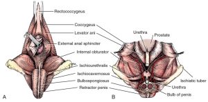
- Male perineum. A, Superficial muscles, caudal aspect. B, Dorsal section through pelvic cavity. The bilobed bulb of the penis is transected, and the proximal portion removed. 1
-
- Schematic left lateral aspect of pelvic structures and a median section of the penis. 1
-
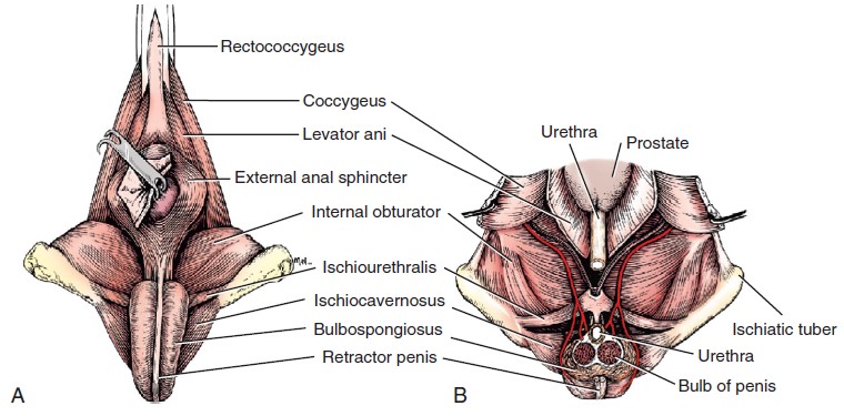
- Upper drawing, a parasagittal section of a penis. A to E, Cross sections at five levels indicated by letters on upper drawing. 1
-

- Vascular supply of the proximal half of the penis. 1
-

- Cat penis, dorsal view. 4
-
- Dog caudal view of pelvic wall mm.

Observe the penile bulb and its relationship to the urethra at the root of the penis. Differentiate between the parts of the root: a. the crura, covered in ischiocavernosus muscles and b. the bulb of the penis, which is covered in bulbospongiosus muscle. Name what arteries supply each part of the root of the penis.
Transect the root of the penis, and observe the corpus spongiosum erectile tissue surrounding the penile urethra and forming the bulb of the penis. Observe the corpus cavernosum erectile tissue which make up the two crura on either side of the bulb of the penis.
Observe the retractor penis muscle extending to the body of the penis.
While identifying the arteries which supply the penis is not necessary at this point, you are responsible for knowing which artery supplies what structure or structures.
Body of the Penis
The body of the penis extends from the root, where the crura blend with each other, to the glans penis covering the os penis in the caudal portion of the prepuce.
Note that the region at the beginning of the body of the penis is compressed from side to side and wrapped by a thick tunic. It is capable of being bent without twisting when the male dismounts during coitus and remains “locked” for a variable period.
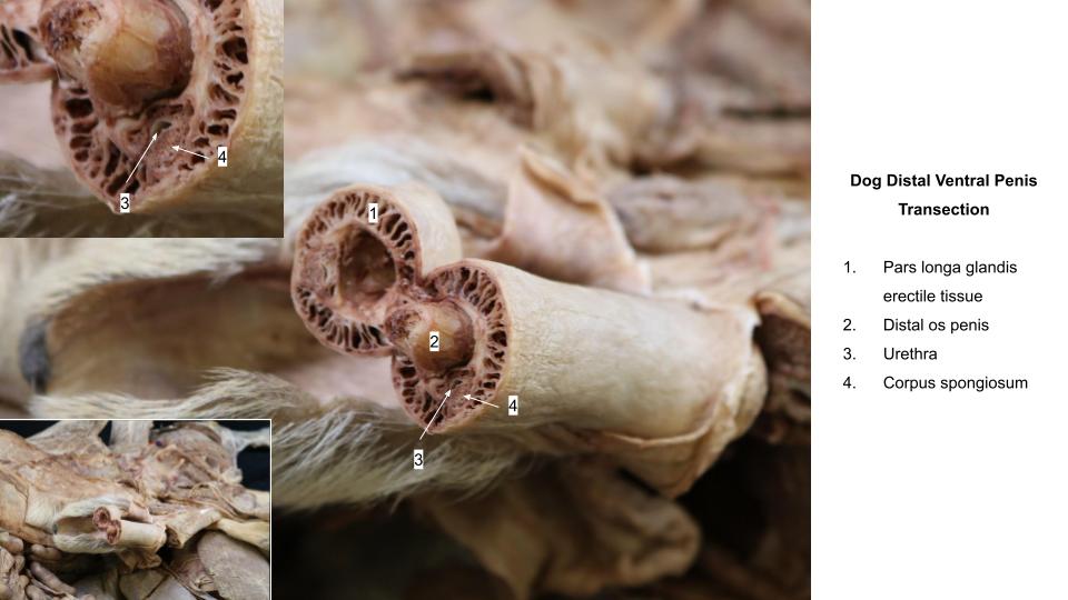
The corpus cavernosum penis of each crus converges toward the midline and the two corpora extend side by side throughout the body to the os penis. A median septum completely separates the two corpora in the carnivore, and each is covered by a thick white capsule (tunica albuginea penis-differentiate this term from tunica albuginea teste) throughout its length. The two corpora cavernosa form a groove ventrally that contains the urethra and the thin corpus spongiosum that surrounds the urethra.
-
- Topographic relations of the penis and other pelvic structures. 1
-
- Dog prepuce and penis
-
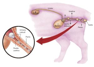
- Male cat urinary organs; inset: enlargement of penile urethra with urinary calculi. 4

-
- Dog penis erectile tissues. 37
Dissect: Partially transect the body of the penis to identify the above structures in cross-section.
Glans Penis
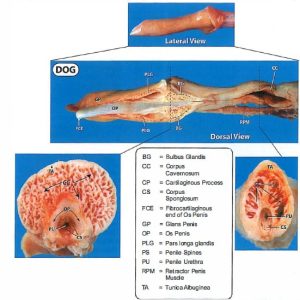
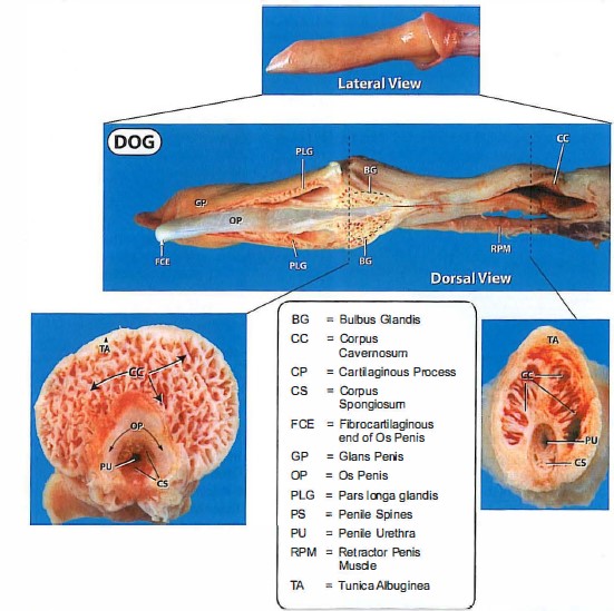
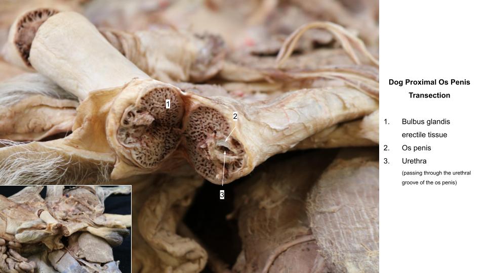
The glans of the penis is composed of corpus spongiosum glandis erectile tissue. In the dog, two subparts of the corpus spongisosum glandis are identified—the proximal bulbus glandis and the distal, more elongate pars longa glandis. The bulbus glandis, surrounding the proximal end of the os penis, is a dorsal extension of the corpus spongiosum. It is an expansile vascular structure that is largely responsible for retaining the penis within the vagina during copulation.
The pars longa glandis is a cavernous tissue structure that overlaps the distal half of the bulbus glandis and continues to the distal end of the penis, partially encircling the os penis and the urethra. The pars longa glandis has no direct arterial communication with the corpus spongiosum penis and is separated from the bulbus glandis by a layer of connective tissue. Venous channels drain the pars longa glandis into the bulbus glandis through this layer.
Note that in the tomcat, the glans penis of the sexually mature penis is covered with numerous highly keratinized, proximally directed penile spines. These are testosterone dependent and will atrophy following castration as will the accessory sex glands (prostate and bulbourethral glands). The cat’s corpus spongiosum glandis is not differentiated into two parts like in the dog.
-
- Topographic relations of the penis and other pelvic structures. 1
-
- Diagram of peritoneal reflections and the male genitalia. 1

-
- Schematic left lateral aspect of pelvic structures and a median section of the penis. 1
-
- Upper drawing, a parasagittal section of a penis. A to E, Cross sections at five levels indicated by letters on upper drawing. 1
-
- Cat penis, dorsal view. 4
-
- Structures common to male cat genital and urinary systems with urethra opened. 4
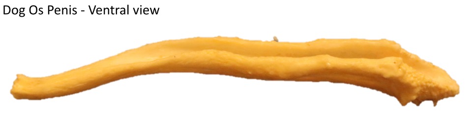
The os penis is a long, ventrally grooved bone that lies almost entirely within the glans penis. It forms about a month after birth as an ossification of the fused distal ends of the corpora cavernosa penis. The body of the os penis extends through the glans penis. The base and body are grooved ventrally by the urethral groove, which surrounds the urethra and the corpus spongiosum on three sides. The bone ends as a long, pointed fibrocartilage in the tip of the glans, dorsal to the urethral opening.
At the level of the collar-like bulbus glandis of the glans of the penis, there is a communication between the corpus spongiosum penis and the bulbus glandis. The dorsal artery of the penis courses to the glans, where it supplies the prepuce, corpus spongiosum penis, and pars longa glandis.
Dissect: Make a longitudinal incision on the dorsum of the distal penis through the glans to observe the pars longa glandis and os penis. Transect the bulbus glandis to visualize the os penis and bulbus glandis erectile tissue.
-
-
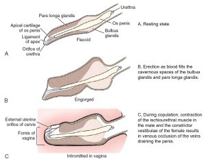
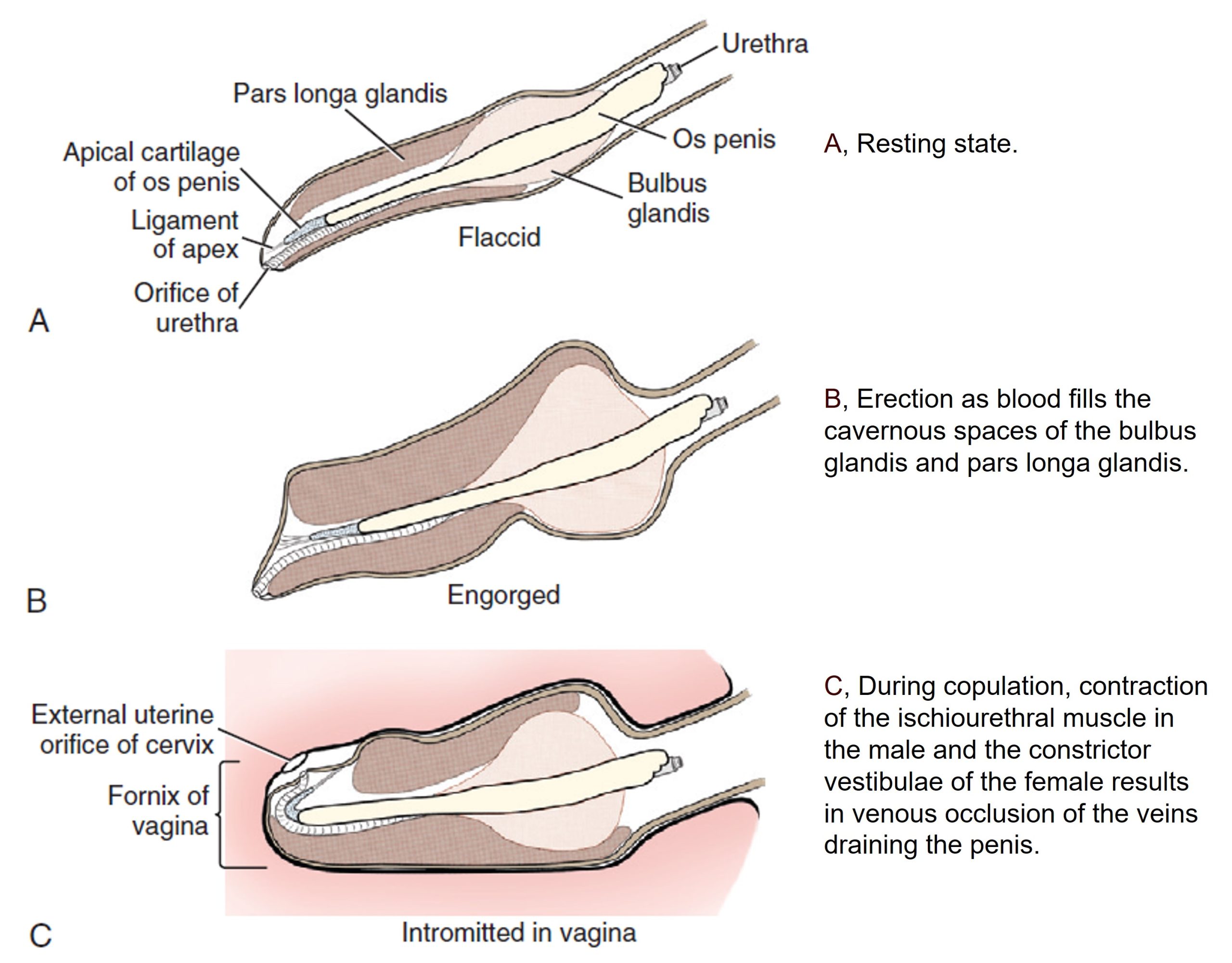
Schematic interpretation of changes in shape of the glans penis
during erection and copulation. 1
-
-
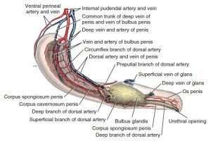
Semidiagrammatic view of penis. The pars longa glandis and the muscles of the root are illustrated
as if transparent. The vessels of only one side are shown. 1

-
- Male genital system of the cat with some muscles and prepuce removed, dorsal view. 4
-
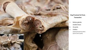
- Transection through bulbus glandis
-

- Transection through distal penis
-

- Dog os penis
Clinical Application: Bulbous glandis and the breeding tie
The “tie” in dog mating occurs when the male’s bulbus glandis and pars longa glandis swells inside the female’s vagina, locking the dogs together for several minutes to a half hour, typically back to back. This natural process helps ensure that the maximum amount of sperm is transferred to the female, increasing the chances of pregnancy. It is important not to try to separate the dogs forcefully, as this can cause injury, but instead to wait calmly until the tie ends naturally.
Normal swelling of the bulbus glandis can at times be mistaken as testicles by clients.
Male Inguinal structures and Testicles
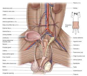
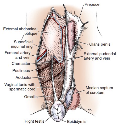
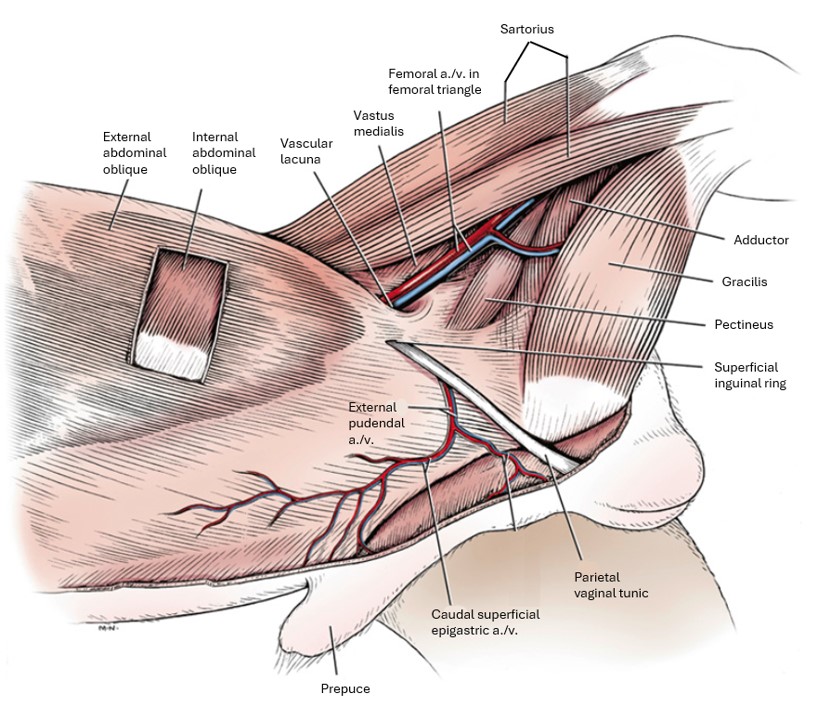
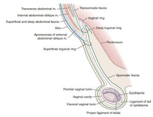
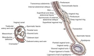
Review the anatomy of the inguinal canal now. Understanding of the inguinal canal will inform the understanding of the layers of tissue surrounding the descended testicle: The inguinal canal is a short fissure filled with connective tissue between the abdominal muscles. It extends between the deep and superficial inguinal rings. It is bounded laterally by the aponeurosis of the external abdominal oblique. It begins at the deep inguinal ring, the bordered cranially by the caudal border of the internal abdominal oblique; caudally by the caudal border of the aponeurosis of the external abdominal oblique (inguinal ligament); and medially, in part, by the superficial surface of the rectus abdominis. It ends at the superficial inguinal ring, which is a slit in the aponeurosis of the EAO. The transversalis fascia and parietal peritoneum pass into the inguinal canal and change name to the internal spermatic fascia and parietal vaginal tunic, respectively. The spermatic cord in the male and the vaginal process and round ligament of the uterus in the female dog pass obliquely caudoventrally through the canal. In the queen, the vaginal process does not extend into the inguinal canal. In both sexes in domestic species, the external pudendal a/v and genitofemoral nerve traverse the canal.
The external pudendal artery and vein leave the superficial inguinal ring caudal and medial to the structures that extend to the testis.
The genitofemoral nerve arises from the ventral branches of the third and fourth lumbar nerves. It is bound by fascia to the external pudendal vein medial to the spermatic cord. It passes through the inguinal canal adjacent to the spermatic cord and innervates the cremaster muscle and the skin covering the inguinal region and proximal medial thigh of both sexes and part of the prepuce in the male.
Recall that although they lack a spermatic cord, the genitofemoral nerve and external pudendal artery still DO extend through the inguinal canal in females of the domestic species.
The internal spermatic fascia, a continuation of the transversalis fascia (which lines the abdominal cavity external to the parietal peritoneum) invested with the parietal vaginal tunic, surrounds the structures emerging from the superficial inguinal ring, including the anatomic spermatic cord. The cremaster m., external pudendal a/v, and genitofemoral n pass through the inguinal canal adjacent to but external to the internal spermatic fascia.
The cremaster muscle is surrounded by the external spermatic fascia (continuation of the fascia of the external abdominal oblique) as it courses along the external aspect of caudal part of the internal spermatic fascia. The cremaster muscle arises from the caudal free border of the internal abdominal m. and attaches to the testes distally. It functions to draw the testicle toward the body.
The external spermatic fascia is a continuation of the loose, subcutaneous connective tissue just exterior to the abdominal muscle walls layers into the inguinal region and then into the testicle.
The internal spermatic fascia is a continuation of the transversalis fascia one it has been pulled through the deep inguinal ring by the descending testicle. The parietal vaginal tunic is adhered to the interior surface of the internal spermatic fascia and is a continuation of the parietal peritoneum starting at the vaginal ring. Recall that the vaginal ring is where the parietal peritoneum turns on itself to enter the deep inguinal ring, where it also name changes to parietal vaginal tunic. The testicle itself, as well as the testicular a./v./n./l., are covered in connecting peritoneum called the mesorchium in the abdominal cavity. Once through the inguinal ring, the mesorchium changes name to the visceral vaginal tunic, the thin serous membrane covering the testicle and accompanying nerves and vasculature outside of the abominal cavity.
-
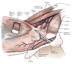
- Abdominal muscles and inguinal region of the male, superficial dissection, left side. 1
-
- Male genitalia, ventral view. 1
-
-
Sagittal view of the scrotum and the caudal abdomen. The image illustrates the relationship between the abdominal muscles and
testicular sheaths. Cranial is to the left in this image. 9
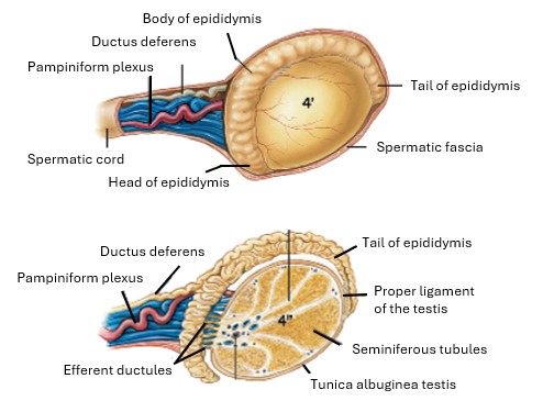
The spermatic cord is carried through the inguinal canal by the descent of the testis and is composed of two distinct parts: the ductus deferens and the testicular artery and vein.
The testicular artery and vein, as well as the testicular lymph vessels and the testicular plexus of autonomic nerves, are closely associated with each other. These vessels and nerves are covered by a fold of peritoneum, the mesorchium, that is continuous with the parietal and visceral vaginal tunics. The artery is tortuous, and woven around it are the nerve plexus and the venous plexus. The venous plexus is the pampiniform plexus, which will be observed with the testicle. The testicular artery and vein are branches of the aorta and caudal vena cava, respectively. They enter the testis at its cranial end. The nerve plexus is autonomic and sensory and contains postganglionic sympathetic axons, which arise from the third to fifth lumbar sympathetic ganglia.
The ductus deferens carries the spermatozoa from the epididymis to the urethra. It arises from the tail of the epididymis at the caudal end of the testis and is attached to the mesorchium by the mesoductus deferens. The mesoductus deferens is the connecting mesentery that attaches the ductus deferens to the body wall proximally and the mesorchium distally. It contains the artery, vein, and nerve of the ductus deferens. Recall that the right and left ductus deferentas are connected to each other by the genital fold.
-
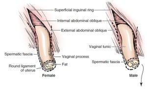
- Diagram of transected vaginal process in male and female. 1
-
- Schema of the vaginal tunic in the male with an inset of a transection.1
-

- Male dog pelvic wall mm.
Dissect: Gently separate and identify the structures in the male that pass through the inguinal canal and the superficial inguinal ring. Find and clean the anatomic spermatic cord as it extends from its emergence through the superficial inguinal ring to the testis. To do so, reflect the external spermatic fascia and incise the combined internal spermatic fascia and parietal vaginal tunic. The genitofemoral nerve and external pudendal artery are adjacent to the spermatic cord, also covered in external spermatic fascia.
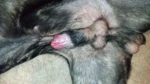
Scrotum and Testicles
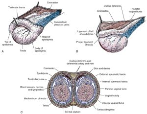
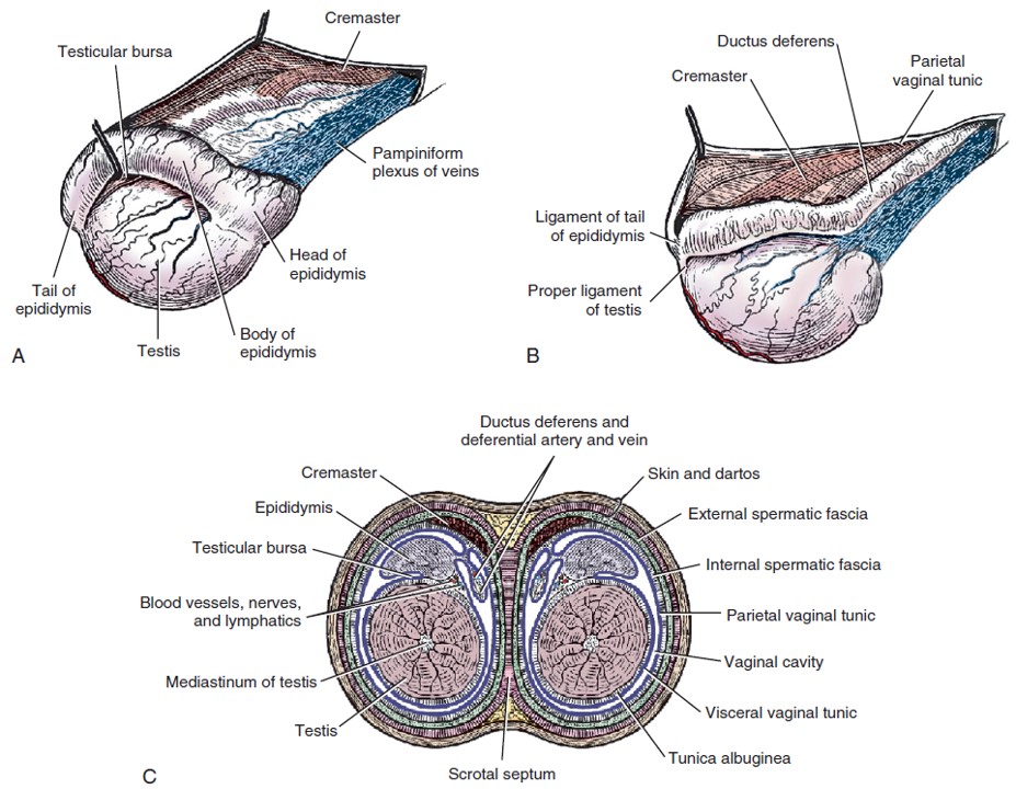
The scrotum is a pouch divided by an external scrotal raphe and an internal median septum into two cavities, each of which is occupied by a testis, an epididymis, and the distal part of the spermatic cord. The tunica dartos, muscle fibers located deep to the skin of the scrotum act in temperature regulation of the testicle. The internal spermatic fascia is connected to the scrotum via the scrotal ligament.
The testis and the associated epididymis and ductus deferens are intimately covered by the visceral vaginal tunic. At the caudal extremity of the epididymis, the visceral vaginal tunic leaves the tail of the epididymis at an acute angle and is continuous with the parietal vaginal tunic. The connective tissue that attaches the epididymis to the parietal vaginal tunic and internal spermatic fascia at this point is the ligament of the tail of the epididymis. The testicular parenchyma is covered by a thick fibrous capsule called the tunica albuginea teste, which itself is covered in visceral vaginal tunic.
-
-
Structures of testes and scrotum. A, Right testis, lateral aspect. B, Left testis, medial aspect. C, Schematic
cross section through scrotum and testes. 1
-

- Left testis of the cat, lateral view and sagittal section. 4
-
- Diagram of peritoneal reflections and the male genitalia. 1
Dissect: Examine the scrotum of the cadaver and note the external raphe. Make an incision over one of the testicles. Pull the testicle from the scrotum. The loose connective tissue in between the testicle and the scrotum is called the external spermatic fascia. Clean it away from the testicle. If not done so already, incise or break the scrotal ligament, which attaches the testicle to the scrotum; it feels like thickened external spermatic fascia and is difficult to differentiate visually. Now, incise the combined internal spermatic fascia (white, fibrous tissue) and parietal vaginal tunic (very thin serous layer adhered to the inside of the internal spermatic fasia). Once through the internal spermatic fascia/parietal vaginal tunic, we have entered the vaginal cavity, a continuation of the peritoneal cavity, which is the space in between the parietal and visceral vaginal tunics. The visceral vaginal tunic is closely fused to the testis and epididymis and surrounds the ductus deferens. The mesorchium is the connecting mesentery of the testis that contains the vessels and nerves of the testis. The visceral vaginal tunic was called the mesorchium in the abdominal cavity and is adhered tightly to the tunic albuginea teste, deep to which is the testicular parenchyma.
The epididymis lies more on the lateral side of the testis than on its dorsal border. For descriptive purposes it is divided into a cranial extremity, or head, where the epididymis communicates with the testis; a middle part, or body; and a caudal extremity, or tail, which is continuous with the ductus deferens. The tail is attached to the testis by the proper ligament of the testis and to the parietal vaginal tunic and spermatic fascia by the ligament of the tail of the epididymis. The ductus deferens passes cranially over the testis medial to the epididymis.
Observe: Identify they head, body, and tail of the epididymis and the ligament of the tail of the epididymis. Identify the proper ligament of the testis.
Clinical Application: Open vs. Closed Castration
Note that in order to perform an open castration, the internal spermatic fascia and parietal vaginal tunic are both incised. Therefore, the vaginal cavity is opened. It is continuous with the peritoneal cavity. In a closed castration, the internal spermatic fascia and parietal vaginal tunic are left intact. Therefore, the vaginal cavity (and peritoneal cavity) are NOT opened.
Ensure that you can tell if these layers have been incised or not on a cadaveric testicle. This concept will be tested in the practical as well as in the lecture portion of the unit assessment.
Review videos
Layers of the testicle – 15 min
Dr. Gallenstein explains a dog neuter (and the layers of the testicle) – 10 min
Male dog penis – 19 min
Terms
Carnivore Penis |
||
| Term | Features | Species differences/comments |
| Prostate gland | ||
| Bulbourethral gland | Not in dog | |
| Prepuce | External lamina | |
| Internal lamina | ||
| Preputial orifice | ||
| Root of penis | Crus – left and right | |
| Ischiocavernosus m | ||
| Corpus cavernosum (penis) | ||
| Bulb of penis | ||
| Bulbospongiosus m | ||
| Corpus spongiosum (penis) | Surrounds penile urethra for entire length of penis | |
| Retractor penis m | ||
| Body of penis | Corpus cavernosum penis | |
| Corpus spongiosum penis | ||
| Tunica albuginea penis | ||
| Glans penis | Corpus spongiosum glandis | |
| Bulbus glandis | Dog only | |
| Pars longa glandis | Dog only | |
| Os penis | Urethral groove | |
| Deep artery of penis | Do not ID. Know what it supplies. | |
| Artery of the bulb of penis | Do not ID. Know what it supplies. | |
| Dorsal artery of penis | Do not ID. Know what it supplies. | |
Carnivore Testicle |
||
| Term | Features | Species differences/comments |
| External spermatic fascia | ||
| Internal spermatic fascia | ||
| Parietal vaginal tunic | ||
| Vaginal cavity | ||
| Visceral vaginal tunic | ||
| ID the tissue layers incised during a closed vs. open castration | ||
| Genital Fold | ||
| Spermatic cord | Ductus deferens | |
| Mesoductus deferens | ||
| Artery of the ductus deferens | ||
| Testicular a/v/n/l | ||
| Mesorchium | ||
| Pampiniform plexus | ||
| Cremaster m. | ||
| Testes | Tunica albuginea testes | |
| Epididymis | Head, body, and tail of the epididymis | |
| Ligament of tail of epididymis | ||
| Scrotal ligament | ||
| Proper ligament of testes | ||
| Scrotum | Tunica dartos (understand concept) | |
| Scrotal raphe | External separation between left and right testicle. Aligns with median septum. | |
| Median septum | Bites of the median septum must be taken when closing a pre-scrotal incision. | |