Lab 11A: Pelvic Cavity and Reproductive Vasculature, Lymph nodes, and Innervation
Learning Objectives
- Identify and describe the iliac aa and branches of the internal iliac artery in the domestic species.
- Identify the superficial inguinal (mammary) lymph node in the domestic species and describe it’s clinical relevance in dairy cattle.
- Identify and describe the pelvic, pudendal, and hypogastric nerves, and state their functions (what structures they innervate and what type of innervation they provide).
- Identify and differentiate between the pelvic plexus and the pelvic plexus ganglia.
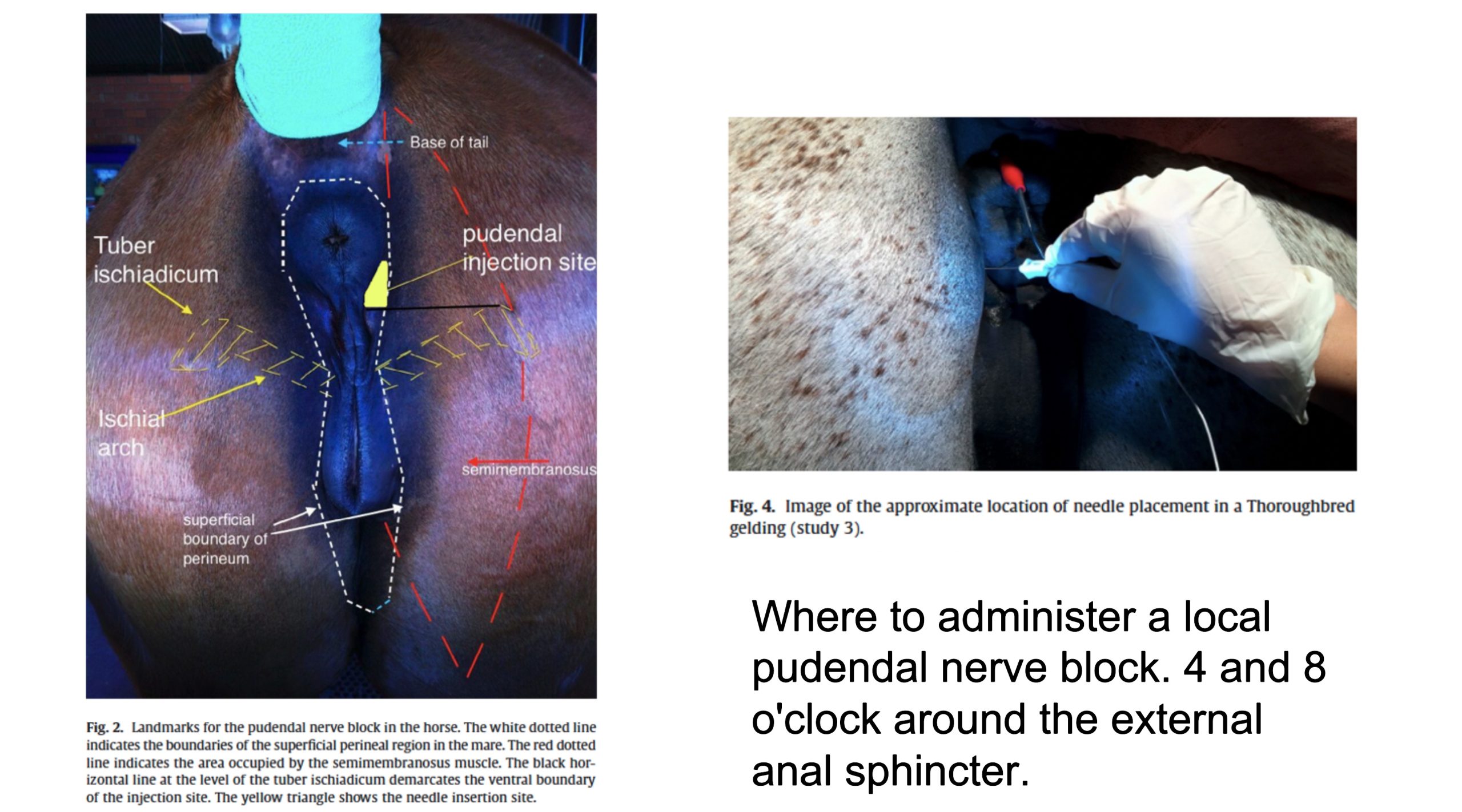
- Describe the pudendal nerve block, where to administer it, and name the structures it desensitizes.
Carnivore Pelvic Cavity and Reproductive Vasculature
Iliac arteries and Branches
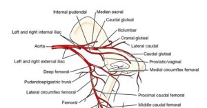
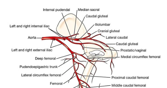
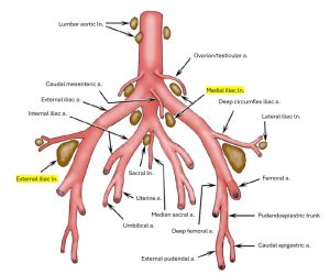
The paired iliac arteries supply the pelvis and pelvic limb. The external iliac arteries run ventrocaudally and are bilateral. Each becomes the femoral artery as it leaves the abdomen through the vascular lacuna (recall that the name-change from external iliac a. to femoral a. is the vascular lacuna.) The internal iliac arteries arise caudal to the external iliac and pass caudolaterally into the pelvis. The left and right internal iliac arteries and the smaller, unpaired median sacral artery terminate the aorta.
-

- Branches of the abdominal aorta, ventral aspect. 1
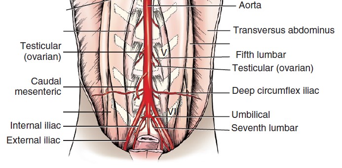
Dissect: Find the origin of the internal and external iliac arteries and the median sacral artery, where it terminates the aorta. To facilitate this dissection and following the vessels into the pelvic cavity the left caudodorsal abdominal wall attachments that remain can be transected. Then the peritoneum and connecting peritonea and fat tissues still in the way can be removed or reflected to further allow visualization of the vessels. Work to also keep the left and right hypogastric nerves extending caudally from the caudal mesenteric ganglion intact. The left one will lead us to the pelvic plexus shortly.
The deep femoral artery is the only branch of the external iliac artery and arises inside the abdomen near the vascular lacuna and courses caudally. Once the external iliac artery exits the vascular lacuna its name changes to the femoral artery.
Typically in the dog, two vessels leave the ventral surface of the deep femoral artery within the abdomen by a short pudendoepigastric trunk. These are the external pudendal and caudal epigastric arteries. In the cat, there is usually no pudendoepigastric trunk; the caudal epigastric a. and external pudendal a. each arise separately from the deep femoral a. Sometimes, this is the case in the canine as well, and at times, the dog will not possess a PE trunk. The external pudendal artery passes through the inguinal canal. Once it exits the inguinal canal, it gives rise to the superficial caudal epigastric artery, which itself courses over the superficial inguinal lymph node.
The superficial caudal epigastric artery primarily supplies the skin and mammary glands in the caudal abdominal region. Additionally, in males, it gives off small branches to the preputial skin.
The superficial inguinal lymph nodes primarily drain the skin of the lower abdomen, inguinal area, perineum, and the external genitalia (scrotum or vulva), as well as the caudal mammary glands. They also receive lymphatic drainage from the pelvic limb, specifically from areas distal to the stifle joint. They drain to the medial iliac lymph nodes.
The caudal epigastric artery arises from the pudendoepigastric trunk and passes cranially on the dorsal surface of the rectus abdominis. It supplies the caudal half of the rectus abdominis and the ventral parts of the oblique and transverse muscles.
-
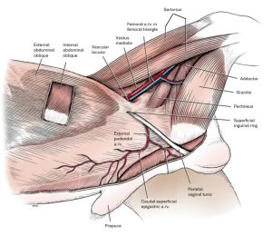
- Abdominal muscles and inguinal region of the male, superficial dissection, left side. 1
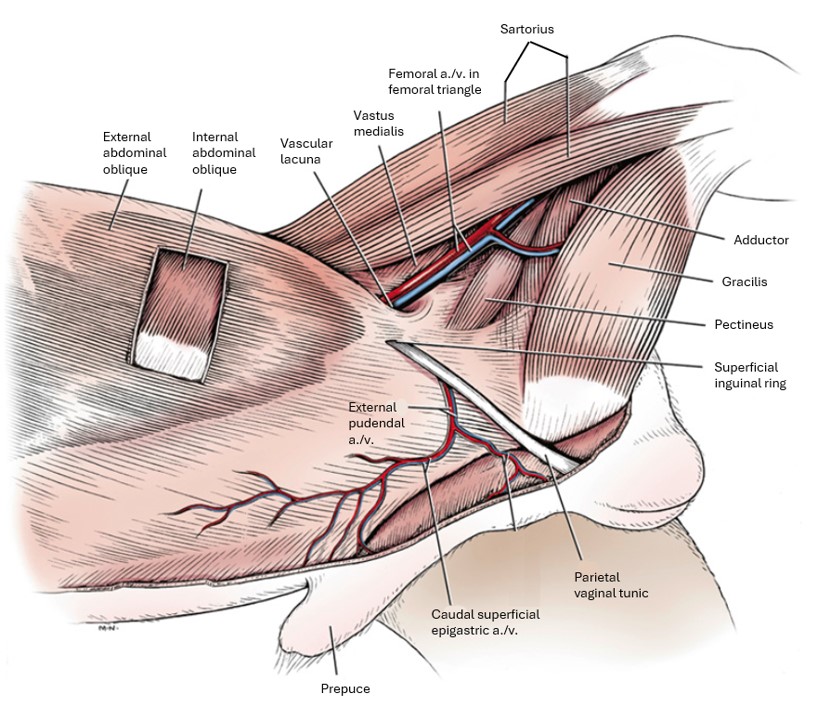
Dissect: Find the deep femoral artery as it branches from the external pudendal artery. Identify the PE trunk (if present), the caudal epigastric artery, the superficial caudal epigastric, and the external pudendal arteries. Recall what structures they supply.
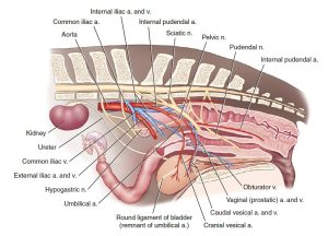
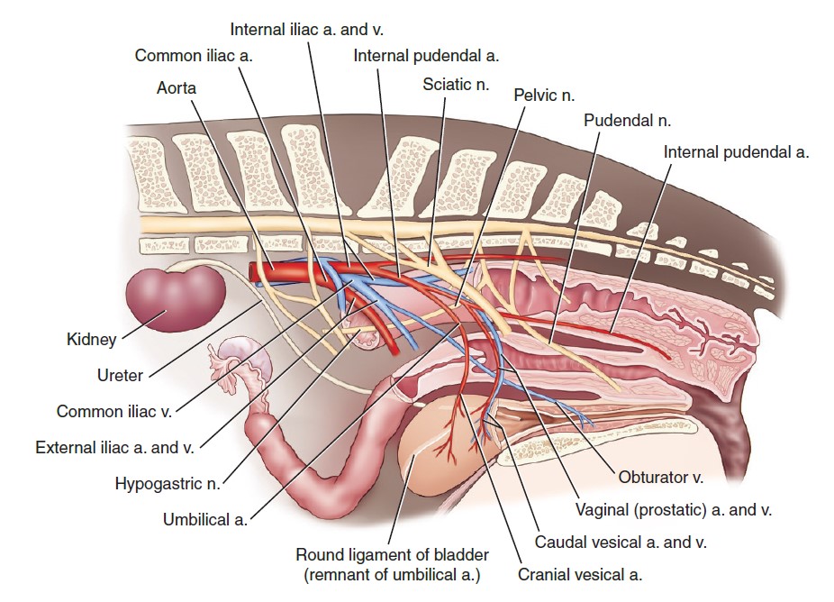
In the carnivore, the internal iliac artery gives off the rudimentary umbilical artery (in the adult, the round ligament of the bladder) and terminates cranial to the sacroiliac joint as the caudal gluteal and internal pudendal arteries. In the fetus, the umbilical artery is a large, paired vessel that carries blood from the aorta to the placenta through the umbilicus. The remnant of this vessel, the round ligament of the bladder, runs along the free edge of the lateral ligament of the bladder. It arises near the origin of the internal iliac artery and courses to the apex of the bladder in its lateral ligament. FYI: In some specimens it remains patent this far and supplies the bladder with cranial vesical arteries. Distal to the bladder the vessel is obliterated. There are no visible remnants in the median ligament of the bladder, which, you will recall, is the connecting peritoneum of the urachus in utero. If the umbilical artery does not remain patent, the caudal vesical arteries supply the entire bladder.
-

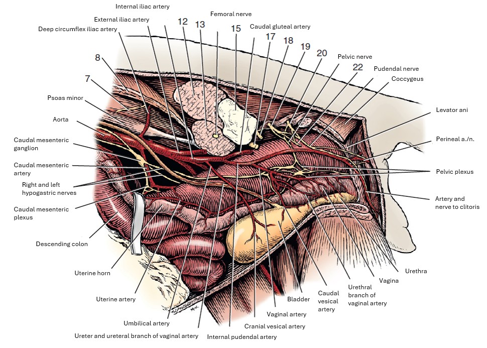
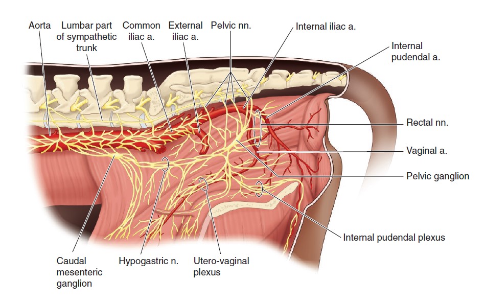
- Autonomic nerves and vessels of pelvic region, left lateral view. 1
-
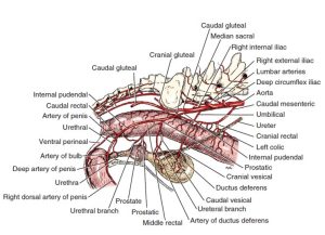
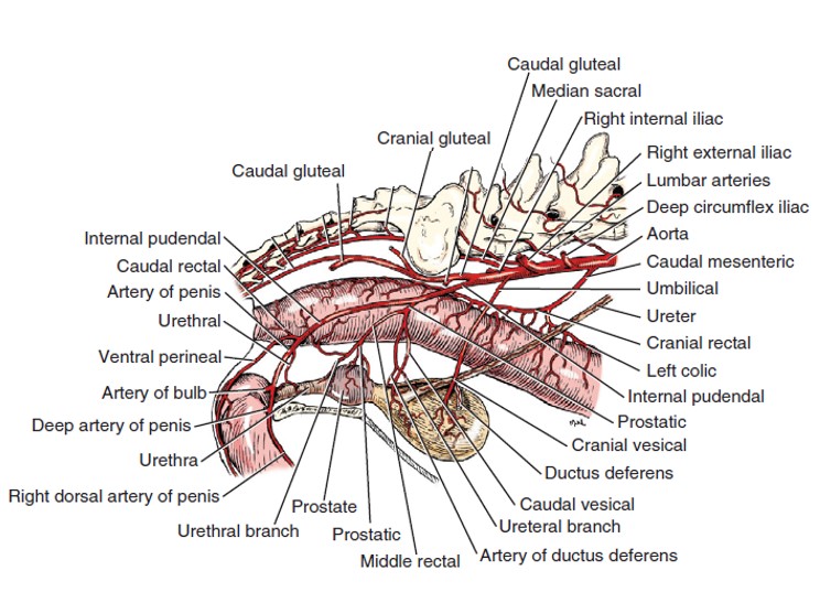
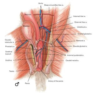
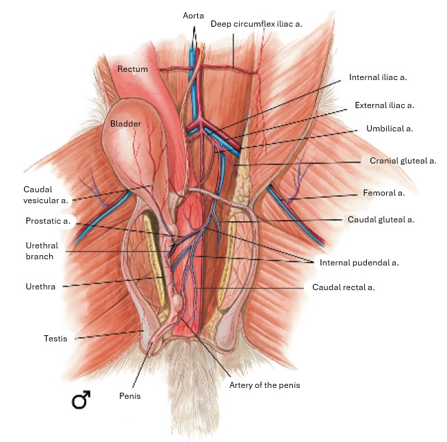
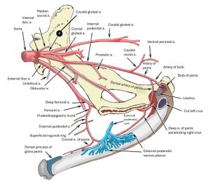
- Arteries of the male pelvic viscera, right lateral aspect. 1
-
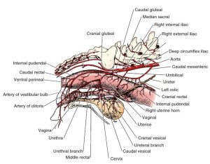
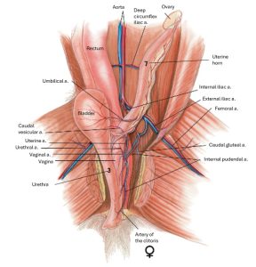
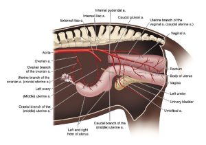
- Arteries of the female pelvis, right lateral aspect. 1
Dissect: Find the left umbilical artery/round ligament of the bladder running along the cranial free edge of the lateral ligament of the bladder. Some specimens may have a patent umbilical artery. In the absence of latex in the vessel, it should be called the round ligament of the bladder. Observe the caudal vesical arteries on the surface of the bladder. The origin of the caudal vesical artery need not be identified; it does not branch directly from the internal iliac a.
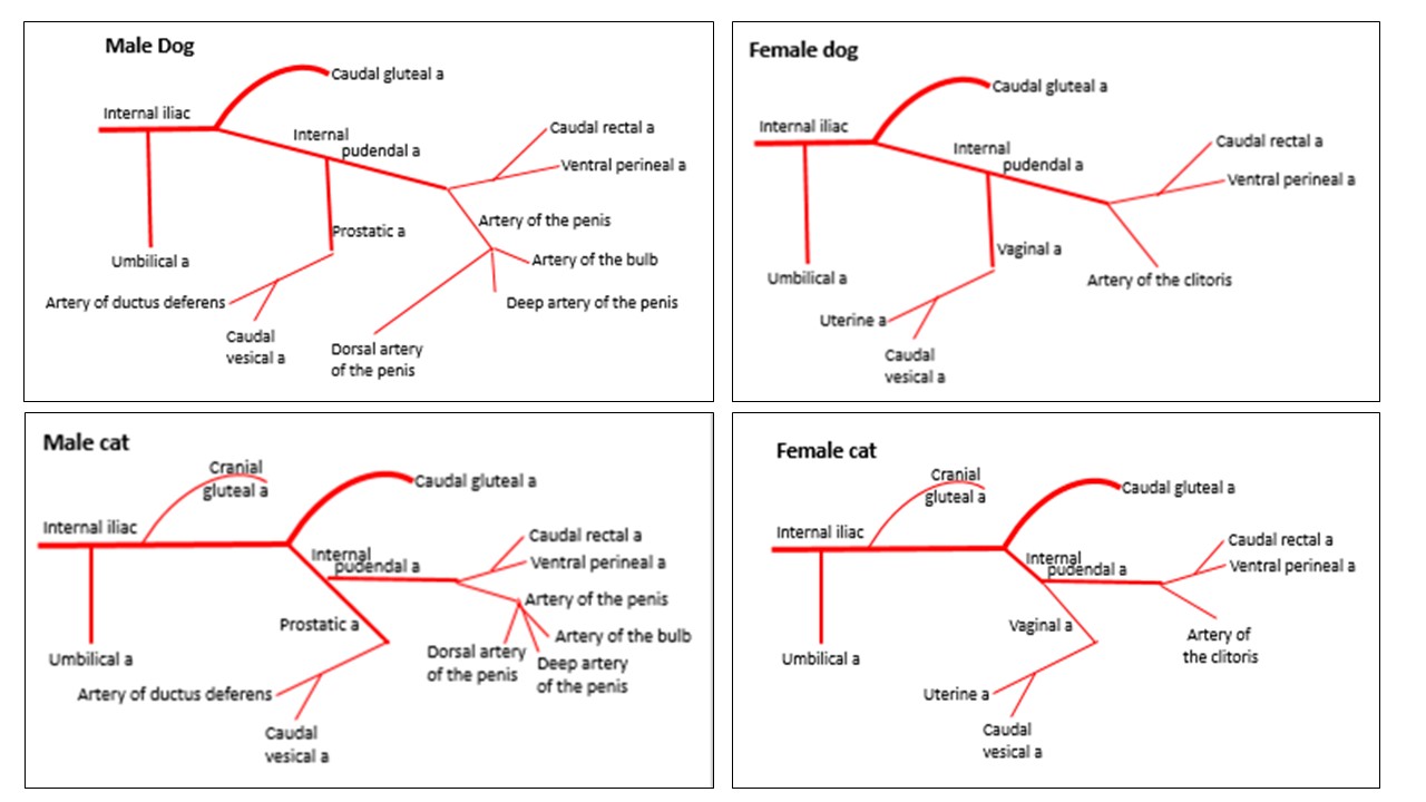
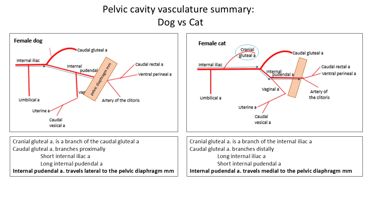
Note that the name change from internal iliac a. to the internal pudendal a. is the caudal gluteal a. In the dog, the caudal gluteal a. arises after a relatively short distance caudal to where umbilical artery branches.
Review question:
The caudal gluteal a. gives rise to the cranial gluteal artery (any chance you remember what muscles these old friends supply)?
In the cat, the first artery to branch from the internal iliac artery after the umbilical artery is the cranial gluteal artery. In the cat, the cranial gluteal artery branches independently from the internal iliac artery. The caudal gluteal a. does not branch from the internal iliac a. until much farther caudally than it does in the dog. Therefore, the internal iliac a. is short in the dog and long in the cat; the internal pudendal a. is long in the dog and short in the cat. See diagrams to better visualize these species differences.
-
-
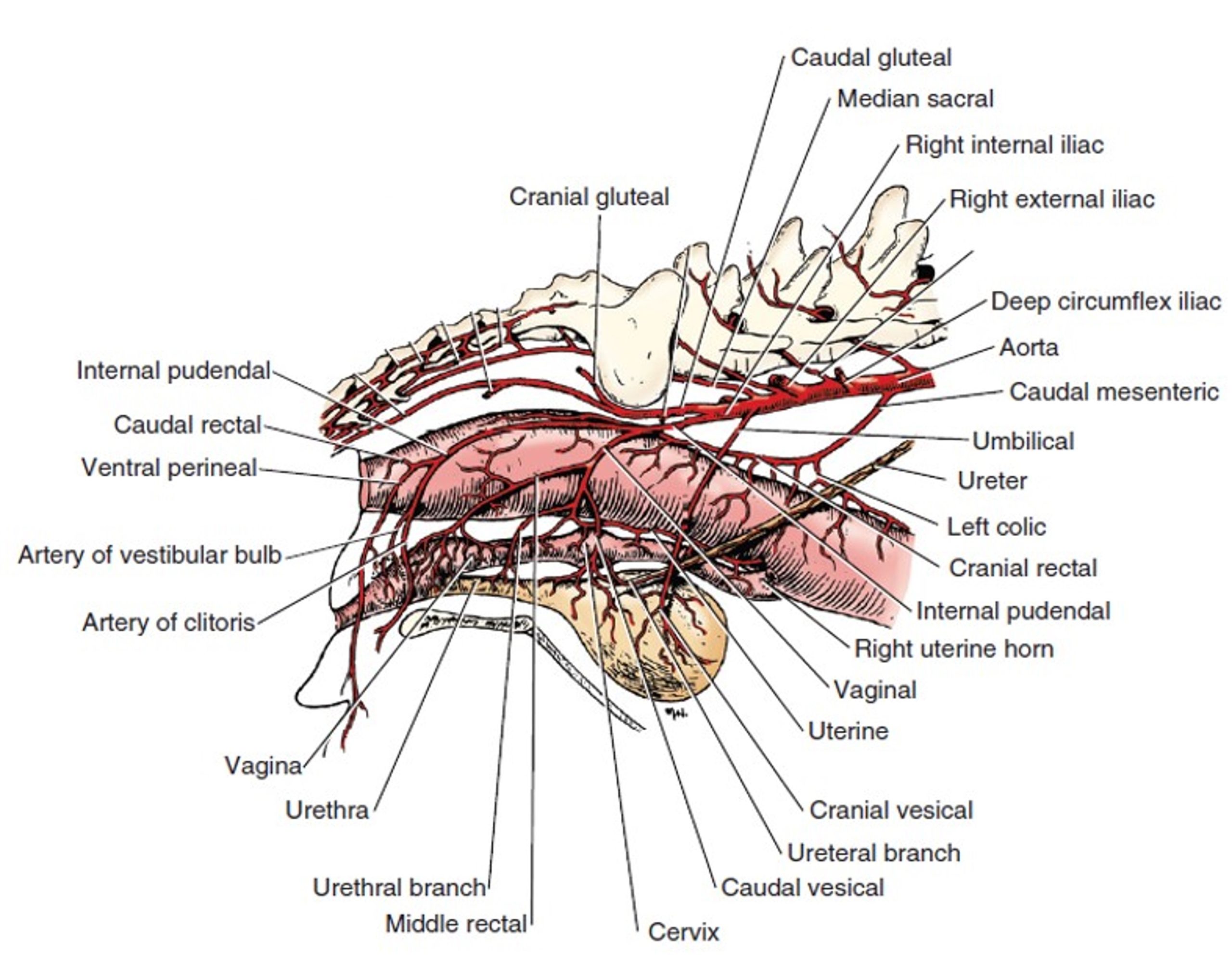
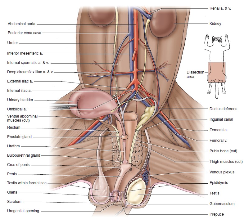
Pelvic cavity of the male cat with many of the viscera removed and urinary bladder
reflected to the right to show vessels and urogenital structures, in ventral view. 5
-
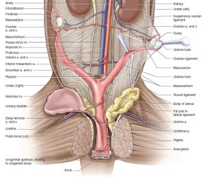
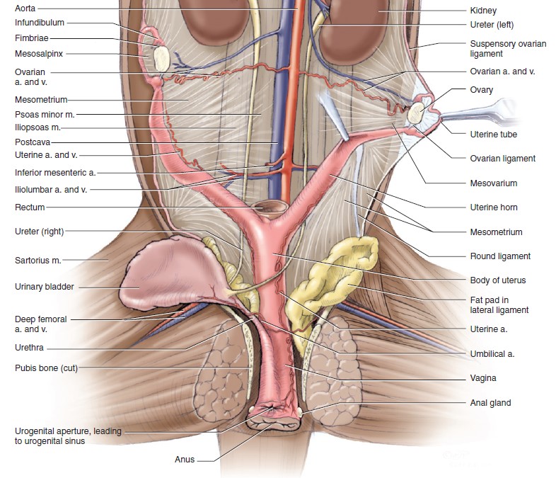
- Pelvic cavity of the female cat, in ventral view. 5
-
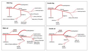
- Dog vs cat vasculature
-

- Dog vs cat vasculature
Dissect: Dissect the following vessels on the left side.
Note that the need to identify every pelvic cavity artery at its origin will not be necessary. However, knowing what artery(ies) supply the pelvic viscera and their structures is need-to-know.
Dog: Locate the caudal gluteal artery branching from the internal iliac artery just caudally to where the umbilical artery branches in the dog.
Cat: First, observe the cranial gluteal artery as it branches from the internal iliac artery. Follow the internal iliac artery more caudally and observe the caudal gluteal artery branching caudodorsally from the internal iliac artery. Caudal to where the caudal gluteal branches, the main artery is the internal pudendal artery. In the cat, the internal pudendal artery takes a sharp turn medially where the caudal gluteal artery branches from the internal iliac before giving rise to the vaginal/prostatic artery. It looks different than it does in the dog. Look at the cat diagrams below to avoid confusion and don’t confuse the internal pudendal artery for the vaginal/prostatic artery.
-
- Arteries of the pelvic limb of the dog. 1
Dissect: Note that arteries which supply reproductive structures in the female have analogous vessels in the male. It may be useful to learn the names of the arteries in one sex first. Then when observing the analogous vessels in the other sex, learn those names.
Dog: Caudal to the caudal gluteal artery, the internal pudendal gives rise to the vaginal/prostatic artery. Following the internal pudendal artery distally from the the caudal gluteal artery, find the vaginal or prostatic artery, which forms an angle of about 45 degrees with the internal pudendal. Note for later that the pelvic nerve runs parallel to the vaginal/prostatic artery.
Cat: If the vaginal/prostatic artery has not yet been located branching from the internal pudendal artery, do so now. Note for later that the pelvic nerve runs parallel to the vaginal/prostatic artery.
Observe: FYI: The vaginal/prostatic artery splits into the uterine a./artery of the ductus deferens (cranially) and the middle rectal artery caudally. The uterine/ductus deferens vasculature then give rise to the caudal vesical aa.
Although the origins of the caudal vesical and uterine artery/artery of the ductus deferens need not be dissected, identifying these vessels on/adjacent to the structures they supply is required.
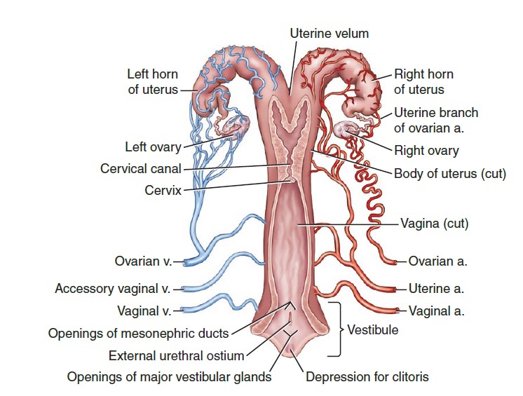
The uterine artery courses cranially along the body and horn of the uterus in the broad ligament and anastomoses with the uterine branch of the ovarian artery in the mesometrium. (This artery must be located on each side and ligated in an ovariohysterectomy procedure). Observe the uterine arteries running lateral to the uterine body and horns.
Recall that the artery of the ductus deferens is within the spermatic cord.
Take another look at the surface of the bladder, on which the branches of the caudal vesical artery are located.
The ovarian artery of the female is homologous to the testicular artery of the male. These paired vessels arise from the aorta about halfway between the renal and external iliac arteries. The ovarian artery varies in size, position, and tortuosity, depending on the degree of development of the uterus. Each ovarian artery divides into two or more branches in the mesovarium just medial to the ovaries. Branches supply the ovary and its bursa and the uterine tube and horn. The branch to the uterine horn anastomoses with the uterine artery, a branch of the vaginal artery that runs cranially in the mesometrium.
The ovarian artery and vein along with surrounding intra-abdominal fat form together form the ovarian pedicle, which is a clinical term. The ovarian pedicles must be isolated and ligated in order to perform an ovariohysterectomy (spay).
The testicular artery leaves the aorta in the midlumbar region and crosses the ventral surface of the ureter. Recall that the testicular artery, vein, and nerve plexus lie in a peritoneal fold, the mesorchium, which can be followed to the level of the vaginal ring, through the inguinal canal and into the scrotum.
The right testicular and ovarian veins enter the caudal vena cava near the origin of the artery from the aorta. However, the left testicular and ovarian veins usually enter the left renal vein. This is important surgically.
-
- Branches of the abdominal aorta, ventral aspect. 1
-
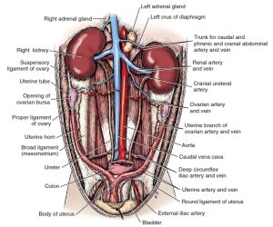
- Female dog urogenital system, ventral aspect. 1

-
- Pelvic cavity of the female cat, in ventral view. 5
Dissect: Locate the ovarian and testicular arteries. Identify the ovarian pedicles in female animals and name the structures located within.
Clinical Relevance: Ovarian Pedicle
The ovarian artery and vein, suspensory ligament, and mesovarium, along with surrounding intra-abdominal fat, together form the ovarian pedicle, which is a clinical term. The ovarian pedicles must be isolated and ligated in order to perform an ovariohysterectomy (spay).

The internal pudendal artery passes obliquely across the greater ischiatic notch. It continues along the dorsal border of the ischiatic spine lateral to the coccygeus muscle and medial to the gluteal muscles and sacrotuberous ligament in the dog. Rather than passing lateral to the coccygeus and levator ani mm, as in most species, the internal pudendal a of the cat travels medial to these mm and near the lateral surface of the rectum.
The internal pudendal artery terminates as a ventral perineal artery, a variable urethral artery, and the artery of the penis or clitoris. The ventral perineal artery may be seen passing caudally. It supplies the caudal rectal artery to the rectum and anus and terminates in the skin of the perineum and the scrotum or vulva.
Note regarding variation in the vasculature at this point: Alternatively, the caudal rectal a. may branch from the internal pudendal a. proximal to and independently from the ventral perineal a. OR the caudal rectal a. may give rise to the ventral perineal a. However these branches arise from the internal pudendal artery, as always, name them for where they are going (not branching patterns). The name change from internal pudendal a. to artery of the penis/clitoris occurs when BOTH the ventral perineal and caudal rectal aa. have branched from the internal pudendal a, in whatever pattern such may occur.
The artery of the penis courses caudoventrally and terminates at the level of the ischial arch as three branches: deep artery of the penis (into corpus cavernosum), artery of the bulb of the penis (into the bulb), and dorsal artery of the penis (not surprisingly, along the dorsal aspect of the penis). In the female the artery of the clitoris courses caudoventrally to supply the clitoris and vestibular bulb.
-
- Arteries of the male pelvic viscera, right lateral aspect. 1
-
- Arteries of the female pelvis, right lateral aspect. 1
-
- Vasculature of the male pelvic region of the cat, ventral view. 4
-
- Vasculature of the female pelvic region of the cat, ventral view. 4

Observe: Dissection and identification of the caudal rectal and ventral perineal arteries, the artery of the clitoris/penis, and the deep artery of the penis, artery of the bulb of the penis, and the dorsal artery of the penis is not required. Need to Know: Recognize that all of these vessels originate from the internal pudendal artery. Be able to state what structures/region the caudal rectal and ventral perineal arteries and the artery of the penis/clitoris supply.
Optional Dissection: If all team members agree (or if someone would like to dissect outside of lab hours), follow the internal pudendal artery caudal from the vaginal/prostatic artery and follow and clean the caudal rectal and ventral perineal arteries, the artery of the penis/clitoris, and, in males, the deep artery of the penis, artery of the bulb of the penis, and the dorsal artery of the penis.
If you are unable to locate these structures on the left, then you may proceed to dissecting the right ischiorectal fossa. Reflect the already transected superficial and middle gluteal mm to uncover the greater ischiatic notch. Transect and reflect the sacral attachment of the sacrotuberous ligament. This exposes the caudal gluteal artery and the sciatic nerve. Deep to these are the internal pudendal artery and pudendal nerve and the ventral branches of the sacral nerves. These ventral branches emerge from the two pelvic sacral foramina and the sacrocaudal intervertebral foramen to form the sacral plexus.
Nerves of the Carnivore Pelvic Cavity
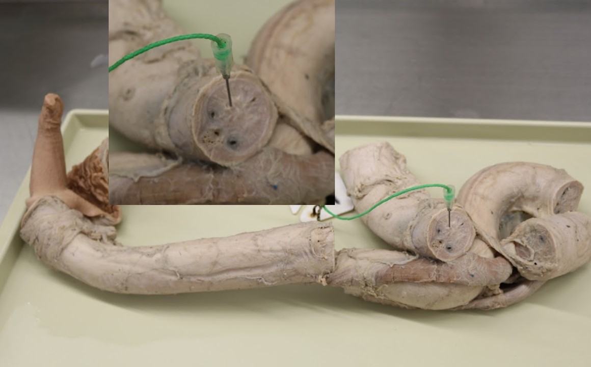
The pelvic plexus lies caudal to a transverse plane passing through the pelvic inlet and dorsal to the prostate. It is closely applied to the surface of the rectum and the vaginal/prostatic artery. It can be identified by tracing the left hypogastric nerve to it. Recall that the left and right hypogastric nerves originate in the caudal mesenteric ganglion and provide sympathetic innervation to the pelvic cavity. Occasionally, ganglia are large enough to be recognized in the plexus. The pelvic plexus contains sympathetic fibers from the hypogastric nerve and parasympathetic fibers from the pelvic nerve.
The pelvic nerve is formed by parasympathetic preganglionic axons that leave the ventral branches of the three sacral spinal nerves. The pelvic plexus ganglia contain the cell bodies of post-ganglionic parasympathetic nerves which ultimately innervate the pelvic viscera. It supplies branches to the urogenital organs, the rectum, and the descending colon. The branches to the urogenital organs join the pelvic plexus and follow the vaginal/prostatic vessels to these organs. The pelvic nerve will extend dorsally from it, parallel to the prostatic/vaginal artery.
-
- Autonomic nerves and vessels of pelvic region, left lateral view. 1
Dissect: Find the hypogastric nerves running caudally from the unilateral caudal mesenteric ganglion. Follow the left hypogastric nerve caudally to the left pelvic plexus and ganglion on the lateral wall of the caudal portion of the rectum. Locate the pelvic nerve running alongside the vaginal/prostatic artery.
The pudendal nerve arises from all three sacral nerves and provides somatic/voluntary innervation to the pelvic cavity and perineum. It passes caudolaterally, where it lies lateral to the levator ani and coccygeus muscles, medial to the superficial gluteal muscle, and dorsal to the internal pudendal vessels. It appears superficially in the ischiorectal fossa after emerging from the medial side of the superficial gluteal muscle and courses caudomedially toward the pelvic symphysis at the ischial arch.
-
- Autonomic nerves and vessels of pelvic region, left lateral view. 1
-

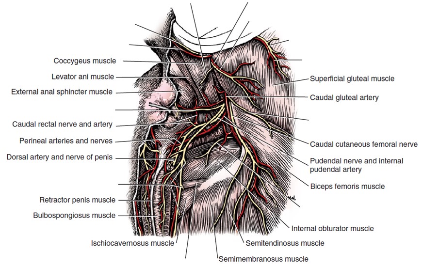
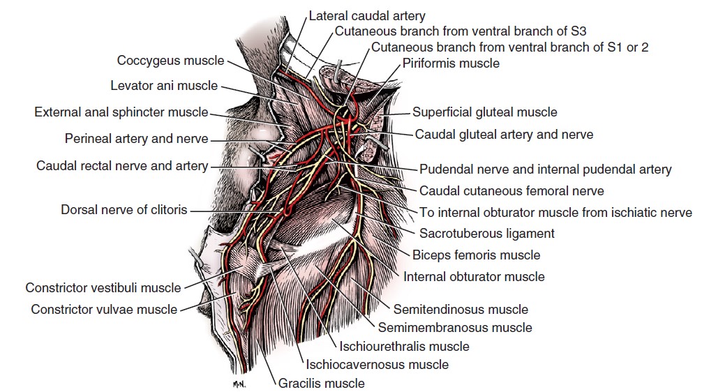
- Nerves, arteries, and muscles of the male perineum, caudolateral aspect. 1
-
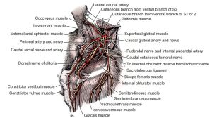
- Nerves, arteries, and muscles of the female perineum, caudolateral aspect. 1
Dissect: Identify the pudendal nerve running alongside the internal pudendal artery. The following branches arise from the pudendal nerve and need not be dissected, but, once again, knowing that they originate from the pudendal nerve, what type of innervation they supply to what structures is mandatory.
a. The caudal rectal nerve may arise from sacral nerves or may leave the pudendal nerve at the caudal border of the levator ani muscle. It innervates the external anal sphincter.
b. The perineal nerves (FYI: superficial and deep) arise from the dorsal surface of the pudendal nerve. They supply the skin of the anus and the perineum and continue to the scrotum or labium. Short nerves from the pudendal or perineal nerves supply the muscles of the penis or the vestibule and vulva.
c. The dorsal nerve of the penis in the male (or of the clitoris in the female) curves around the ischial arch and reaches the dorsal surface of the penis, where it courses cranially. It continues through the glans penis and ends in the skin covering the apex of the penis. It provides sensory nerves to the skin of the glans. In the female the smaller dorsal nerve of the clitoris runs ventrally to the ventral commissure of the vulva, where it terminates in the clitoris.
-
- Nerves, arteries, and muscles of the male perineum, caudolateral aspect. 1
-
- Nerves, arteries, and muscles of the female perineum, caudolateral aspect. 1
Clinical Application: Anal Sac Resection/Perineal Hernia

Ungulate Pelvic Cavity and Reproductive Vasculature
Lymph nodes and Aortic termination
Numerous lymph nodes are associated with the terminal part of the abdominal aorta. Those located along the aorta, caudal to the kidneys, are designated lumbar aortic nodes.
Observe: Horse, ruminant, pig: Observe the termination of the abdominal aorta to identify the origins of the external and internal iliac arteries and to locate the medial iliac lymph nodes in this area (note, those found at the junction of the cranial and caudal branches of the deep circumflex iliac a. are the lateral iliac lymph nodes). FYI – clinically, the medial and lateral iliac lymph nodes are collectively the “iliac lymph nodes”. The median sacral a., found in ruminants, is not present in the horse.
Ruminant and pig: Follow a little further out along the external iliac a. to identify a large node. This is the external iliac lymph node, found consistently in the ox, sheep and pig and maybe less so in the goat. FYI: This lymph node corresponds to the proximal femoral lymph node of the horse, which is located in the femoral triangle and will not be identified.
-
- Termination of bovine aorta with associated lymph nodes, ventral view. 2

External iliac a. and its branches
Observe: Horse and goat and pig: Observe the external iliac a. and identify the following bolded branches:
1. Deep circumflex iliac a. (in the horse this vessel may arise from the aorta or at the origin of the external iliac artery; recall that its branches out in the abdominal wall are a good cause for hemorrhage during body wall incisions in the flank!)
2. Uterine a. (artery of the ductus deferens in male) – observe the uterine artery adjacent to the uterus. No need to observe its origin. FYI: In the HORSE the uterine artery branches from the external iliac a. In ruminants and pig the uterine a. branches off umbilical a.). You need not identify the artery of the ductus deferens a. but know that it is analogous to the uterine a.
3. Deep femoral a. (recall that after the external iliac a. exits the vascular lacuna, it becomes the femoral a.)
a. Pudendoepigastric trunk (in horse this may arise from external iliac a.!)
i. Caudal epigastric a. – courses cranially on deep surface of RA m.
ii. External pudendal a. – passes through deep inguinal ring into inguinal canal.
-
- Termination of bovine aorta with associated lymph nodes, ventral view. 2
-
- Blood supply to the reproductive tract of the mare. 9

-
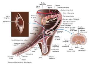
- The blood supply of the bovine penis. Inset: Illustration showing a penile hematoma. 9

Ungulate Pelvic Vessels and Innervation
Observe: While identifying the following nerves and vessels in the ungulate is not mandatory, except as noted, it is required to understand what nerves innervate which structures and what type of innervation they supply to these structures.
Innervation
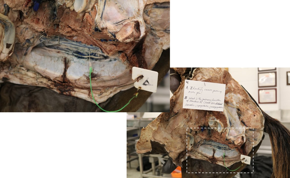
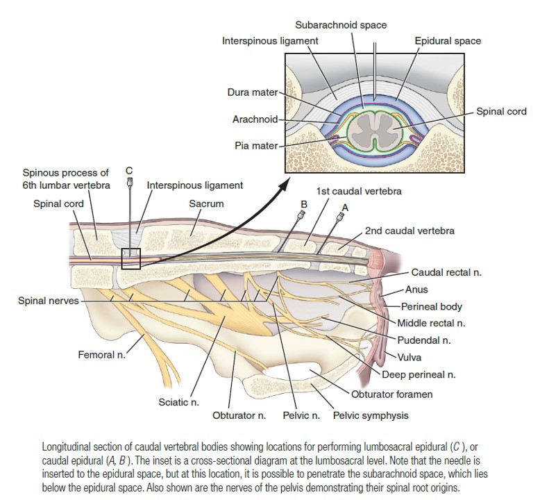
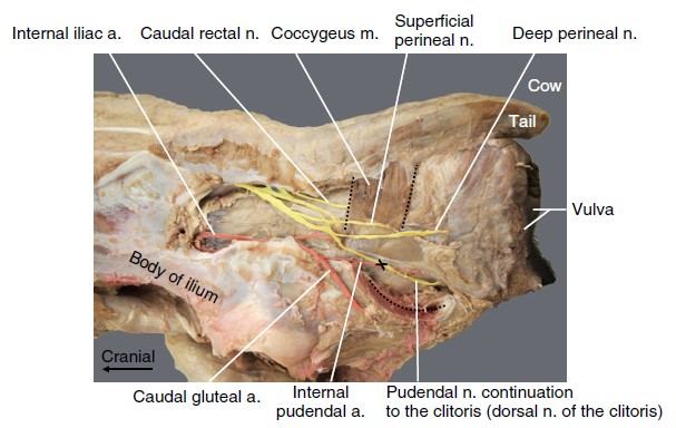
The pudendal nerve arises rom various sacral nerves in the ungulate (S2-S4 in ruminants; S2/3-S4 in the horse) and courses caudoventrally on the medial surface of the sacrosciatic ligament, where it can be palpated rectally in the ox. Note that the pudendal nerve is more invested in the sacrosciatic ligament in the horse and we see a portion of it from the lateral side. Of note is that that external anal sphincter m. is provided somatic innervation by the caudal rectal nerve, which is NOT a branch of the pudendal nerve in the ungulate. The pudendal nerve supplies sensory innervation to the rectum, perineal skin, and internal and external reproductive organs and motor to much of the striated perineal musculature. Deep and superficial perineal nerves in addition to various cutaneous branches originate from the pudendal nerve, which finally continues as the dorsal nerve of the penis/clitoris. The dorsal nerve of the penis supplies innervation to the following structures: the skin and fascia of the penis, including the prepuce, and to the glans penis. The dorsal nerve of the penis also innervates the following structures:
- Penile shaft: The dorsal nerve of the penis provides sensory innervation to the skin of the penile shaft.
- Glans penis: It also innervates the glans penis, contributing to its sensory function and the reflex ejaculatory activity.
- Anterior urethra: The dorsal nerve has fibers that extend into the corpus spongiosum to innervate the urethral lumen, relaying sensory information during micturition (urination) and ejaculation.
-
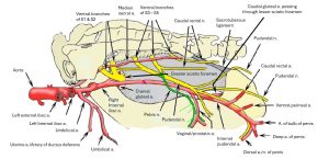
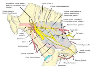
- Branches of right internal iliac artery and sacral plexus nerves of ox, medial view. 2

-
- Lateral view of the equine female caudal abdomen and pelvis focused on the urogenital system anatomy. 9
The pelvic nerve lies medial to the pudendal nerve and adjacent to the vaginal/prostatic aa. and carries preganglionic parasympathetic fibers from the sacral segments of the spinal cord to the pelvic plexus, from which they are distributed to the pelvic viscera. Note that unlike in the thorax and abdomen, the pelvic ganglion contains nerve cell bodies of post-ganglionic parasympathetic nerves. Recall that in the head, thorax, and abdomen, ganglions contain nerve cell bodies of post-ganglion sympathetic nerves.
Recall that the pelvic plexus also receives the hypogastric nn. which supply sympathetic innervation to the pelvic viscera.
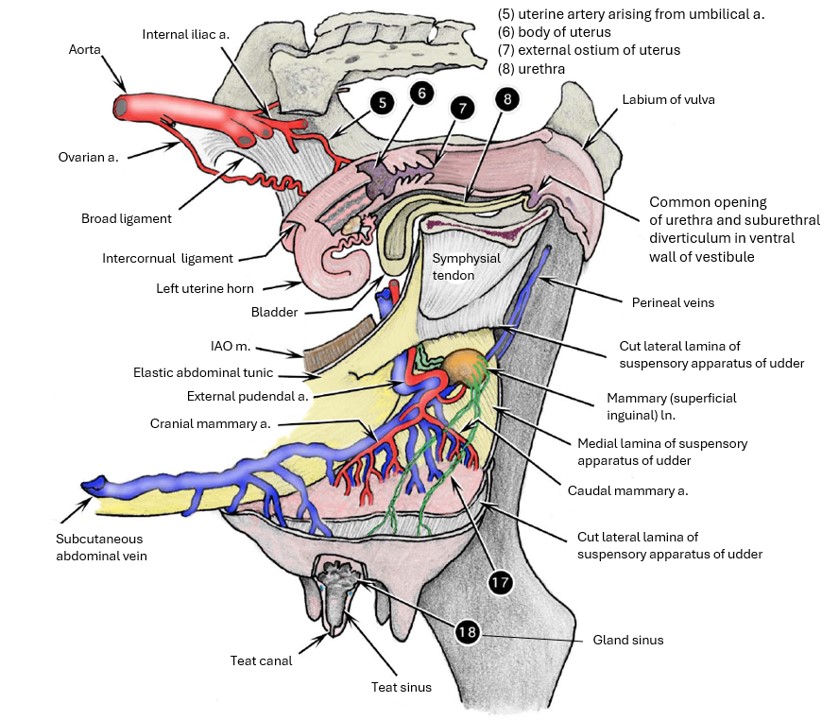
The udder of the cow is supplied by sensory and autonomic nerves. The sensory innervation is derived from the ventral cutaneous branches of L1 and L2, the genitofemoral nerve (L3 and L4), and the mammary (superficial perineal) branch of the pudendal nerve. Of these, the genitofemoral nerve is of major importance, because it innervates much of the skin, the teats, and most of the glandular tissue.
The autonomic innervation is supplied by sympathetic fibers from the caudal mesenteric ganglion, which accompany the genitofemoral nerve through the inguinal canal.
-

- Blood supply and innervation to the bovine perineal area. 9
-

- Pelvic nerves of the cow. 9
Clinical Application: Pudendal Nerve Block
Pudendal Nerve Block
- The pudendal nerve mainly supplies somatic sensory and motor innervation to the pelvic cavity viscera. Therefore, it may be blocked to provide surgical analgesia for common perineal and reproductive procedures in the standing animal. Administration desensitizes the pudendal nerve and its branches and the dorsal nerve of the penis/clitoris. Therefore, the perineal region and the penis/clitoris are desensitized as is the penis.
- Mammary tissue and the prepuce are supplied, mostly, by the genitofemoral nerve; these structures must be desensitized locally if procedures are performed on them.
- The pudendal nerve block is administered at the four and eight o’clock locations ventrolateral to the external anal sphincter muscles.
TO KNOW: Which nerves and structures are/are not desensitized by the pudendal nerve block. What is the main type of innervation supplied to the pelvic cavity viscera by the pudendal nerve and its branches. Where to administer the nerve block.
-

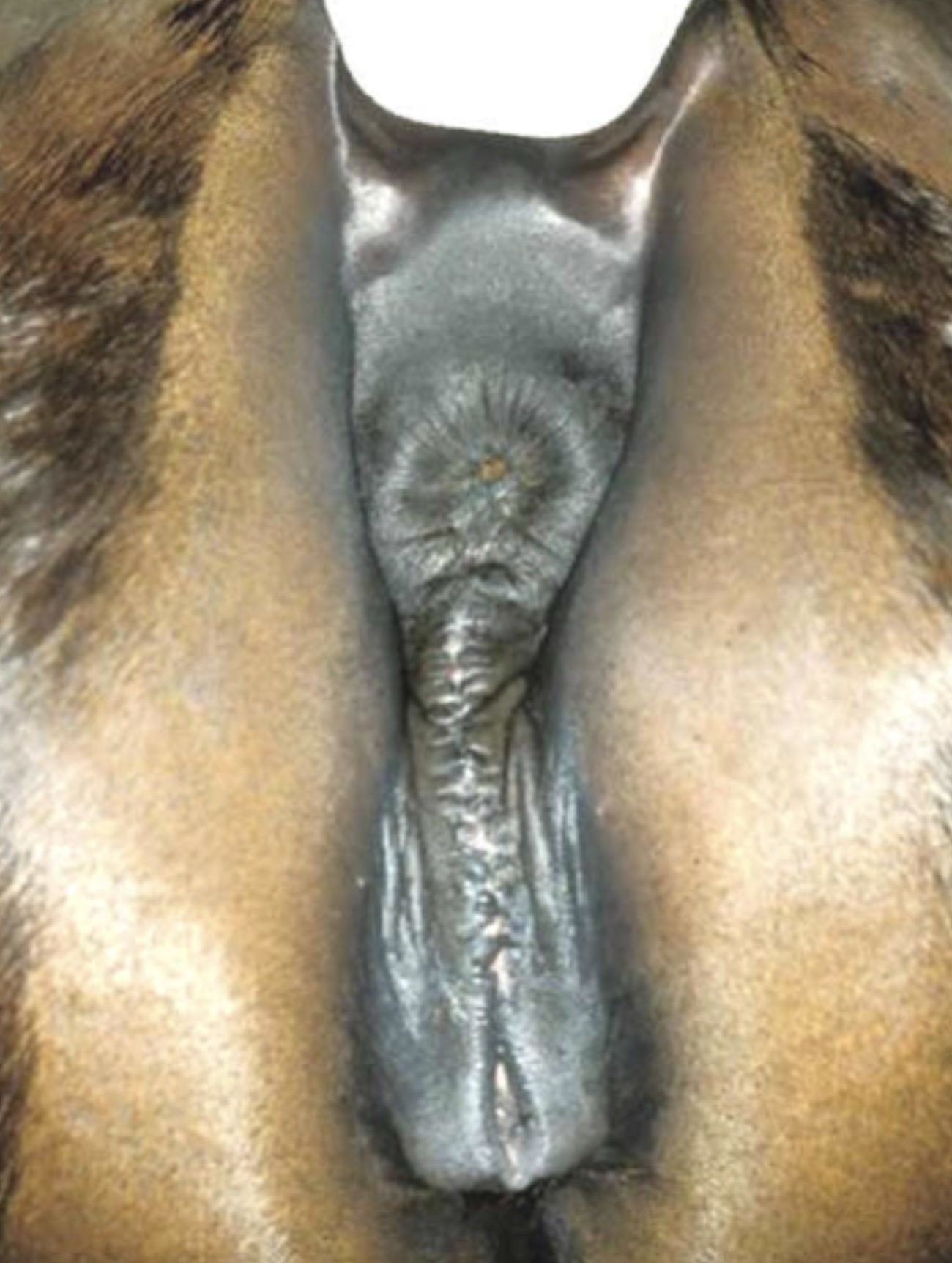
- Caslick’s Procedure
-
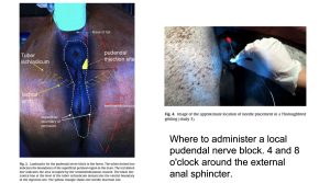
- pudendal nerve block application
-
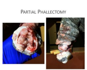
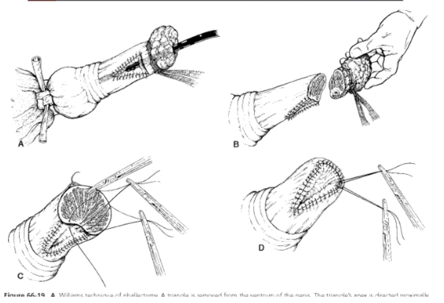
- partial phallectomy

-
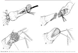
- partial phallectomy surgical diagram
Ungulate Pelvic Cavity and Reproductive Vasculature
In the mare, the uterine artery is a branch of the external iliac artery. In ruminants and the pig, the uterine artery typically arises as the first branch of the umbilical artery, which is a branch of the internal iliac artery.
-
- Branches of right internal iliac artery and sacral plexus nerves of ox, medial view. 2
-
- Lateral view of the equine female caudal abdomen and pelvis focused on the urogenital system anatomy. 9
-

- Nerves and vessels located medial to the sacrosciatic ligament of the cow. 12
Observe: Identify the uterine artery as it courses along the uterine horns in all species.
Clinical Application: Palpation of the uterine artery
The uterine artery of the cow is palpated rectally during a pregnancy examination; its size can be used as an indication of the stage of gestation.
The internal iliac a. branches into the internal pudendal a. and caudal gluteal a. In the horse, this branching occurs cranially not much beyond the origin of the internal iliac a. (a ‘high splitter’ like the canine) and the caudal gluteal a. subsequently emerges through the dorsal aspect of sacrosciatic ligament. In artiodactyls, the division of the internal iliac a. occurs quite caudally (a ‘low splitter’ like the feline) and the caudal gluteal a. branches off at the level of the lesser ischiatic foramen, through which it exits in the ruminant. In the pig, the internal iliac a. exits the pelvic cavity through the greater ischiatic foramen to course along the lateral aspect of the sacrosciatic ligament. At the level of the lesser ischiatic foramen, the caudal gluteal a. branches off, remaining external, and the internal pudendal a. continues by re-entering the pelvic cavity through the lesser ischiatic foramen.
The vaginal/prostatic artery (which supply their namesakes) and then the caudal rectal artery (supplies the external anal sphincter muscle) and the ventral perineal a. (supplies, you guessed it, the ventral perineum) branch from the internal pudendal a. distal to the origin of the caudal gluteal artery.
Once they have branched, the internal pudendal artery changes name to the artery of the penis/clitoris. The artery of the penis then splits into the dorsal artery of the penis, artery of the bulb, and deep artery of the penis.
Recall that the the stallion has developed an extensive external pudendal venous plexus to receive blood from the cavernous spaces of the penis immediately following dismounting. The venous plexus is drained by the large accessory external pudendal vein, which does not return through the inguinal canal but rather perforates the origin of the gracilis muscle and joins the deep femoral vein. The much smaller external pudendal vein enters the abdominal cavity through the inguinal canal, along side the external pudendal artery, as is typical.
-
- Blood supply to penis of horse, left lateral view. 2

-
- Pelvic wall of horse, lateral view. 2

-

- Dorsal view of the blood supply to the bovine reproductive tract (cow). 9
Superficial Inguinal (Mammary) Lymph Node
Observe: Identify the superficial inguinal lymph node (also called mammary ln in dairy animals), as described below.
Numerous large, superficial lymphatics drain the mammary tissue to the mammary ln. (superficial inguinal ln.).
Efferent lymphatics in the cow (and goat?) then continue through the inguinal canal, to the external iliac ln.(and in the mare to the proximal femoral ln.).
The superficial lymphatics of the cow udder can be observed in the living animal, and their direction is primarily caudodorsal. These lymphatics can be distinguished from the superficial veins, which course in a more
craniodorsal direction.
-
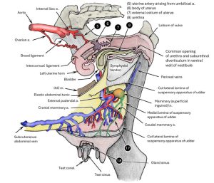
- Genitalia and udder of cow with much of left wall of genitalia and udder removed, left lateral view. 2
Review videos
Dog pelvic cavity a./v./n., Gerard – 8 min
Dog pelvic cavity a./v./n., Gerard – 30 min
Dog pelvic cavity a./v./n., Gallenstein – 21 min
Mare pelvic a./v./n. – 7 min
Mare PE Trunk – 4 min
Buck pelvic a./v./n. – 10 min, watch until 20:30
Calf pelvic cavity – 14 min
Terms
| Iliac arteries and Branches in the Carnivore | ||
| Term | Features | Species differences/comments |
| External iliac a. | ||
| Femoral a. | ||
| Vascular lacuna | ||
| Internal iliac a. | ||
| Median sacral a. | Unpaired | |
| Deep femoral a. | ||
| Pudendoepigastric (PE) trunk | Absent in cat. Know from where it branches and what it splits into in all species. ID in large animal. | |
| External pudendal a. | Know that this is the artery that passes through the inguinal canal in all species and both sexes. ID in large animal. | |
| Caudal epigastric a. | Observe along the rectus abdominus in all species. | |
| Superficial caudal epigastric a. | Know that it supplies caudal mammary tissue. | |
| Superficial inguinal lymph node | ||
| Umbilical a. | Round ligament of the bladder (in adult) | |
| Caudal gluteal a. | ID in carnivore only. | |
| Internal pudendal a. | ID in carnivore only. | |
| Lateral ligament of the bladder | ||
| Caudal vesical a. | No need to ID. Know what it supplies. | |
| Cranial gluteal a. | No need to ID. Inconsistently present (when umbilical artery is still patent). Know what it supplies, when present. | |
| Vaginal/prostatic a. | ID in carnivore only. | |
| Uterine a. | ID in all adjacent to uterus. | |
| Artery of the ductus deferens | ID in spermatic cord or next to ductus before it enters deep inguinal ring. | |
| Ovarian/testicular a. | ||
| Ovarian pedicle | Clinical term in small animal. Ovarian a./v., intra-abdominal fat, mesovarium, suspensory ligament. | |
| Ventral perineal a. | No need to ID. Know what region it supplies. | |
| Artery of the penis/clitoris
ID in carnivores |
Deep artery of the penis (FYI) | Know what the artery of the penis/clitoris supply (should be pretty straight forward). |
| Artery of the bulb of the penis (FYI) | ||
| Dorsal artery of the penis (FYI) | ||
| Caudal rectal a. | Do not ID. Know what it supplies. | |
| Nerves of the Pelvic Cavity in the Carnivore | ||
| Term | Features | Species differences/comments |
| Pelvic plexus | ID in carnivore only. The pelvic plexus ganglia contain the cell bodies of post-ganglionic parasympathetic nerves. The plexus itself has both sympathetic and parasympathetic fibers. | |
| Pelvic nerve | ID in carnivore only. Parasympathetic. | |
| Hypogastric nerves | ID in carnivore only, coming from ca. mesenteric gangion and plexus. Sympathetic. | |
| Perineal nerve(s) | FYI: Deep and superficial | No need to ID. Know from what main nerve they branch and what region they supply. |
| Pudendal nerve | ID in carnivore only. Somatic. Apply knowledge of this nerve to pudendal nerve block. | |
| Caudal rectal nerve | No need to ID. Somatic to external anal sphincter. | |
| Dorsal n of the penis/clitoris | Know what they innervate. | |
| Ungulate Lymph nodes and Aortic termination | ||
| Term | Features | Species differences/comments |
| Medial iliac lymph nodes | ||
| External iliac lymph node | Follow the external iliac a. caudally, and this will be the large ln lateral to it. | ID in calf only. Bovine and pigs possess these. |
| External iliac a. | ||
| Internal iliac a. | ||
| Median sacral a. | ||
| Deep circumflex iliac a. | ||
| Uterine/artery of the ductus deferens | ID uterine artery adjacent to uterus and a. of ductus deferens alongside ductus deferens. | |
| Femoral a. | ||
| Deep femoral a. | ||
| Pudendoepigastric (PE) trunk | ||
| External pudendal a. | ID within abdominal cavity. | |
| Caudal epigastric a. | ||
| Nerves in the Ungulate Pelvic Cavity | ||
| Term | Features | Species differences/comments |
| Pudendal nerve | ID in carnivore only. Know what it supplies. Somatic innervation to PC. | |
| Caudal rectal nerve | NOT a branch of the pudendal nerve in the ungulate. ID in carnivore only. What does it innervate? Type of innervation? | |
| Perineal nerve(s) | FYI: Deep and superficial | No need to ID. Know what structures innervated and what kind of innervation. |
| Dorsal nerve of the penis/clitoris | No need to ID. Know what structures innervated and what kind of innervation. | |
| Pelvic plexus | ID in carnivore only. Know what type of innervation is present. Which nerve cell bodies synapse here? | |
| Pelvic nerve | ID in carnivore only. What does it innervate and what type of innervation does it supply in all species? | |
| Hypogastric nerve | ID in carnivore only. What does it innervate and what type of innervation does it supply in all species? | |
| Genitofemoral nerve | ID as it enters deep inguinal ring and, where possible, coursing to the prepuce/mammary glands. What does it innervate and what type of innervation does it supply in all species? | |
| Caudal mesenteric ganglion | ID in carnivore only. What type of innervation does it supply in all species? | |
| Ungulate Pelvic Cavity and Reproductive Vasculature | ||
| Term | Features | Species differences/comments |
| Uterine a. | ID adjacent to uterus only. | |
| Internal iliac a. | ||
| External iliac a. | ||
| Internal pudendal a. | ID in carnivore only. Know what it supplies. | |
| Caudal gluteal a. | No need to ID. Know what it supplies. | |
| Vaginal/prostatic a. | ID in carnivore only. Know what it supplies. | |
| Caudal rectal a. | No need to ID. Know what it supplies. | |
| Ventral perineal a. | No need to ID. Know what it supplies. | |
| Artery of the penis/clitoris | Artery of penis/clitoris: ID in carnivore without following further branches | |
| Deep artery of the penis | FYI. | |
| Artery of the bulb of the penis | FYI. | |
| Dorsal artery of the penis | FYI. | |
| External pudendal venous plexus | ID on stallion penis. Recall drains the penis post-copulation. | |
| Accessory external pudendal vein | Male horse only. Know conceptually, ie external pudendal venous plexus drains through this vessel. The external pudendal v. is present but is comparatively much smaller. | |
| Superficial inguinal ln | Also called mammary ln in dairy animals in particular. | ID in carnivores where still present and adjacent to mammary tissue in large animals in cadavers or wet specimens. |
EXAMPLE PRACTICAL EXAM QUESTIONS