Lab 4A: Male Ungulate Urethral and Reproductive Anatomy
Learning Objectives
- Identify the major features of the male reproductive tract, comparing between ungulate species.
- Review urinary bladder and urethral anatomy.
- Articulate how penile structures are vascularized.
Male Ungulate Genitalia
Scrotum, spermatic fascia, testicles
Adhered to the dermis of the scrotum is a modification of the subcutaneous tissue layer known as the tunica dartos, which consists of smooth muscle bundles mixed with elastic and collagenous fibers. Contraction of the tunica dartos wrinkles (shrivels) the scrotal skin, thereby reducing the exposed surface area of the scrotum and holding the testes closer to the body wall, reducing heat loss. The tunica dartos also forms the septum of the scrotum, which separates the right and left testes. The location of the septum is indicated externally on the skin by the raphe of the scrotum.
-
-
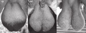
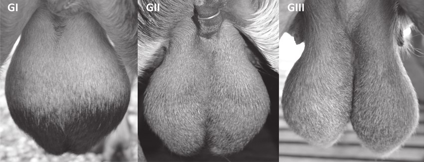
Scrotal region of goats of the different Groups: (GI) Animals presenting no bipartition; (GII) animals with bipartition at 50%;
(GIII) animals with bipartition up to 50% of the testicular length. Influence of the bipartite scrotum on the testicular and scrotal temperatures in goats
-
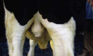
- Scrotum of a bull. The musculature in the tunica dartos is contracted, causing elevation of the scrotum. 8

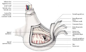
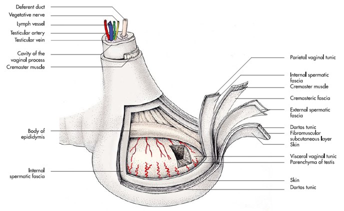
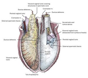
The external spermatic fascia is continuous proximally with the external fascia of the abdominal wall. This relatively loose fascia is what allows the vaginal tunic to move within the scrotum (and enveloped testis and epididymis). The internal spermatic fascia is continuous proximally with the transversalis fascia of the internal abdomen and is adhered to the parietal vaginal tunic. The internal spermatic fascia accounts for the toughness and opacity of the vaginal tunic, a clinical name for the combined internal spermatic fascia and the parietal vaginal tunic.
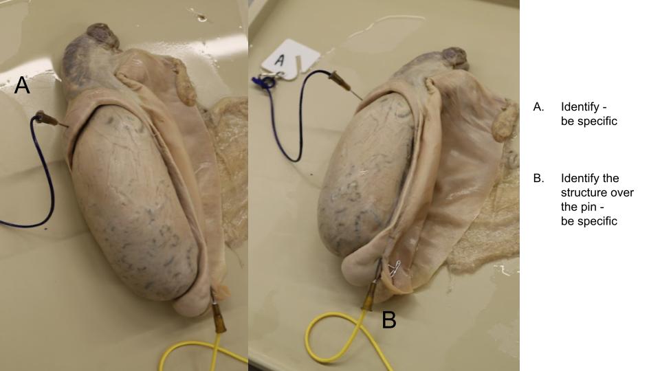
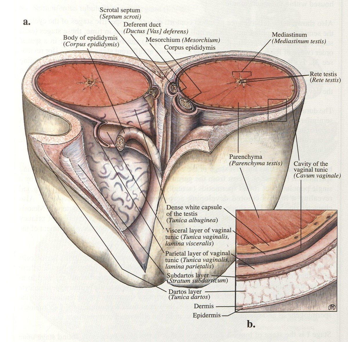
Incision of the internal spermatic fascia/parietal vaginal tunic unit along the free border of the testis and proximally over the spermatic cord opens the vaginal cavity. The testis, epididymis and spermatic cord structures can be examined, all closely invested on their surface by the visceral vaginal tunic. A shallow cut made into the testis deep to the vaginal cavity exposes the parenchyma of the testes and supporting tissues. This incision will have gone through both the external visceral vaginal tunic and the underlying thicker, fibrous capsule, called the tunica albuginea testis.
-
- Testis of the stallion. 7
-
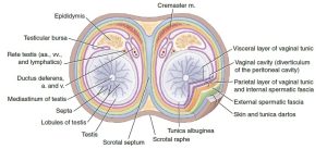
- Transverse section through the scrotum, testes, epididymis. 9
-
-
Cranial view of the opened scrotum of a bull;
the investments of the testis have been partly dissected. 8

-

- Ruminant testis and scrotum. 38
-
- Testis and spermatic cord of the bull. 7
Observe: Note the orientation of the testes relative to the body, there are species differences between the stallion, bull, and boar. Check an image before doing your first castration in any species!
Reference wet specimens and prosected cadavers to identify the following structures: septum of the scrotum, raphe of the scrotum, tunica dartos (know conceptually), external spermatic fascia, internal spermatic fascia, parietal vaginal tunic, vaginal cavity, visceral vaginal tunic, tunica albuginea testis, and testicular parenchyma.
Epididymis
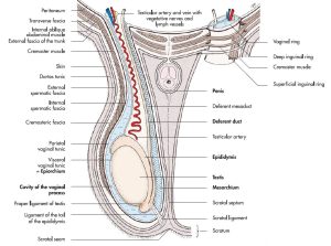
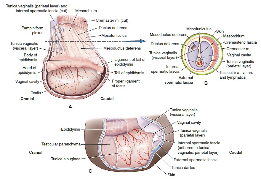
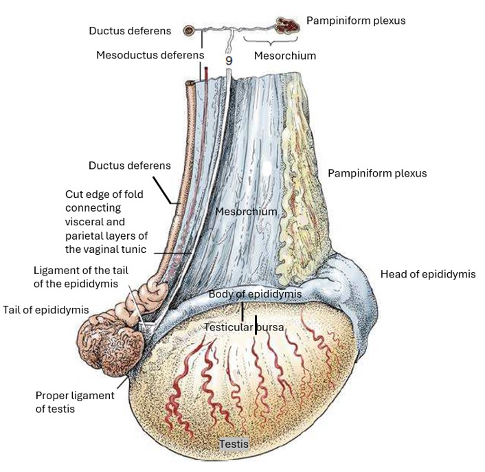
The head of the epididymis is fused to the testis, where testicular vessels enter the testis. The tail of the epididymis is attached to the testis by the proper ligament of the testis. The body of the epididymis is only attached along the medial side, leaving a space between the epididymis and testis, the testicular bursa, which is open laterally. The ligament of the tail of the epididymis attaches the tail of the epididymis to the parietal vaginal tunic. At the tail of the epididymis the ductus epididymis becomes continuous with the ductus deferens. The scrotal ligament represents a third ligamentous tissue and attaches the internal spermatic fascia to the inner scrotal wall.
-

-
Equine testis, epididymis, and associated tissues. (A) Lateral view—parietal part of tunica vaginalis and internal spermatic fascia cut.
(B) Cross-sectional view of spermatic cord, vaginal cavity, and surrounding tissues. (C) Tissue layers covering testis. 9
-

- Lateral view of the right testis of a stallion. 8
-

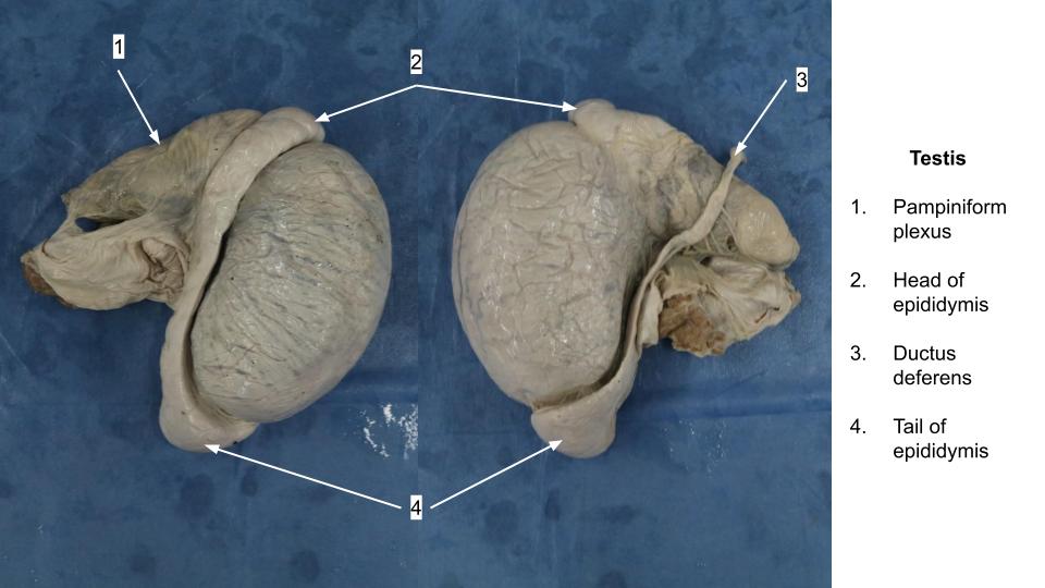
- Testis
Observe: the epididymis and its attachments: head, body, and tail of epididymis, testicular bursa, proper ligament of the testes, ligament of the tail of the epididymis, scrotal ligament. If the testicle is still connected to the scrotum, identify the scrotal ligament as the attachment between the scrotum and the internal spermatic fascia. If it has been broken, recognize that in order to exteriorize the testicle from the scrotum during a castration, the scrotal ligament must be broken in order to exteriorize the testicle to be resected. Know what structures each ligament attaches.
Spermatic cord and cremaster muscle
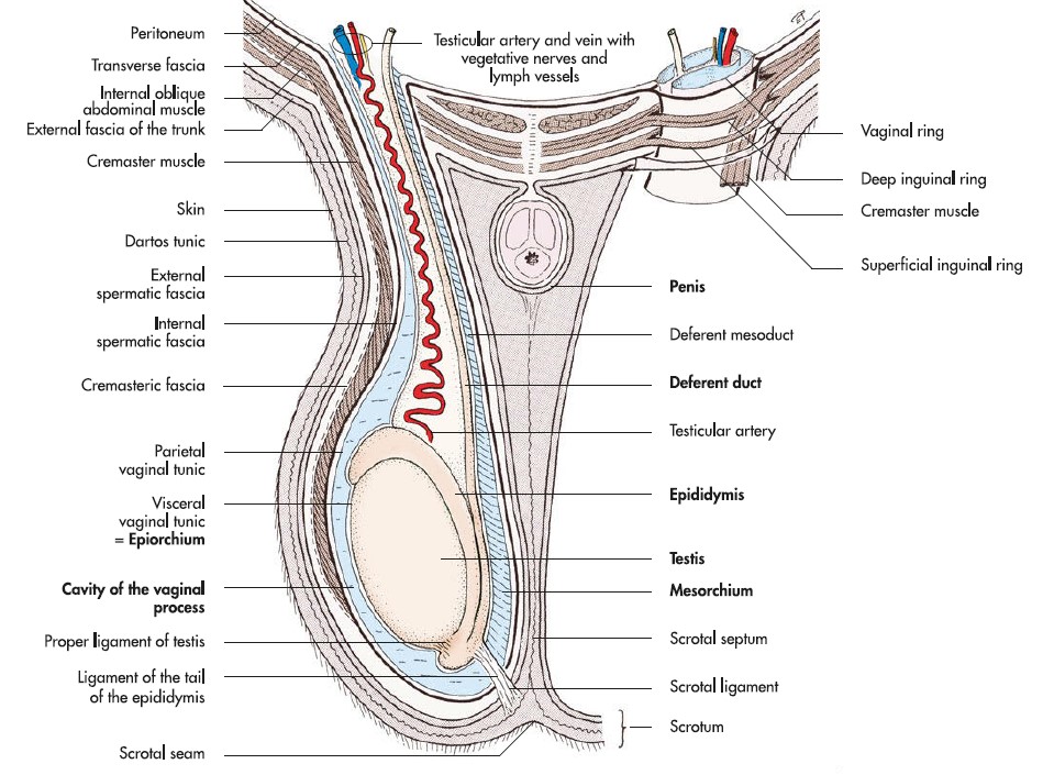
There are varying definitions of what structures make up the spermatic cord amongst anatomy texts (who knew!:)). We will go with the following – the ductus deferens; the a/v of the ductus deferens; and their connecting fold, the mesoductus deferens; the testicular a/v; smooth muscle fibers; lymphatics and testicular (sympathetic) nerves; and their connecting fold, the mesorchium. The cremaster m. (from the IAO m.) lies outside the parietal vaginal tunic and is thus not part of the anatomic spermatic cord. However, clinically, the cremaster m. and vaginal tunic are usually included as part of the ‘spermatic cord’.
As the testicular artery approaches the testis, it becomes highly tortuous, forming a spring- like configuration. Surrounding that part of the artery, the testicular vein forms an elaborate, mesh-like plexus, the pampiniform plexus.
-
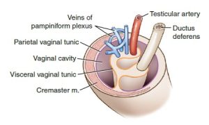
- Spermatic cord including vessels and deferent duct within the vaginal tunic. 9

-
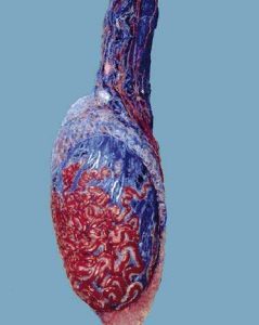
- Corrosion cast of bull’s vessels, arteries in red and veins in blue, within and on the testis and the pampiniform plexus. 8

-
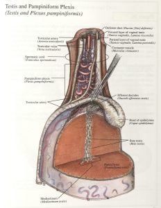
- Ruminant testis and pampiniform plexis. 38

Observe: ductus deferens, a/v of the ductus deferens (may not actually visualize), mesoductus deferens, testicular a/v (know that testicular n and lymph drainage is present), pampiniform plexus, mesorchium, spermatic cord (clinical vs. anatomic), cremaster m.
The testis and epididymis drain directly via the testicular lymphatics to the medial iliac lnn. (ie not to the superficial inguinal lnn., which the scrotum and external spermatic fascia drain to).
Clinical relevance:
Unless there is concurrent involvement of the scrotum, orchitis does not produce enlargement of the superficial inguinal lnn.
Pelvic portion of ductus deferens, Accessory sex glands
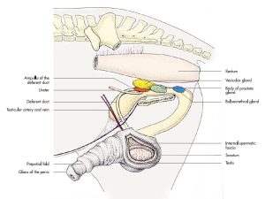
Follow the ductus deferens, attached to its mesoductus deferens into the pelvic cavity and to where it meets its opposite number in the genital fold, between the rectum and bladder (identified back when we discussed peritoneal pouches). Within the genital fold and further caudally each ductus deferens (in the stallion and ruminants) widens considerably due to ampullary glands located in their walls, and this part is referred to as the ampulla of the ductus deferens. The ampullary glands are 1 of the 4 accessory sex glands in the stallion, bull and buck.
The other three accessory sex glands are found in all the ungulates and are the vesicular glands, the prostate, and the bulbourethral glands.
FYI – Not far into the pelvic urethra there is a ridge of tissue projecting from the dorsal wall, the colliculus seminalis. Ejaculatory ducts have openings on this ridge, and provide a common outflow duct for the ductus deferens and vesicular gland. On the lateral sides of the colliculus seminalis, there are numerous openings of the prostatic ducts of the compact portion of the prostate (stallion, bull, boar).
Clinical application:
Because complete development and maintenance of the accessory sex glands are androgen-dependent in the domestic mammalian species, size of these glands (as determined by rectal palpation or ultrasound) may be used to distinguish between a castrated male and a testosterone-producing cryptorchid male.
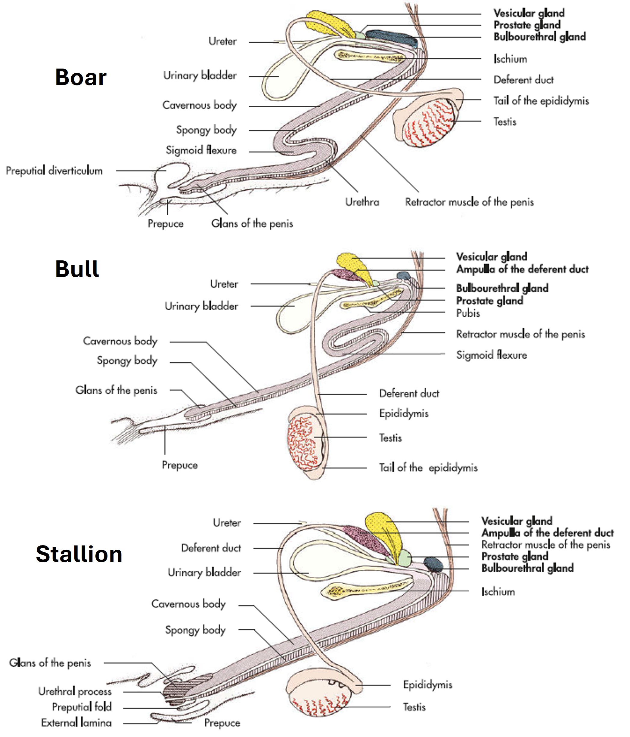
Comparative
-

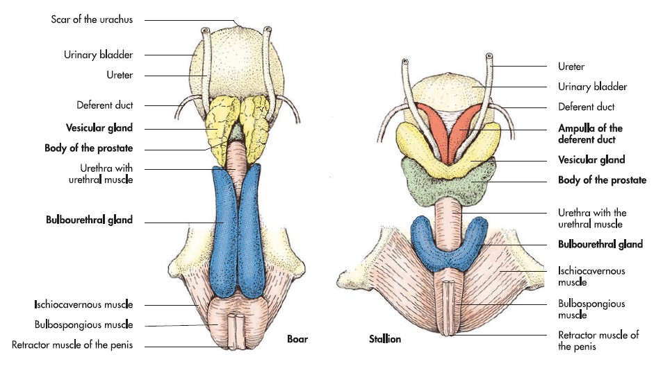
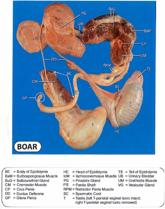
- Accessory genital glands of the boar and stallion.7
-
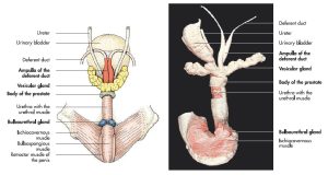
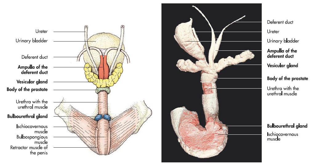
- Accessory genital glands of the bull. 7
Bull & Buck
-
- Accessory sex glands of the bull. 37
-
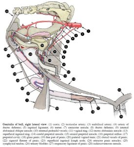
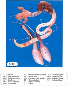
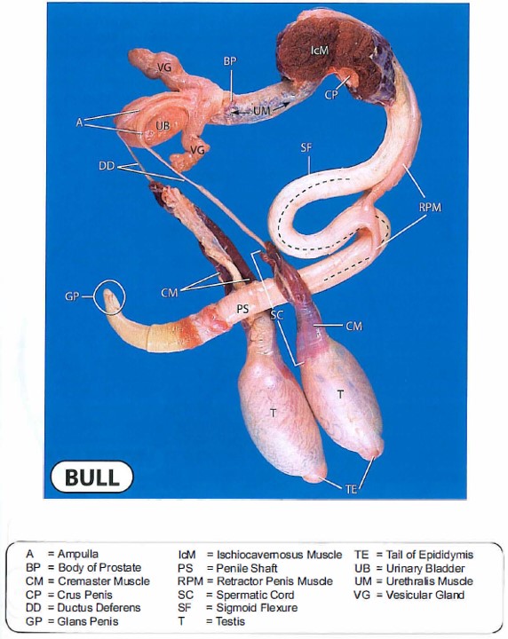
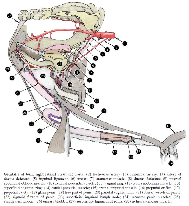
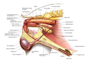
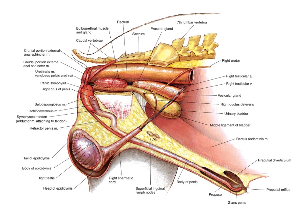
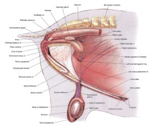
- Genitalia of bull, right lateral view/ 2
-

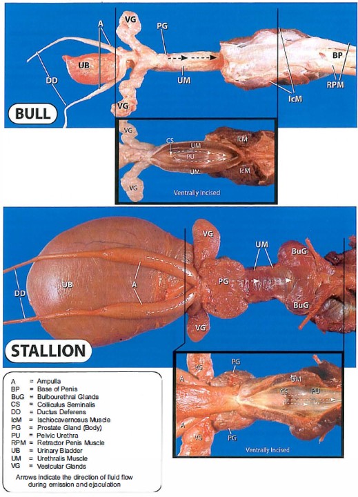
- Dorsal view of the accessory sex glands of the bull and stallion. 37
-
- Accessory sex glands of the bull. 38

-
- Prostate of the bull. 38
-

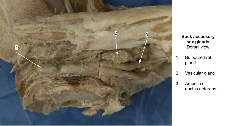
- Buck accessory sex glands
Stallion
-
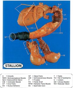
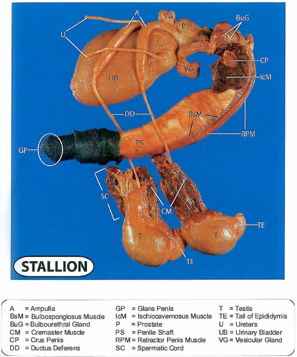
- Male genital organs of the stallion. 7

-
- Accessory sex glands of the stallion. 37
-
-
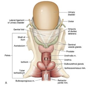
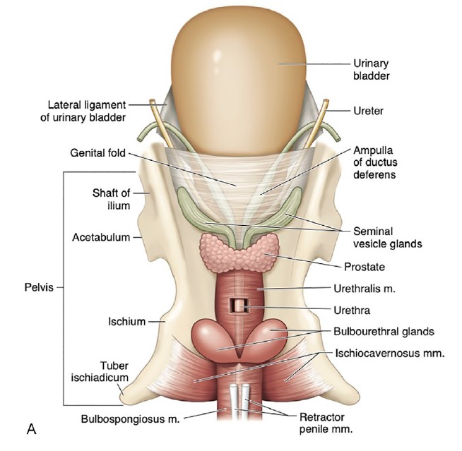
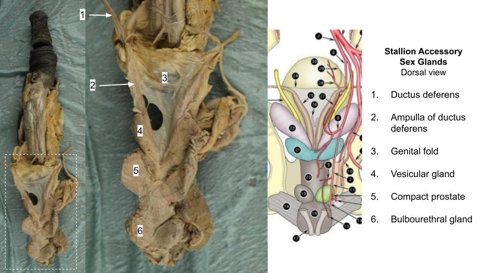
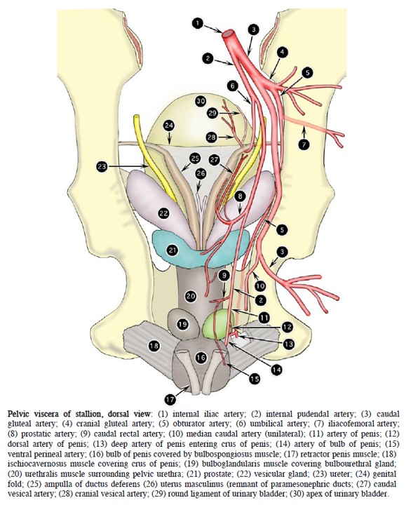
Genitourinary tract of the stallion (dorsal view), emphasizing the relationship of the accessory sex glands to
the urinary bladder and pelvic canal. 9
-

- Stallion accessory sex glands
Boar
-
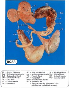
- Accessory sex glands of the boar. 37
-
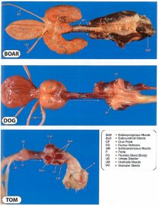
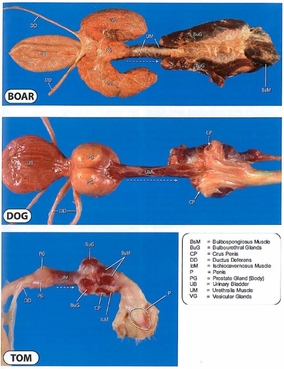
- Dorsal view of the accessory sex glands of the boar, dog and tom. 37
-
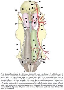
- Pelvic viscera of boar, dorsal view. 2

-
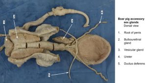
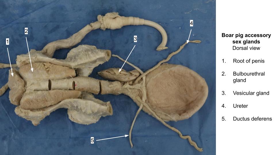
- Boar accessory sex glands
Observe: For the following, focus on identifying the accessory sex glands in wet specimens and instructor prosections.
Ampulla of the ductus deferens: Within the genital fold and further caudally each ductus deferens (in the stallion and ruminants) widens considerably due to ampullary glands located in their walls
Vesicular glands: Identify the vesicular glands lying lateral and caudal to the ampullae of the ductus deferens. In the stallion, the vesicular glands are smooth, bladder-like structures and therefore clinically are often called the seminal vesicles. The vesicular glands of the bull consist of firm, highly folded tubular structures that appear to be lobulated. In the mature boar, the vesicular glands are represented by two large, pyramid-shaped masses.
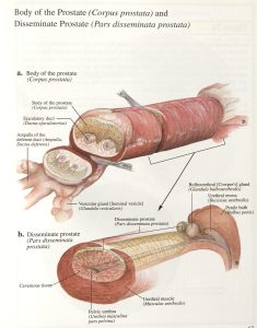
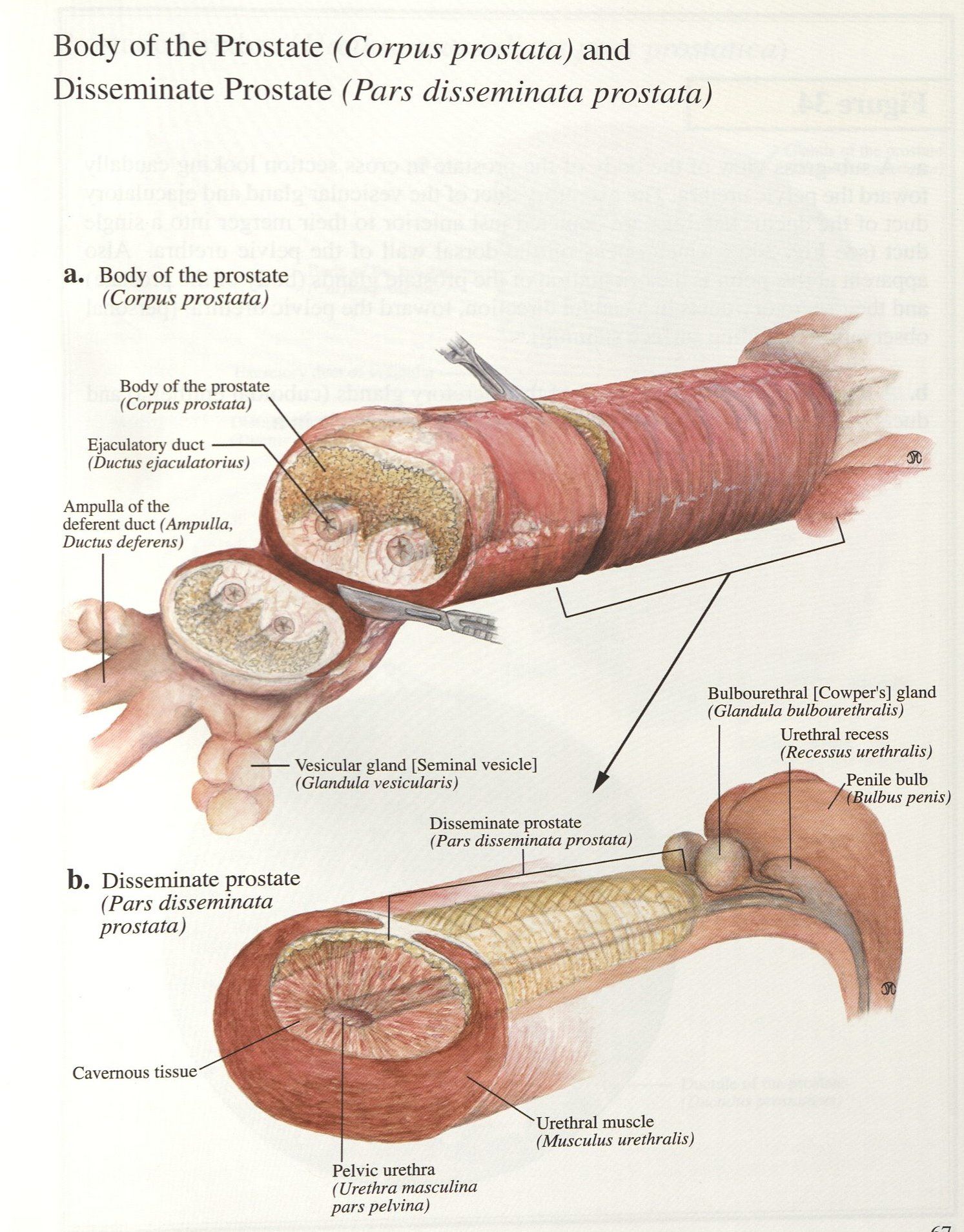
Prostate gland: Identify the prostate located immediately caudal to the vesicular glands. The prostate of the stallion is distinctly bilobed, complete with a connecting isthmus, and consists entirely of a compact part which flanks the junction of bladder and urethra. The bovine prostate consists of both a compact part (body) and a disseminate part. The body is not divided into distinct lobes. The disseminate part is spread along the pelvic urethra. Small ruminants only have a disseminate prostate gland. In the buck, the disseminate prostate completely surrounds the pelvic urethra, whereas in the ram, it is limited to the dorsal and lateral aspects. In the boar, the compact part (body) of the prostate is small, with most of the gland being formed by the disseminate part.
Bulbourethral glands: Identify the paired bulbourethral glands, located dorsolaterally to the urethra near the ischial arch. FYI: the bulbourethral glands are covered by the bulboglandularis m. in the stallion. The bulbourethral glands are spectacularly developed in the mature boar, representing large cigar-shaped structures either side of the pelvic urethra.
Review of the Internal bladder, pelvic urethra
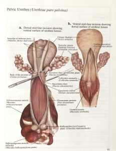
Observe: Before we continue over the ischial arch into the penis and penile urethra, let us back up briefly to confirm a few bladder and pelvic urethra things. Review wet specimens with opened urinary bladder and incised pelvic urethra to study the following anatomy. Recall that the outer wall of the urinary bladder is lined by a muscle called the detrusor muscle, which is under parasympathetic control via the pelvic nerves. Locate the openings of the ureters in the dorsal wall of the bladder neck and the internal urethral orifice at the junction of bladder and pelvic urethra. This opening is controlled by the internal urethral sphincter innervated by the hypogastric nerves (sympathetic control). Join these three holes (two ureteral openings and the internal urethral orifice by an imaginary triangle on the dorsal bladder wall – you have outlined the trigone of the bladder (vesicular trigone). Note the striated urethralis m./external urethral sphincter surrounding the pelvic urethra which is caudal to the neck of the bladder. The urethralis muscle is innervated by the pudendal nerves (voluntary somatic innervation).
-
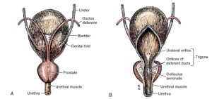
- Bladder and prostate. A, Dorsal aspect. B, Ventral aspect, partially opened on midline. 1

-
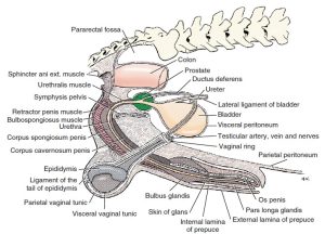
- Diagram of peritoneal reflections and the male genitalia. 1

-
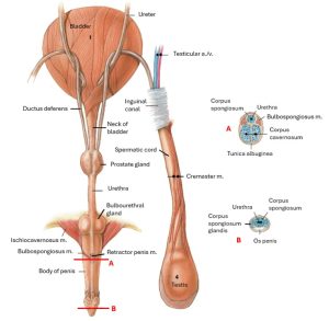
- Male genital system of the cat with some muscles and prepuce removed, dorsal view. 4

Penis, associated muscles and penile urethra
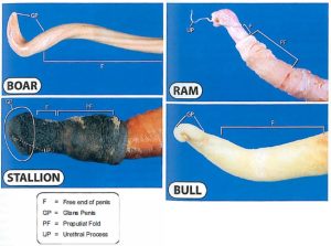
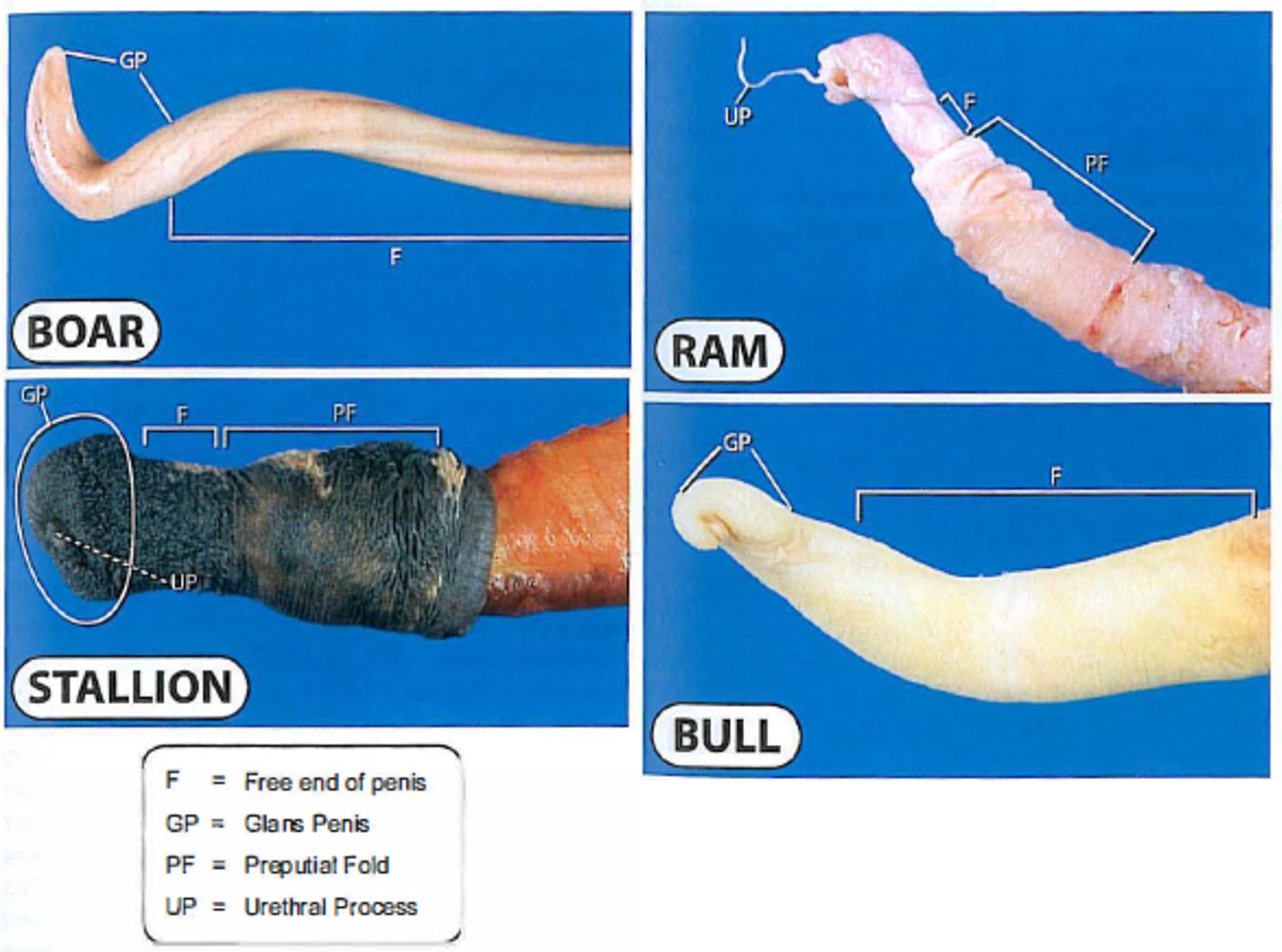
Before dissection, consider the following broad classification of penes:
1. Fibroelastic type: pig and ruminant; highly fibrous structure; it is firm, even in the quiescent state.
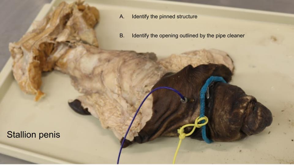
2. Musculocavernous type: stallion; contains more smooth muscle in its wall and is soft in the quiescent state.
The penis has 3 subparts – the root, the body, and the glans. The free part of the penis is that part of the penis which extends beyond the prepuce when erect. Understanding and identification of cross sectional penis anatomy is critical (successful phallectomy requires that one knows the appropriate tissues to suture together to prevent postoperative hemorrhage and to leave a urethral opening!).
-

-
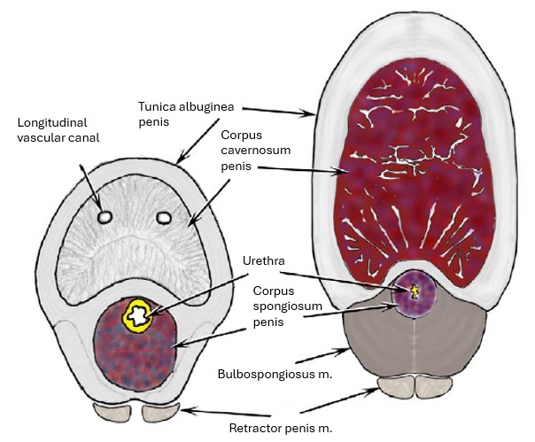
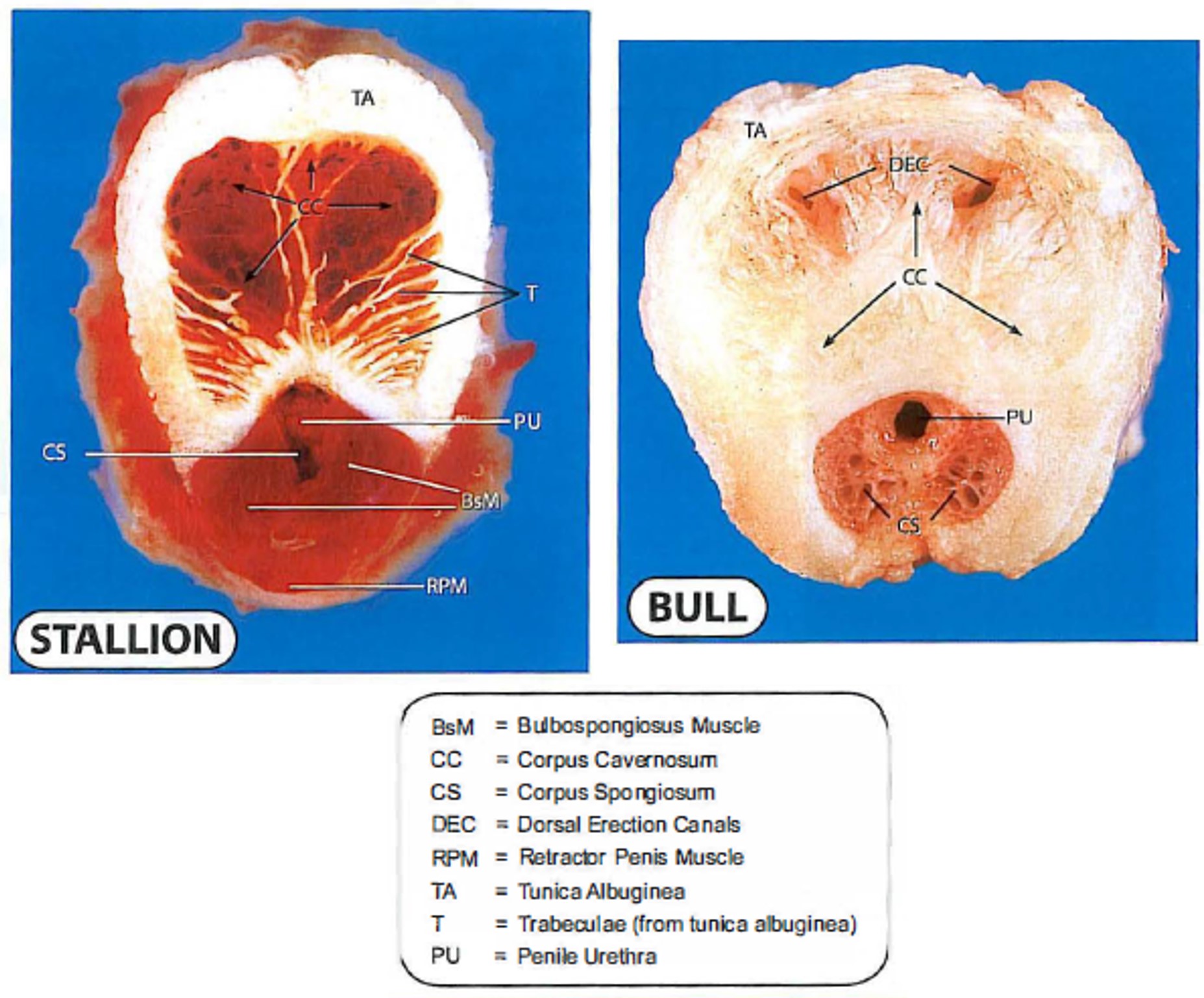
Cross section through body of penis of bull (left) & stallion,
showing comparison of fibroelastic (bull) and musculocavernous (stallion) types. 2
-

- Comparison of fibroelastic and musculocavernous penises. 37
-

- Bull penis cross-sections 38
Observe: For all penis anatomy, be sure to know the structures on cross-section throughout the length of the penis, as well as what is seen on the surface of the intact structure. Refer to wet specimens for identification of all structures in this section, in addition to what is studied on the cadavers.
Root of penis
Just caudal to the level of the bulbourethral glands, the pelvic urethra curves around the ischial arch and continues into the root of the penis as the penile urethra. At this same location, the urethralis m. continues as the bulbospongiosus m.
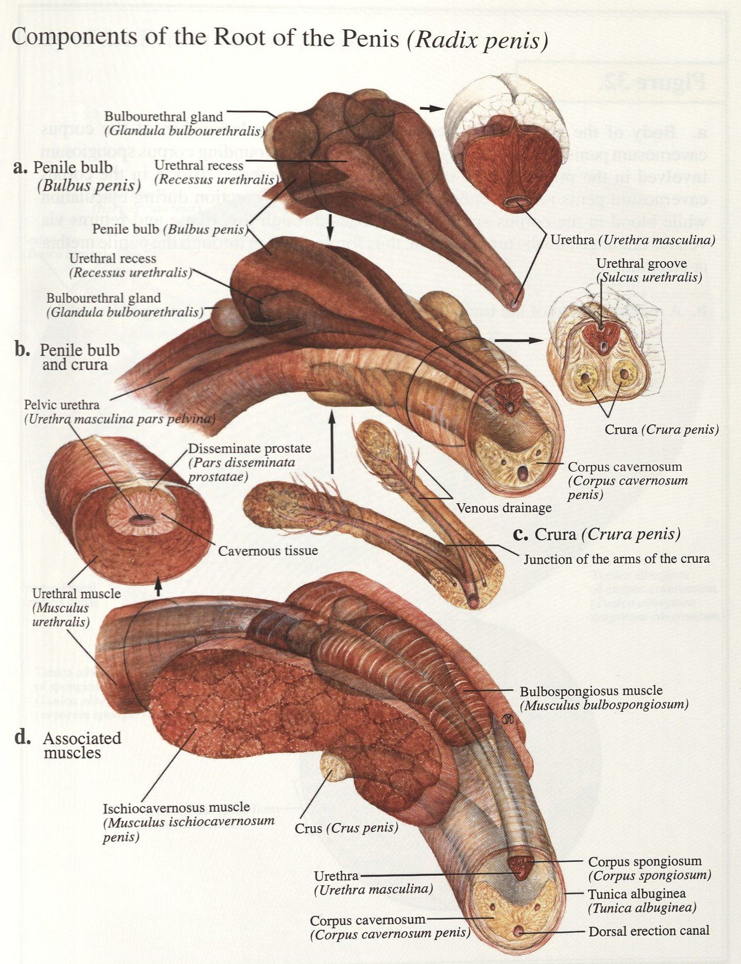
The root of the penis consists of a trio of converging structures:
1) bulb of the penis – on midline, surrounded by the bulbospongiosus m. and composed of corpus spongiosum penis erectile tissue enveloping the urethra. Note, the bulbospongiosus m. of the ruminant and pig does not continue very far distally onto the body of the penis; however, in the horse, it extends all the way to the free part of the penis.
2) and 3) right and left crura of the penis. Each crus originates directly from the lateral ischial arch on each side. A crus is composed of corpus cavernosum penis erectile tissue encapsulated by the dense, strong connective tissue layer, the tunica albuginea penis, and the crus is covered by ischiocavernosus m.
-
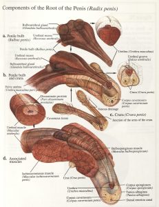
- Root of the penis of the bull. 38
-
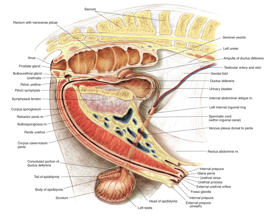
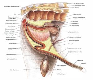
- Reproductive organs of the boar. (Note: sigmoid flexure inaccurately located too caudally here.)30
-
- Reproductive organs of the buck. 30

Observe: Identify the structures described above in the ungulate species: root of penis, bulb of penis, corpus spongiosum penis, bulbospongiosus m., left and right crus (pl. crura) of the penis, corpus cavernosum penis, tunica albuginea penis, ischiocavernosus mm.
-
- The bovine penis and its muscles: caudolateral view. 8

-
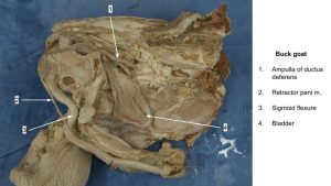
- Buck sigmoid flexure

Observe: Identify the paired retractor penis muscles (smooth muscle). These muscles originate from one or more caudal vertebrae, pass around the rectum and anus and then converge and continue down the caudal and ventral surface of the bulbospongiosus m. and penis. In ruminants and the pig, the retractor penis mm. insert on the ventral surface of the distal (i.e. second) bend of the sigmoid flexure (small ruminants) and further beyond on the free part of the penis (bull-see below). In the horse, these muscles insert at the free part of the penis.
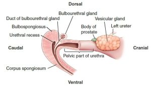
Instructor cadaver dissection only: In bull, buck and boar, consider removing the ischiactic tuberosity first of all, then make a lateral incision starting at the caudal end of the pelvic urethra and on into the penile urethra, i.e. cut through the bulbospongiosus m. and spongiosum erectile tissue to open the urethra to about 5-10 cm distal to the ischial arch. At the ischial arch, probe the dorsal urethral wall to locate the blind diverticulum, the urethral recess. Duct openings of the bulbourethral glands may be identified in the wall of the recess.
Observe: The urethral recess is present in the bull, buck, boar, ram, and male llama. In a prosected cadaver, at the ischial arch, probe the dorsal urethral wall to locate the blind diverticulum that is the urethral recess.
Clinical Application: Urinary catheter placement and the urethral recess
The urethral recess complicates urinary catheterization in ruminants and pigs; the catheter may lodge in the recess.
-
- The urethral recess prevents retrograde passage of a catheter in cattle, small ruminants, and pigs. 9

Body of penis
The body of the penis begins at the midline convergence of the left and right crura and continues to the glans. Also at the body, the corpus spongiosum erectile tissue has tapered from the bulb and continues through the body (and glans) directly surrounding the urethra. The body of the penis is anchored to the ventral pelvis by a pair of short ligaments, the suspensory ligaments of the penis. Recall, in the horse, bulbospongiosus m. is present through to the free part of the penis (see below). In ruminants and the pig, the body is characterized by the presence of a sigmoid flexure. During erection, the sigmoid flexure straightens out, and the fully extended penis becomes more rigid. In ruminants, the sigmoid flexure lies caudal to the neck of the scrotum (i.e. post-scrotal) whereas in the boar it is located cranial to the scrotum (i.e. pre-scrotal).
-

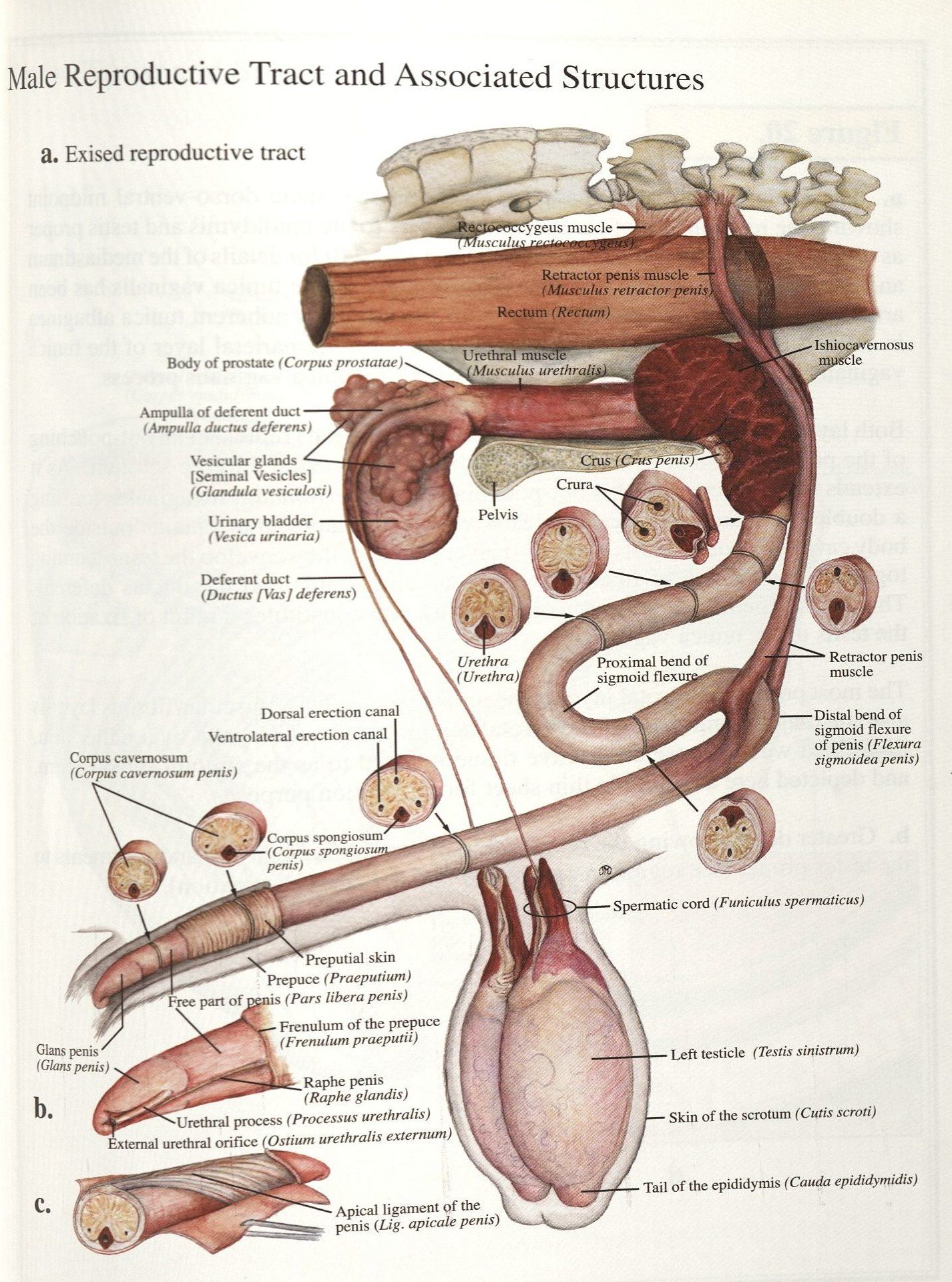
- Reproductive organs of the stallion. 30
-
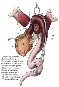
- Reproductive organs of the bull. 30

-
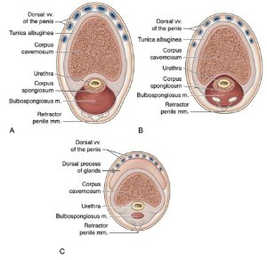
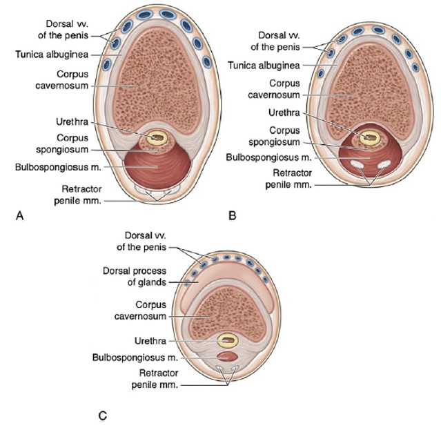
- Cross-sectional anatomy of the horse penis: (A) distal to the root; (B) midshaft; and (C) free end. 9
-
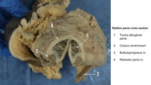
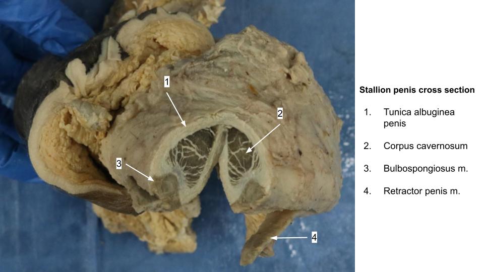
- Stallion penis cross section
Observe: Identify the body of the penis in the ungulate species, both on the intact penis and in cross-section. Also identify the sigmoid flexure in the ruminants and pig.
Clinical Relevance: Urinary calculi and the sigmoid flexure
The sigmoid flexure is a frequent site for urinary calculi to obstruct, particularly in small ruminants. In breeding bulls, penile rupture and hematoma may occur at the sigmoid flexure (distal bend). A perineal urethrostomy is performed in the body of the penis due to the accessible location of the urethra in the perineal region. Urethrostomy allows for urination through a newly created opening. This may be necessary to treat urethral obstruction further distal e.g. a stone caught at the sigmoid flexure.
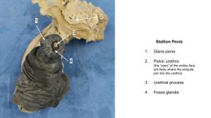
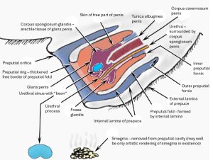
Glans penis and terminal urethra
The glans penis is the distal portion of the penis, consisting of the corpus spongiosum glandis erectile tissue. In the horse, this large erectile body overlaps (caps) the distal part of the corpus cavernosum penis and forms a blunt head with a circumferential ridge, the corona glandis. In older stallions, there may be numerous papillae associated with the corona glandis. Immediately proximal to the corona glandis, the glans is constricted, forming the collum glandis (recall colli refers to ‘neck’). The glans is relatively small in the cat, pig, and ruminant. In the ruminant, the glans is an asymmetrical cap cushioning the tip of the penis. Recall that corpus spongiosum erectile tissue surrounds the penile urethra for its full length. Differentiate the corpus spongiosum glandis from the corpus spongiosum.
-
- Extended penis of the stallion and its relationship to the prepuce. Inset: free end of the penis with the skin removed. 9

-
- Glans penis by species. 37
-
- Stallion penis

Instructor Equine Cadavers Only– assuming the embalmed penis is extrudable or already extruded, make a full thickness vertical midline cut along the shaft of the free part of the penis, for about 5-8 cm. This incision should divide the urethra.
Observe: On an equine penis which has had the glans penis incised longitudinally, spread apart the two cut halves and identify the three erectile tissues, plus the tunica albuginea penis and the urethra. Examine these structures on cadavers and wet specimens.
On all ungulates identify the glans penis, corpus spongiosum glandis, and cross sectional and longitudinal anatomy through the glans penis.
Urethral Process (part of the glans penis)
The penile urethra terminates at the external urethral orifice. In the horse and ruminants (not pig) the urethra ends as a distal extension, the urethral process. In the horse, the urethral process is in the center of the glans, and is surrounded by a recess, the fossa glandis. In the dorsal wall of the fossa glandis an opening leads to a bean-shaped diverticulum, the urethral sinus. Secretions and cellular debri of the prepuce, called smegma, can accumulate in the urethral sinus of the horse and form a hard ‘bean’. The urethral process is narrow and short in the bull, and narrow and longer in the buck and ram.
-
- Glans penis by species. 37
-

- Free part of the penis of a stallion. 7
-
- Prepuce and glans of the penis of the stallion. 7
-
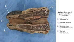
- Stallion penis transection
-
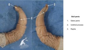
- Bull penis
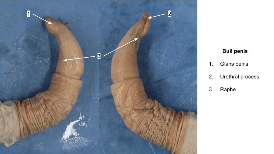
Observe: Identify these structures on cadavers and in wet specimens: external urethral orifice, urethral process (not present in pig), fossa glandis (horse), urethral sinus (horse), smegma (when hardened forms a “bean’) in horse (may not identify but be aware of).
Clinical Relevance: Bean formation and urinary obstruction
When a very large bean is present in the urethral sinus, it may compress the urethral process and interfere with urination; this may be a cause of dysuria in the male horse. Beans are removed as part of a general preputial cleaning in male horses.
Note from Lindsey: “Beaning” is an unusual colloquial way to describe removing a bean from the urethral sinus during sheath cleaning, but this video had the best view. Also, please wear gloves; the smell of smegma is difficult to remove.
Prepuce and Free Part of the Penis
Ruminants and Boars
The external lamina of the prepuce is the hair-covered skin. At the preputial orifice, it is continuous with the internal lamina of the prepuce (hairless skin that lines the preputial cavity). The internal lamina continues caudally to a preputial fornix, where, except in the horse, it immediately reflects back onto the penis for a short distance. Here it is loosely attached to the underlying penis and the free part of the penis (that part of the penis distal/cranial to where the internal lamina connects to the penis) is generally protected in the created preputial cavity.
Instructor Dissection Only: On the bull, boar and buck, beginning at the preputial orifice, open the preputial cavity using a lateral incision to expose the free part of the penis.
Observe: In the newborn calf and in males that were castrated prior to puberty, the internal lamina of the prepuce typically adheres to the skin of the free part of the penis. In the neonatal bull calf (likely more notable) and buck (rudimentary), note the frenulum of the prepuce, which is the fold of skin connecting the raphe of the prepuce with the raphe of the penis.
Clinical Relevance: Persistant Penile Frenulum
A persistent preputial (often referred to as penile) frenulum, where the internal lamina did not separate from the penis, results in the penis being anchored to the prepuce and, upon erection, the penis is strongly deviated by this tie. Surgical transection of the persistent frenulum solves the problem.
-
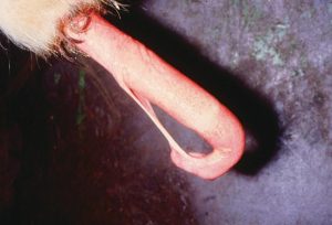
- Persistent penile frenulum of a bull. veteriankey.com

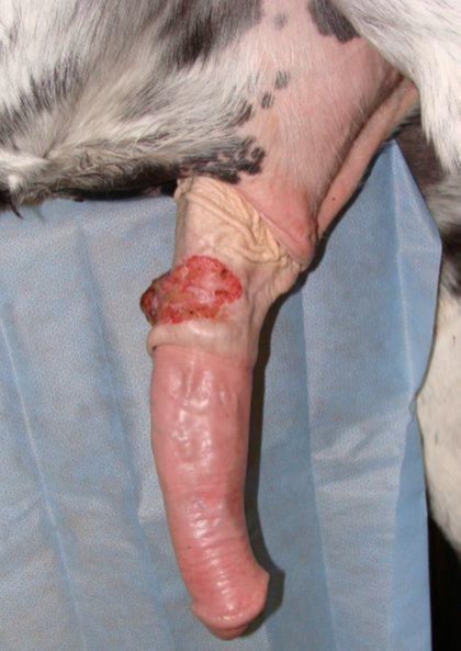
Clinical Relevance: Excess internal lamina/preputial prolapse (bulls)
The prepuce of some bulls has excess internal lamina. When the internal lamina everts out through the preputial orifice, this is called preputial prolapse, and the tissue is vulnerable to trauma. Surgical removal of excess internal lamina is corrective.
Read more: Modified_partial_posthectomy_surgery_for_chronic_p
-
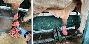
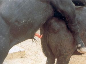
- Close examination of prolapsed prepuce; note edema and sunburnt preputial skin. 39

-

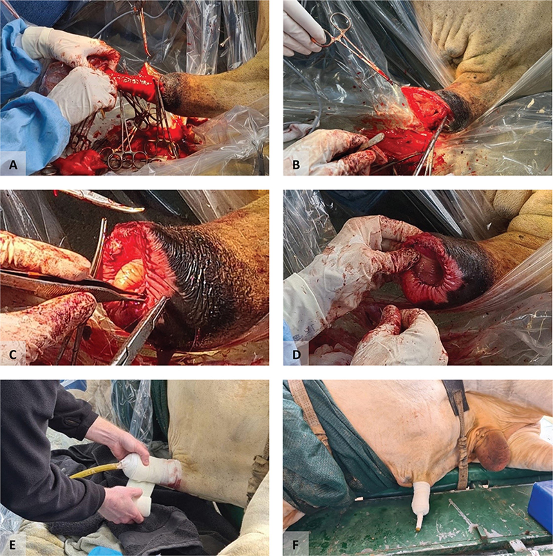
- Partial posthectomy surgery for chronic preputial prolapse in a bull. 39
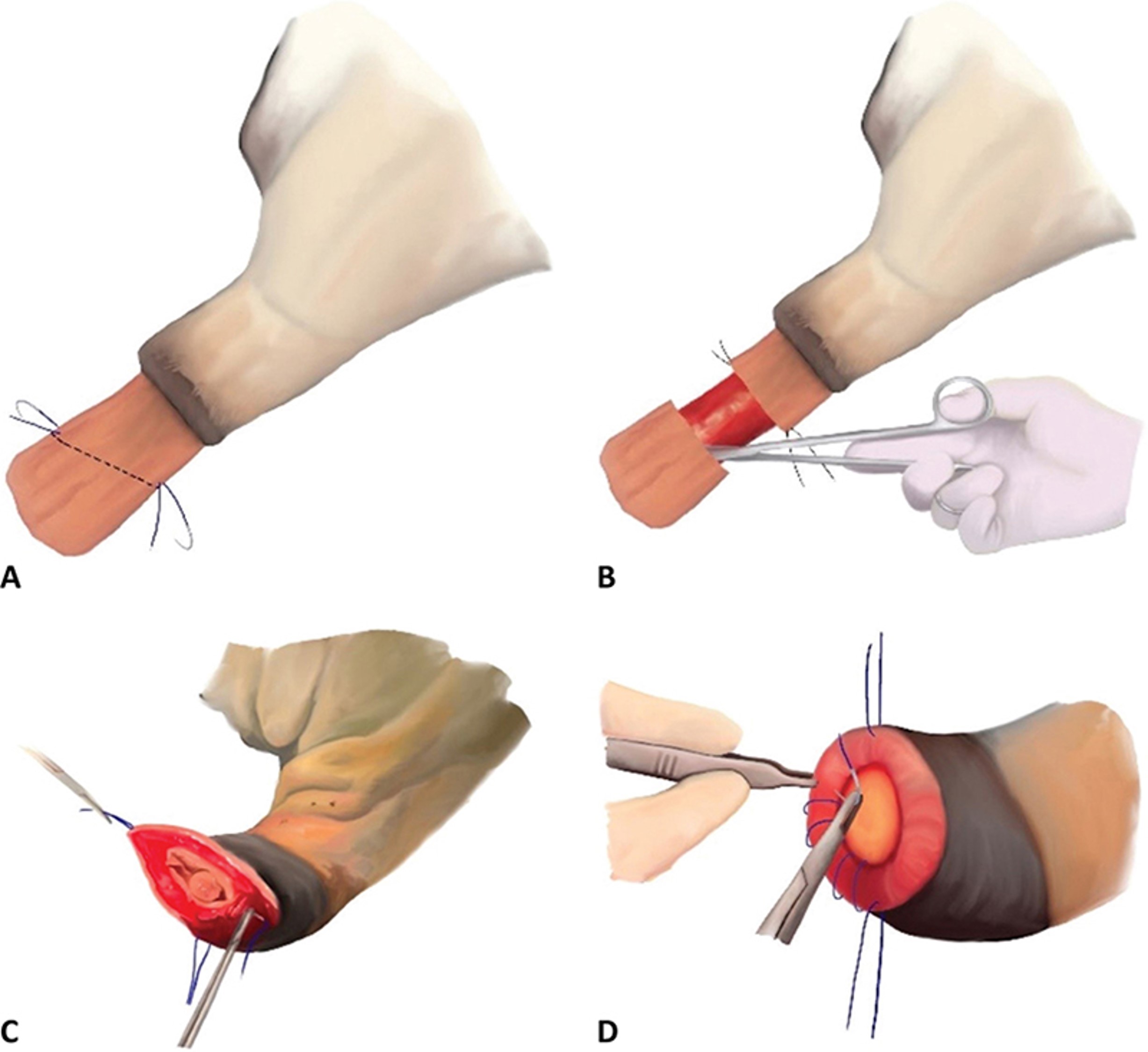
-
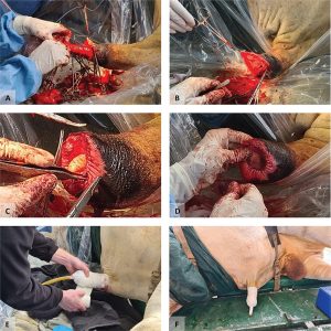
- Surgery to repair a chronically prolapsed prepuce in a bull. 39
Clinical Relevance: Spiral or Corkscrew Deviation
Note the characteristic twisting of the bovine penis that is accentuated further during erection. Although the bovine urethral process is on the left side of the penis, it is seen from the right side of the animal because of this normal left-hand spiral. The left hand (or counter clockwise) spiraling of the bovine penis increases at intromission. Premature spiraling of the penis can prevent intromission and is a recognized clinical problem known as spiral or corkscrew deviation.
In the boar, note that the free part of the penis is naturally twisted in a left-hand spiral, similar to that which occurs in the bovine penis at intromission. This spiral also becomes more distinct during erection.
-
- Spiral (corkscrew) penile deviation is not uncommon in mature bulls. nadis.org.uk

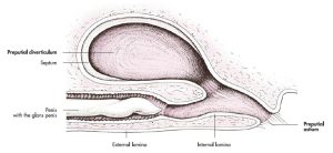
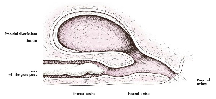
Preputial diverticulum of the boar
Observe: Examine the boar prepuce (prosected cadaver and/or wet specimen) along its dorsal wall and identify an out pouch. This is the preputial diverticulum. It is partially divided by a septum.
-
- Reproductive organs of the boar. (Note: sigmoid flexure inaccurately located too caudally here.)30
-
- Prepuce and glans of the penis of the boar. 7
Instructor cadaver dissection: Dissect and isolate the preputial diverticulum of the boar.
Clinical Relevance: Preputial diverticulum
The preputial diverticulum stores a secretion that lubricates the penis during copulation. It can become infected and need to be opened laterally for drainage. The combined debris (e.g., urine, desquamated epithelial cells) that accumulates in this diverticulum is thought to be responsible for the strong odor associated with the adult boar. Surgical removal of the preputial diverticulum has been used to reduce the odor and semen contamination in boars used for artificial insemination, and in pot-bellied pigs kept as pets. The penis may become lodged in the preputial diverticulum during erection, which inhibits protrusion of the penis. This is another reason why the preputial diverticulum may be removed in boars used for artificial insemination.
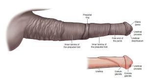
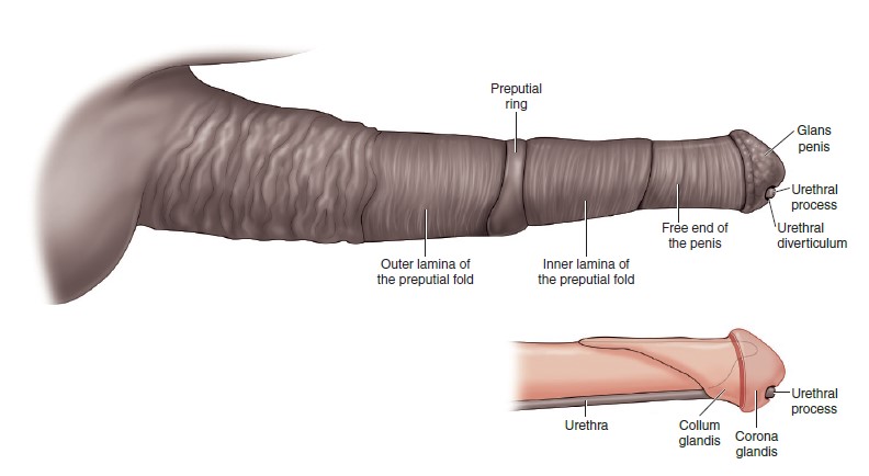
Horse
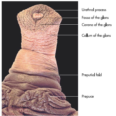
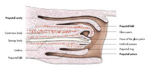
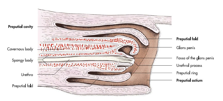
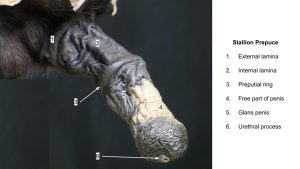
In the horse, there is a second fold of internal lamina, the preputial fold. This arrangement forms a telescopic prepuce. On reaching an outer preputial fornix, the internal lamina is reflected cranially almost back to the preputial orifice; it then turns caudally again and at this turn forms a thickened free border, the preputial ring. From here it continues caudally to an inner preputial fornix and then reflects cranially for a short distance onto the penis becoming continuous with the more tightly adherent skin of the free part of the penis. During erection, the entire preputial fold and initial part of the internal lamina reflect onto the body of the extended penis.
Like in the ruminant and pig, the free part of the penis is that part that lies within the preputial cavity and is covered by tightly adherent skin, i.e. skin that is not specifically part of the more loosely attached internal lamina of the prepuce. Therefore, the distal body of the penis and the glans of the penis contribute to the free part. Commonly, the glans and free part of penis are considered the same structure and they are not.
-

-
Extended penis of the stallion and its relationship to the prepuce. Inset: free end of the penis
with the skin removed. 9
-
- Prepuce and glans of the penis of the stallion. 7
-
-
Telescopic prepuce of horse, left lateral view with “accessories”
included at no extra charge. 2

-
- Stallion prepuce
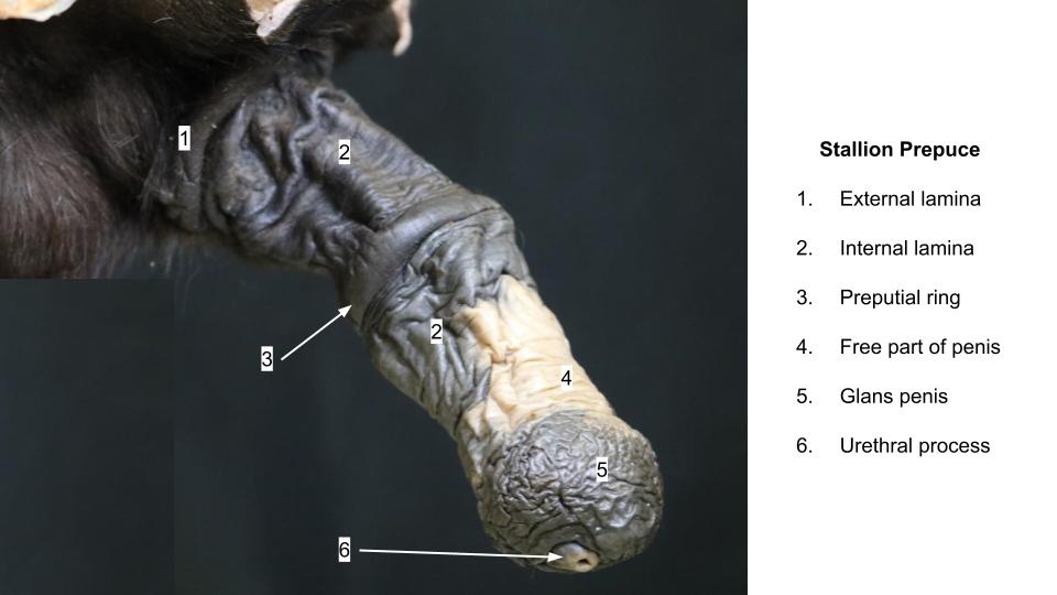
Observe: Identify the following parts of the prepuce and free part of the penis for all species: free part of penis, external lamina of the prepuce, internal lamina, preputial orifice, preputial fornix (outer and inner for horse). In the horse, also recognize that the prepuce is telescoping (horse), and identify the preputial fold (horse) and preputial ring (horse). Wet specimens will be particularly useful in the horse as the cadaveric prepuce is often stiff and hard to manipulate.
Clinical Relevance: Preputial Neoplasia/Squamous Cell Carcinoma in Geldings
Preputial and penile neoplasia (often SCC) is not uncommon in aged geldings.
Question: On what part of the prepuce is the squamous cell carcinoma located in this gelding?
Vasculature of the penis
Clearly, a considerable blood supply to the penis is required for the various erectile tissues to function physiologically. You might also guess that the musculocavernous penis of the stallion, in particular, requires extensive vascular input, and equally a relatively quick method of vascular drainage, to facilitate detumescence and avoid unnecessary duration of an extended penis (and consequently a possible increase risk of injury). Let us review and identify what we can of the vasculature.
Observe: We won’t focus on dissecting out all the branches of the artery of the penis but you should be familiar with their names and what they supply. These are the same for the ruminant and boar as they are for the carnivore. Although you will not be asked to identify all of the arteries of the penis, you may be asked what artery supplies which erectile tissue, and from what arteries they originate on any species, as follows: The continuation of the internal pudendal artery is the artery of the penis, which then gives rise to the artery of the bulb (to supply the bulb and CSP), the deep artery of the penis (to supply the crus and CCP), and then continues as the dorsal artery of the penis (to supply body, glans and prepuce). The dorsal a. of the penis travels with the dorsal v. and dorsal n. of the penis.
On the ruminant cadavers, identify the dorsal artery of the penis.
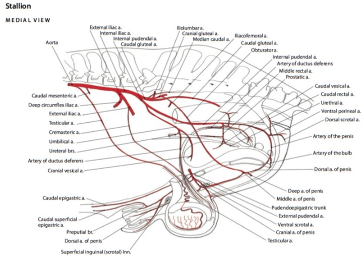
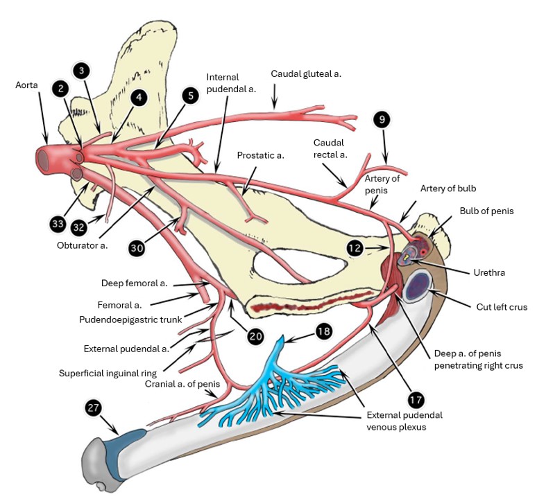
The horse has a more elaborate set up and its penis is supplied with blood from three major sources:
1) The internal pudendal artery continues as the artery of the penis, which gives rise to the artery of the bulb and continues as the small dorsal artery of the penis.
2) The obturator artery, a branch of the cranial gluteal artery, courses with the obturator nerve along the body of the ilium, exits the pelvic canal through the obturator foramen, and becomes the middle a. of the penis. The middle a. of the penis gives rise to the deep artery of the penis and also anastomoses with the dorsal a. of the penis traveling along the dorsal aspect of the penis.
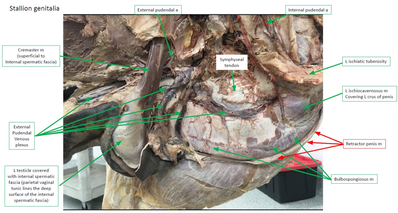
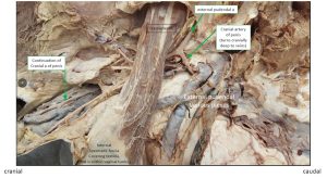
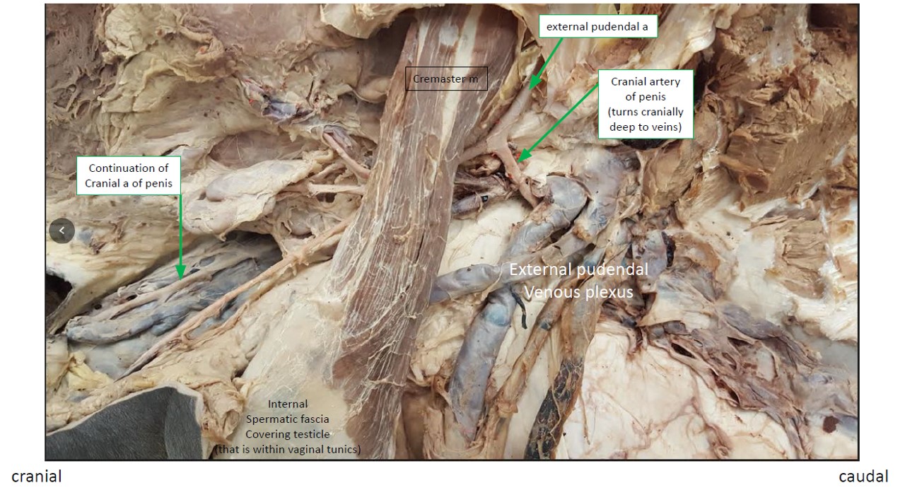
3) The external pudendal artery passes out through the inguinal canal and gives rise to the cranial artery of the penis, which also forms an anastomosis with the dorsal a. of the penis to boost blood supply to the cranial (distal) portions of the penis.
For rapid venous outflow from the erect penis, the stallion has developed an extensive external pudendal venous plexus to receive blood from the cavernous spaces of the penis immediately following dismounting. The venous plexus is drained by the large accessory external pudendal vein, which does not return through the inguinal canal but rather perforates the origin of the gracilis muscle and joins the deep femoral vein. The much smaller external pudendal vein enters the abdominal cavity through the inguinal canal, alongside the external pudendal artery, as is typical.
-
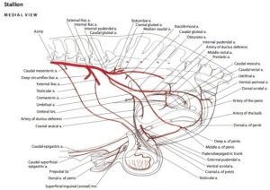
- Arteries of the reproductive tract of the stallion. 6
-
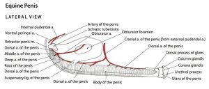
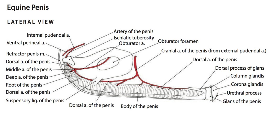
- Arteries of the penis of the stallion. 6
-
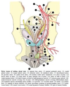
- Pelvic viscera of stallion, dorsal view. 2
Instructor Cadavers Only: On male equine cadavers dissect and isolate the dorsal artery and nerve of the penis and the cranial a. of the penis.
Observe: On the stallion, identify the dorsal artery of the penis, external pudendal a., and the cranial artery of the penis. Also identify the external pudendal venous plexus.
-
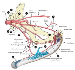
- Schematic representation of blood supply to penis of horse, left lateral view. 2
-
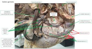
- Stallion blood supply
-
- Stallion blood supply closeup
Innervation of the penis
In all species, the dorsal nn. of the penis are the principal continuations of the right and left pudendal nerves.
-
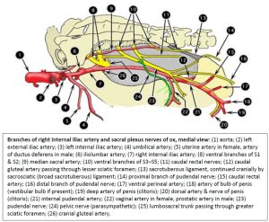
- Branches of right internal iliac artery and sacral plexus nerves of ox, medial view. 2
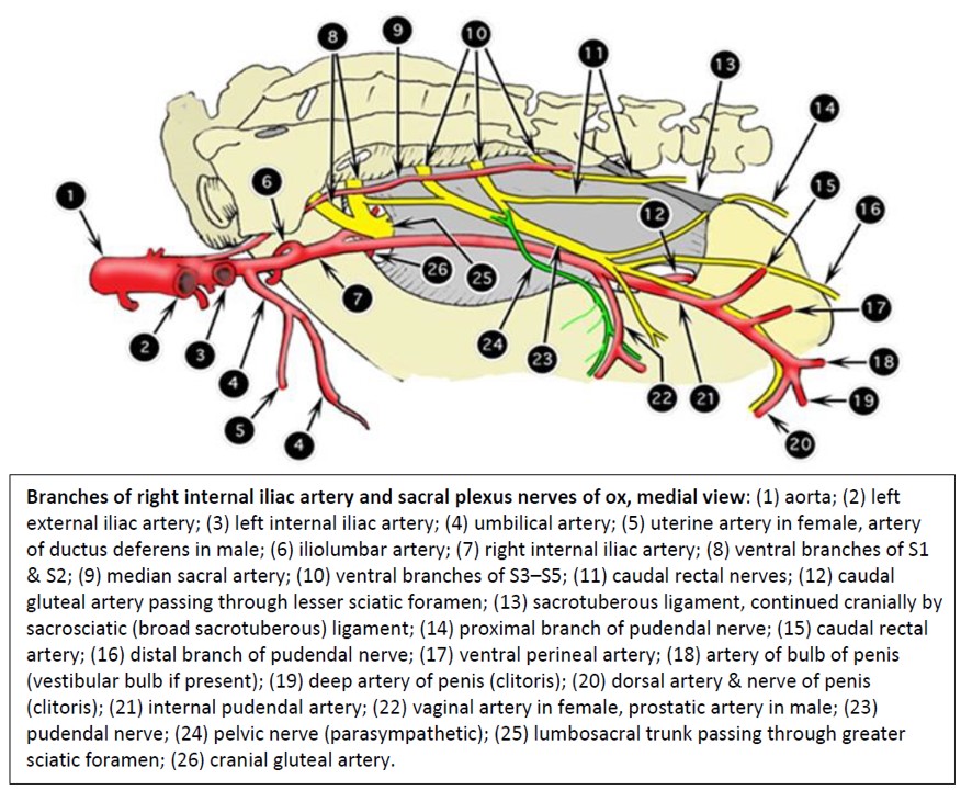
Observe: Identify the dorsal n. of the penis adjacent to the dorsal a. of the penis. Know that the dorsal nerve of the penis is a branch of the pudendal nerve and my be desensitized by performing a pudendal nerve block (see clinical relevance in the vasculature and innervation lab).
Review videos
Tracing the path of sperm – 3 min
Comparing accessory sex glands across species – 6 min
Comparing fibroelastic penises to musculocavernosus penises – 5 min
Stallion (horse) penis cross-section – 2.5 minutes
Buck (goat) pelvic cavity – 9 min
Bull calf pelvic cavity – 15 min
Urethral recess – ox – 2.5 minutes
Boar (pig) pelvic cavity – 30 min
Fresh Boar Tract – 7 min
Stallion (horse) prepuce – 10 min
Terms
Scrotum, spermatic fascia, testicles |
||
| Term | Features | Species differences/comments |
| Tunica dartos | ||
| Septum of the scrotum | ||
| Raphe of the scrotum | ||
| External spermatic fascia | ||
| Internal spermatic fascia | ||
| Parietal vaginal tunic | ||
| Vaginal cavity | ||
| Visceral vaginal tunic | ||
| Tunica albuginea testes | ||
| Testis (parenchyma of testes) | ||
| Epididymis | Head of the epididymis | |
| Body of the epididymis | ||
| Tail of the epididymis | Continues as the ductus deferens | |
| Proper ligament of the testis | The tail of the epididymis is attached to the testis by the proper ligament of the testis. | |
| Ligament of the tail of the epididymis | The ligament of the tail of the epididymis attaches the tail of the epididymis to the parietal vaginal tunic. | |
| Scrotal ligament | Attaches the internal spermatic fascia to the inner scrotal wall. | |
| Testicular bursa | ||
| Spermatic cord | Ductus deferens | |
| a/v of the ductus deferens | ||
| Mesoductus deferens | ||
| Testicular a/v/n | ||
| Mesorchium | ||
| Pampiniform plexus | ||
| Cremaster m. | Caudal slip of the internal abdominal oblique m. ID in the abdomen and adjacent to the anatomic spermatic cord. Note that many clinicians will include the cremaster m. as part of the spermatic cord (clinical term) but that anatomically it is outside of the internal spermatic fascia surrounded by external spermatic fascia and therefore not part of the anatomic spermatic cord. | |
Accessory sex glands |
||
| Term | Features | Species differences/comments |
| Ampulla of the ductus deferens | Horse and ruminants | |
| Vesicular glands | Ungulates only | |
| Compact prostate gland | All but small ruminant | |
| Disseminate prostate gland | Ruminants and porcine | |
| Bulbourethral gland | Not in dogs | |
Internal bladder, pelvic urethra |
||
| Term | Features | Species differences/comments |
| Urethralis m./external urethral sphincter | ||
| Pelvic urethra | ||
| Internal urethral orifice | Opening between trigone and prostate gland, in neck of bladder. | |
| Trigone of the bladder | ||
Penis, associated muscles and penile urethra |
||
| Term | Features | Species differences/comments |
| Fibroelastic type | ||
| Musculocavernous type | ||
| Penis | Root | Composed of bulb and crura |
| Body | ||
| Glans | ||
| Free part of the penis | ||
| Penile urethra | ||
| Bulb of the penis | Bulbospongiosus m. | |
| Corpus spongiosum | ||
| Crura of the penis | Corpus cavernosum penis | |
| Tunica albuginea penis | ||
| Ischiocavernosus m. | ||
| Retractor penis muscles | ||
| Urethral recess | Ruminants and Boar | |
| Body of the penis | Sigmoid flexure | Ruminants and Boar |
| Corpus cavernosum & Corpus spongiosum | ||
| Glans penis | Corpus spongiosum glandis | |
| External urethral orifice | Urethral process | |
| Fossa glandis | ||
| Urethral sinus | ||
| Smegma/bean | ||
Prepuce and Free Part of the Penis |
||
| Term | Features | Species differences/comments |
| External lamina | ||
| Preputial orifice | ||
| Internal lamina | ||
| Preputial fornix | Only one preputial fornix in all but the stallion (see below). | |
| Free part of the penis | ||
| Frenulum of the prepuce | Know Conceptually. Will have regressed in our cadavers. May still be present in calf. | |
| Raphe of the prepuce | Most readily observed on ruminants but present in all. | |
| Raphe of the penis | Most readily observed on ruminants but present in all. | |
| Preputial diverticulum | Boar | |
| Preputial fold | Horse only | |
| Outer preputial fornix | Horse only | |
| Preputial ring | Horse only | |
| Inner preputial fornix | Horse only | |
Vasculature and innervation of the penis |
||
| Term | Features | Species differences/comments |
| Internal pudendal artery | No ID – Know as origin of A. of Penis | |
| Artery of the penis | Artery of the bulb | No ID – understand what it supplies |
| Deep artery of the penis | No ID – understand what it supplies | |
| Dorsal artery of the penis | No ID – understand what it supplies | |
| External pudendal venous plexus | ||
| Dorsal nn. of the penis | ||