Lab 10A: Placenta and Mammary glands
Learning Objectives
- Identify the parts of a placenta and differentiate among the types of placentation of the domestic species.
- Identify the major features of mammary glands, comparing between species.
- Describe and identify the major blood supply, innervation and associated lymph nodes of the mammary glands.
Placentation
Observe: Refer to wet specimens and any available pregnant cadavers to identify placental structures of the carnivore and ungulate species.
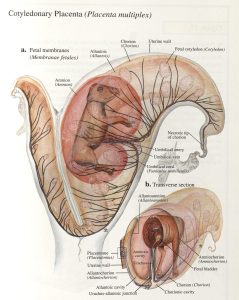
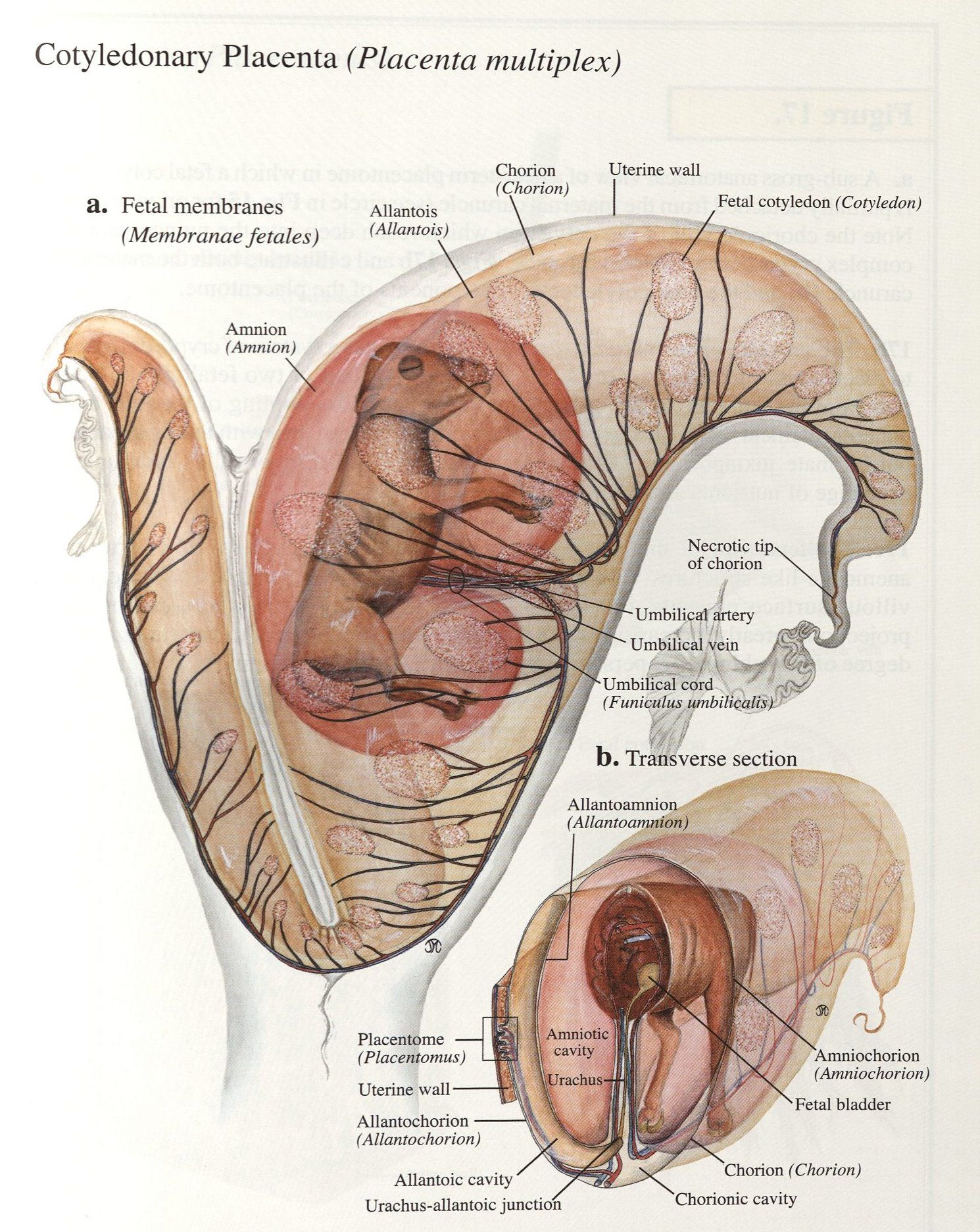
Fetal membranes include the chorion and allantois (i.e. the chorioallantois) and amnion, with the chorioallantois attached diffusely to the endometrium in the mare and sow and at cotyledons in the ruminant; the amnion directly surrounds the amniotic cavity containing the fetus bathed in amniotic fluid.
The placenta is a transient organ of metabolic interchange between the conceptus and the dam. It is also a transient endocrine organ. The placenta is composed of a fetal component derived from the chorion and a maternal component derived from modifications of the uterine endometrium. The discrete regions of contact between the chorion and the endometrium form specific zones of metabolic exchange. The placenta also produces a variety of hormones. This transient endocrine function is important for the maintenance of pregnancy and the induction of parturition (giving birth).
Chorionic villi are small, finger-like projections that are on the surface of the chorion. These tiny villi protrude away from the chorion toward the uterine endometrium. Placentas are classified according to the distribution of chorionic villi on their surface, giving each placental type a distinct anatomical appearance. Placentas may also be classified by number of tissue layers separating maternal and fetal blood.
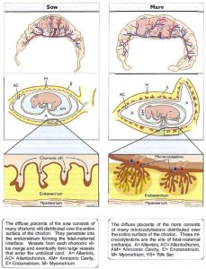
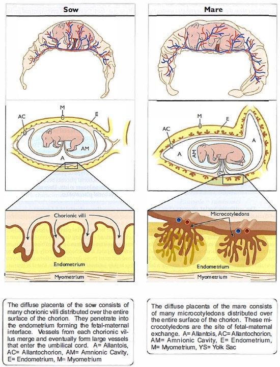
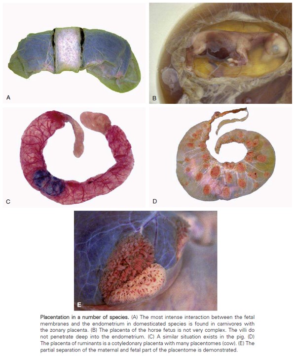
The epitheliochorial placenta is the least intimate among the placental types. In the epitheliochorial placenta, both the endometrial epithelium (maternal side) and epithelium of the chorionic villi are intact, i.e. there is a complete, intact layer of epithelium in both the maternal and fetal components. The epitheliochorial placenta is found in the sow and the mare.
Ruminants have a synepitheliochorial placenta. The endometrial epithelium transiently erodes and then regrows, causing intermittent exposure of the maternal capillaries to the chorionic epithelium.
The endotheliochorial placenta is characterized as having complete erosion of the endometrial epithelium and underlying interstitium. Thus, maternal capillaries are directly exposed to epithelial cells of the chorion. Dogs and cats possess endotheliochorial placentation.
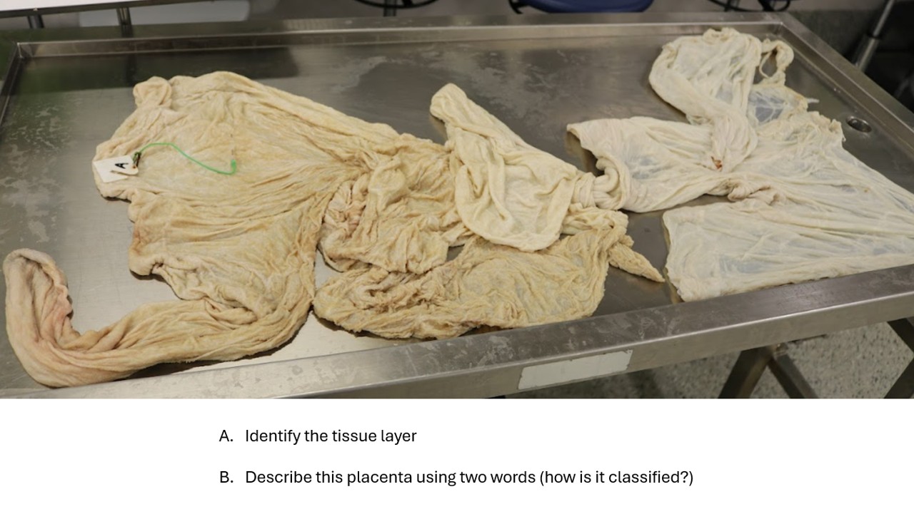
The diffuse placenta of the pig and horse has a velvet-like surface with many closely spaced chorionic villi that are distributed over the entire surface of the chorion. The cervical star of the mare’s placenta is a radial pattern created on the chorion that represents the part of the placenta that contacted the closed cervix and provides a good landmark when examining a mare’s placenta post-foaling. The horse and pig have a diffuse, epitheliochorial placentation.
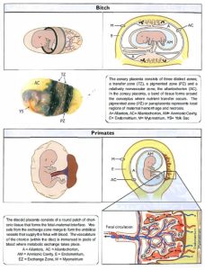
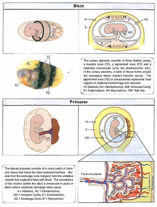
Zonary placentas have a band-like zone of chorionic villi. They are found in dogs and cats and include a prominent region of exchange that forms a broad zone around the chorion near the middle of the conceptus. A second region consists of a highly pigmented ring at either end of the central zone consisting of small hematomas. The function of this zone is not well understood. A third region is the transparent zone on the distal ends of the chorion that has poor vascularity. This zone may be involved in absorption of materials directly from the uterine lumen. Dogs and cats have zonary, endotheliochorial placentas.
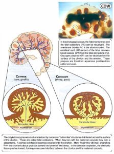
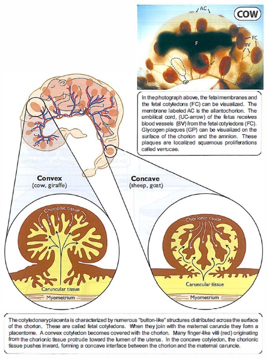
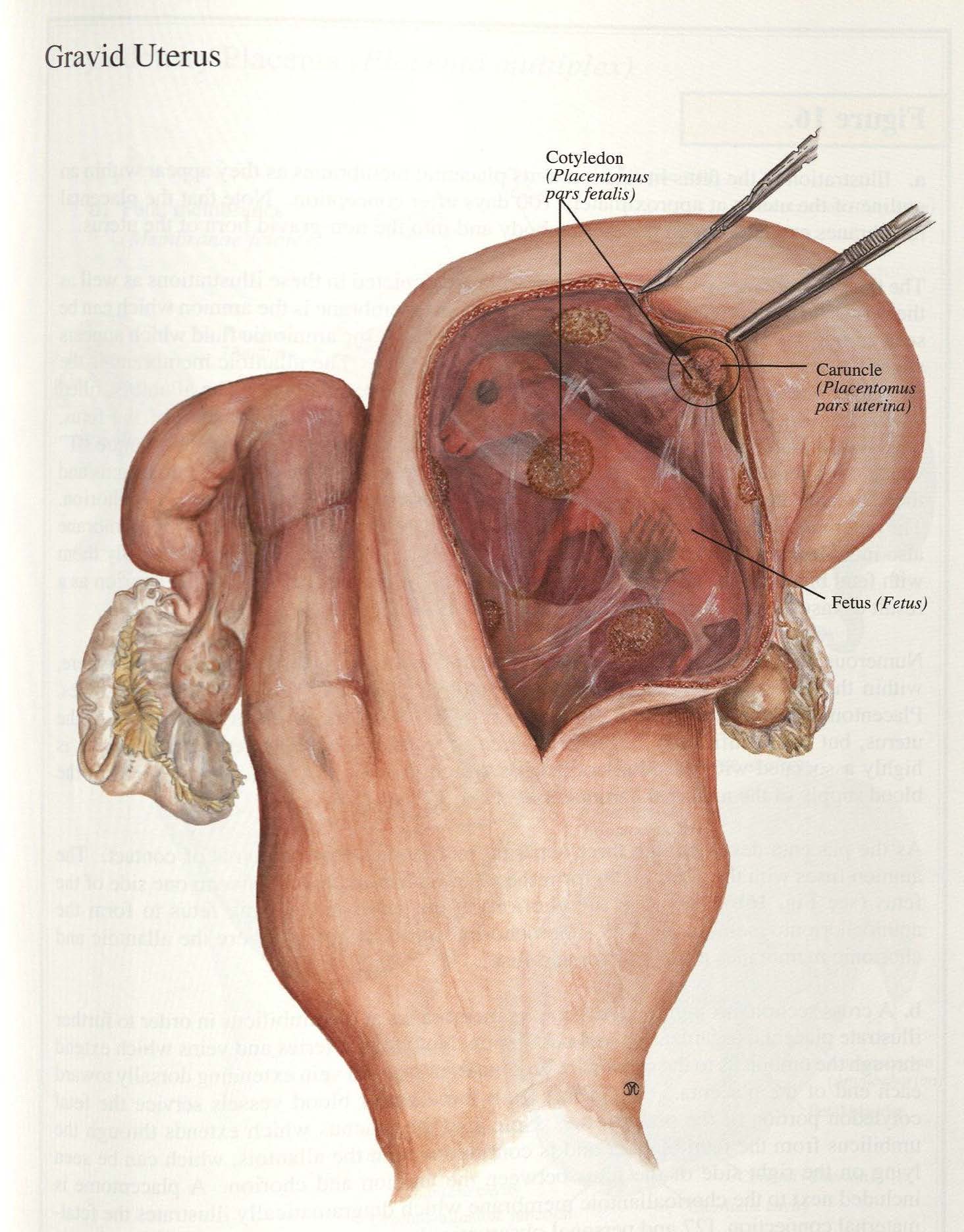
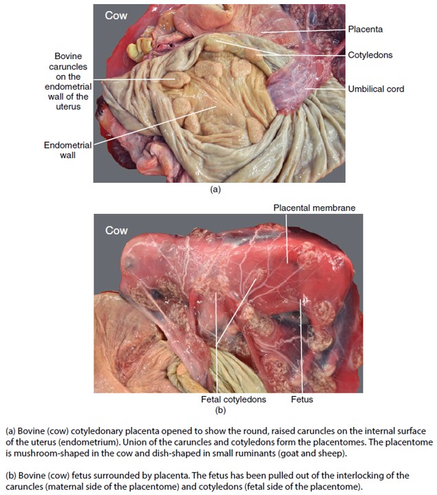
Cotyledonary placentas have numerous, discrete button-like structures called cotyledons. Ruminants have a cotyledonary placenta. A cotyledon is defined as a placental unit consisting of abundant blood vessels and connective tissue. As the placenta expands, fetal cotyledons develop over endometrial caruncles to form the combined unit known as placentomes. During the formation of the placentomes, chorionic villi protrude into crypts in the caruncular tissue. In the cow, the placentomes from a convex structure, while in the ewe they are concave. Ruminants possess a cotyledonary, epitheliochorial placenta.
The discoid placenta is found in rodents and primates. It is characterized by having one or two distinct adjacent discs. These discs contain chorionic villi that interface with the endometrium and provide the region for nutrient and metabolic waste exchange, i.e. they are hemochorial.
-
- Zonary and discoid placentas. 37
-
- Diffuse placentation. 37
-
- Cotyledonary placenta. 37
Observe: Identify the endometrium, chorion, allantois, and amnion of the domestic species. Recognize the placentation of the sow and mare as diffuse epitheliochorial and that of the ruminant as epitheliochorial, cotyledonary placentation. Identify caruncles, cotyledons, and placentomes of the ruminant placenta. Differentiate the carunlces of small ruminants from that of the bovine. Identify the cervical star on the mare placenta and explain what forms it.
-
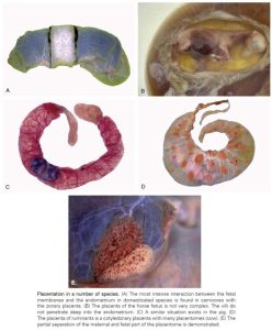
- Placentation in a number of species. 8
-
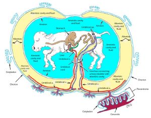
- Bovine fetus and placenta. 2

-
- Ruminant placenta 38
-
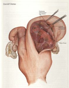
- Gravid bovine uterus38
-
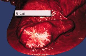
- Cervical star on chorioallantois of the mare. LORI

-
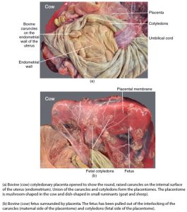
- Bovine placenta and fetus. 12
Clinical Application: FYI – Freemartinism*
The below extracted from: A. Esteves, R. Båge and R. Payan-Carreira. Freemartinism in Cattle. In: Ruminants: Anatomy, Behavior and Diseases, 2012, chapter 7, Editor: R. Evandro Mendes, Nova Science Publishers Inc: pp 99-120.
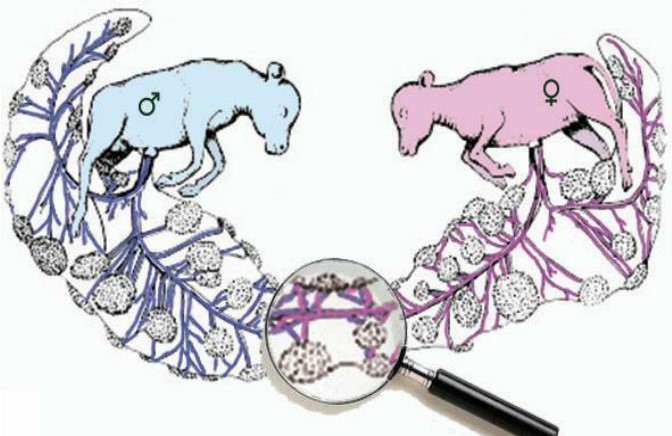
Freemartinism is a particular form of intersexuality in cattle, and represents one of the most common forms found. This pathology can also be observed in buffalo and small ruminants, although in sheep and goats other forms of intersexuality seems to be more frequent than the freemartin syndrome. A bovine freemartin is usually defined as a sterile female calf, born co-twin with a male fetus that shows underdeveloped or misdeveloped genital tract as a result of early development of vascular anastomoses between fetuses of different sex. As consequence of placental anastomoses between the heterozygotic twins, blood chimaerism occurs (60, XX/XY) and passage of male gonad determinants or hormones (such as Anti-Müllerian hormone and androgens) are responsible for disrupted differentiation of the female embryonic gonads and disturbed genital tract development. Comparing to the dramatic changes observed in genital differentiation in the freemartin heifer, the male co-twin only evidence minimal gross defects, though a decrease in male fertility have been reported.
The external genitalia of the freemartin females is usually feminine in appearance, though some minor differences may be perceived at closer examination, and the animal is commonly raised as female. However, the internal genitalia is masculinised in some extent, impairing reproduction and fertility. Absence of anatomical continuity between the vagina and the uterus, hypoplastic or absent uterus, and hypoplastic or streak gonads, co-existing with vesicular glands, are common findings in freemartin heifers. As a rule, heifers born twin to a bull have to be considered sterile and should be identified as early as possible to cull them from replacement stock. However, a small number of females born from twinning pregnancies are not freemartin, although blood chimaerism may be detected. Furthermore, some freemartin animals have been identified that were born as singleton due to the death in uterus of its co-twin.
-

- Schematic representation of chorion-allantois anastomoses between two cattle fetus of different sex. Freemartinism in Cattle
-
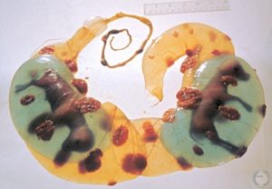
- Twin placentation – 75 days. Amniotic sacs injected with methylene blue. M. Drost

*From Wikipedia:
The etymology of the term “freemartin” is uncertain: speculations include that “free” may indicate “willing” (referring to the freemartin’s willingness to work) or “exempt from reproduction” (referring to its sterility, or to a farmer’s decision to not bother trying to breed a freemartin, or both), or that it may be derived from a Flemish word for a cow which gives no milk and/or has ceased to be capable of bearing offspring; “martin” is generally held to derive from an Irish or Gaelic word for “cow” or “heifer”, although connections to Martinmas have also been posited.
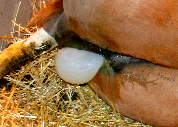
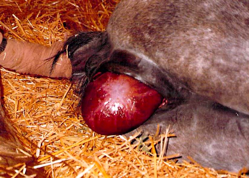
Clinical application: Premature placental separation, or “red bag” foaling
A “red bag” foaling is a critical emergency in equine birthing where the placenta separates prematurely from the mare’s uterine wall, presenting as a red, velvety membrane before the foal. This situation restricts oxygen and nutrient flow to the foal, making immediate intervention crucial.
-
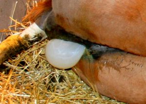
- Amnion protruding through the vulva. CSU
-
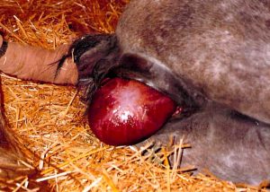
- Chorioallantoic membrane protruding through the vulva (red bag) CSU
Fetal Circulation and Fetal Structures
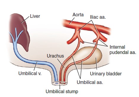
Observe the large umbilical arteries of the newborn calf. During fetal life, these vessels extended cranially through the umbilical opening into the umbilical cord. Deoxygenated blood from the fetus was carried through these arteries to the cotyledons of the placenta. The umbilical arteries are located to either side of the elongated urinary bladder and urachus of the fetus. The umbilical aa. become the round ligaments of the bladder, located in the lateral ligaments of the bladder.
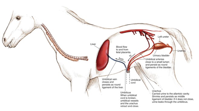
The umbilical vein carries oxygenated blood from the placenta to the fetus. The remnant of the umbilical vein in the adult is the round ligament of the liver, located in the free edge of the falciform ligament.
The urachus extends cranially from the definitive urinary bladder apex to the umbilical opening, where it drains urine from the fetal bladder into the allantoic cavity. The urachus may close before birth at which point fetal urine will accumulate in the amniotic cavity. Following birth, the urachus should completely atrophy, leaving only a small scar at the apex of the bladder. The peritoneal fold that enclosed the urachus persists as the median ligament of the urinary bladder.
The umbilical cord composed of the urachus, the umbilical arteries and veins, and ensheathing connective tissue.
-

- Umbilicus and umbilical remnant structures. 9
-
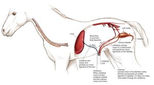
- Neonatal organs of the foal. Left lateral view. 30
-
- Bovine fetus and placenta. 2
Observe: Describe the fetal circulation, including vessels carry oxygenated vs. deoxygenated blood. Identify the umbilical arteries and lateral ligaments of the bladder in the calf cadavers. Know that umbilical arteries regress to form the round ligaments of the bladder in the adult. Know that the umbilical veins regress to form the round ligaments of the liver in the adult. Describe where the urachus is located, what it does in the fetus, where urine drains before and after the urachus closes pre-parturition, and what structure is formed when the urachus regresses.
Fetal Circulation – 14 min
Clinical Relevance: Patent Urachus
Patent urachus is not an uncommon condition in foals and an ascending infection at the site with abscess formation or systemic septicemia is a concern. Ruptured bladder/urachus, most commonly in the male foal, is also well recognized clinically with development of uroperitoneum and consequent electrolyte and acid-base disturbances. Surgical and medical therapies are utilized to manage these conditions.
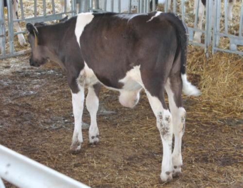
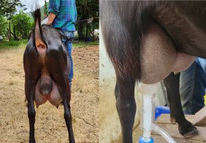
Q4: What does not appear normal? What might be a cause?
Mammary Glands and Suspensory Apparatus
Observe: Refer to the wet specimens and pro-sections available to identify the following bolded structures.
As the mammary glands of the different species are examined, special emphasis should be placed on those of the cow and goat. The first point to be made is that mammary glands are modified sweat glands. The second point is that mammae (all the tissues associated with one teat) are very commonly referred to as ‘mammary glands’ clinically and this language will be used in the lab guide.
The four ‘mammary glands’ (aka quarters) of the cow are closely applied to one another, forming the udder. The pair of glands on the right and left sides are separated on midline by the intermammary groove. Each ‘mammary gland’ possesses a single teat (mammary papilla); however, accessory (supernumerary) teats may be present.
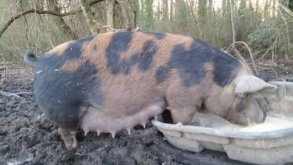
Does and ewes have two ‘mammary glands’ (aka halves); sows have 14-16 ‘mammary glands’ (a chain of 7-8 each side) and mares have two ‘mammary glands’.
Male mammals typically have rudimentary or vestigial mammae, with teats visible (but not always! Stallions/geldings don’t normally have teats, but jack donkeys frequently do, bucks and bulls do, and bucks may develop functional glands).
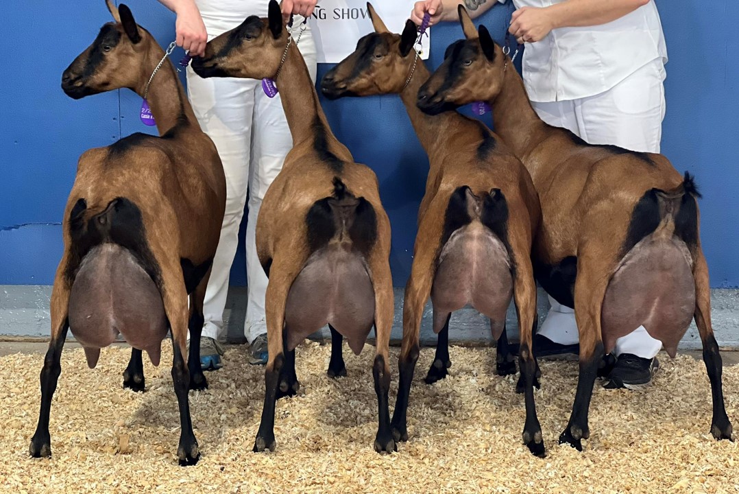
-
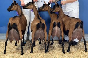
- Oberhasli dairy goats. L. Cobb
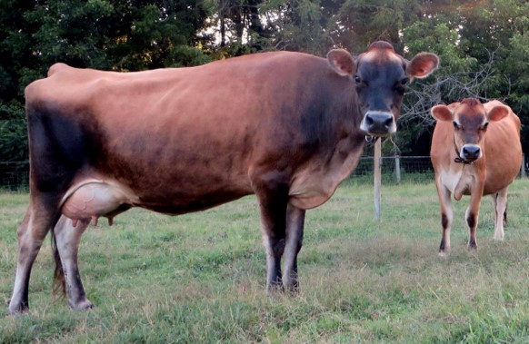
-

- Jersey dairy cow. L. Cobb
-
- East Friesian dairy sheep. L. Cobb
-
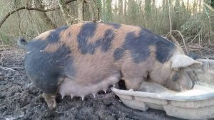
- Lactating Tamworth sow. L. Cobb
-

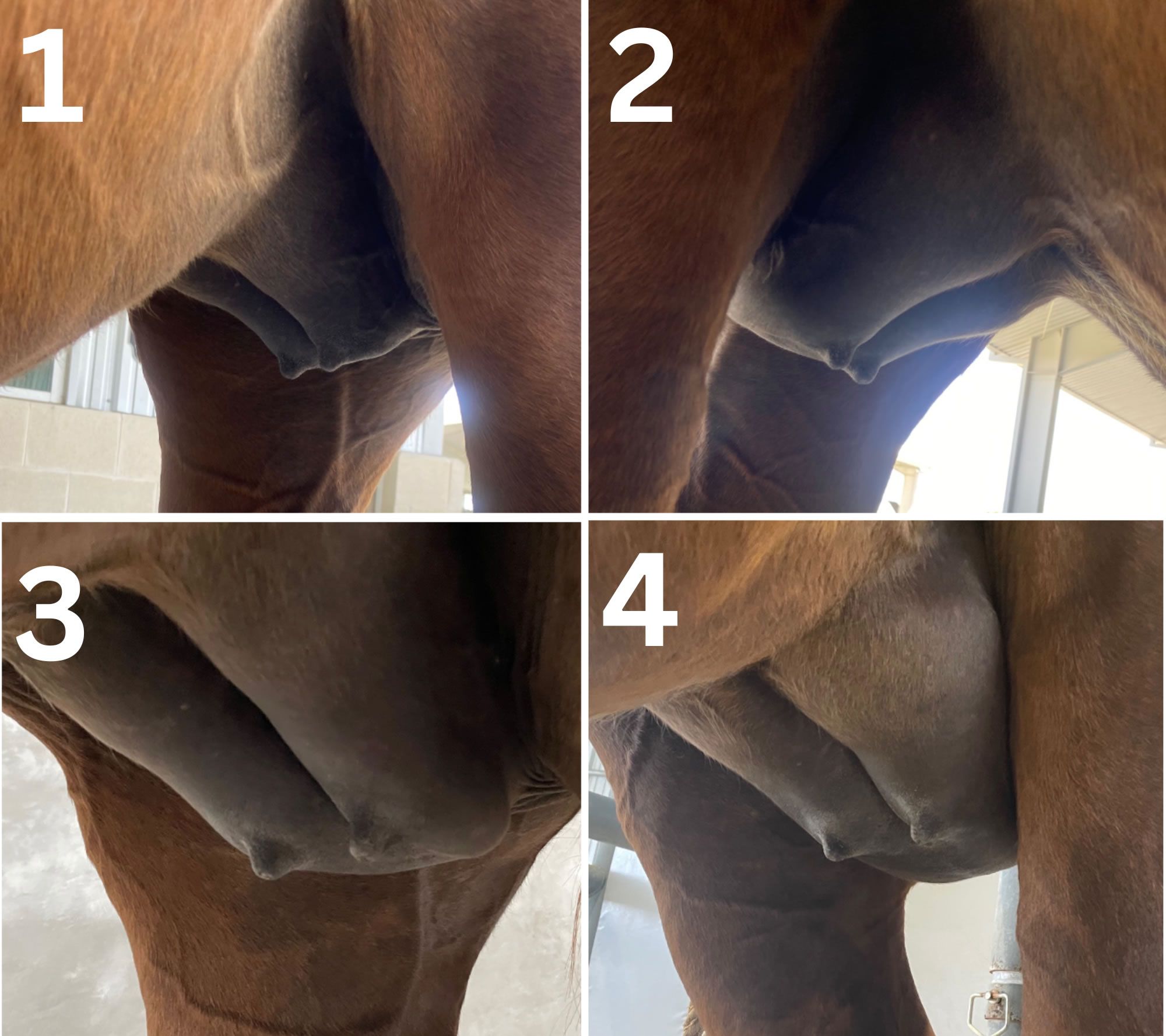
- Weekly progression of the mammary gland as foaling approaches. Mad Barn
-
- Lactating Lamancha buck, Forrest-Pride Blak Falcon. Cathlin Williams
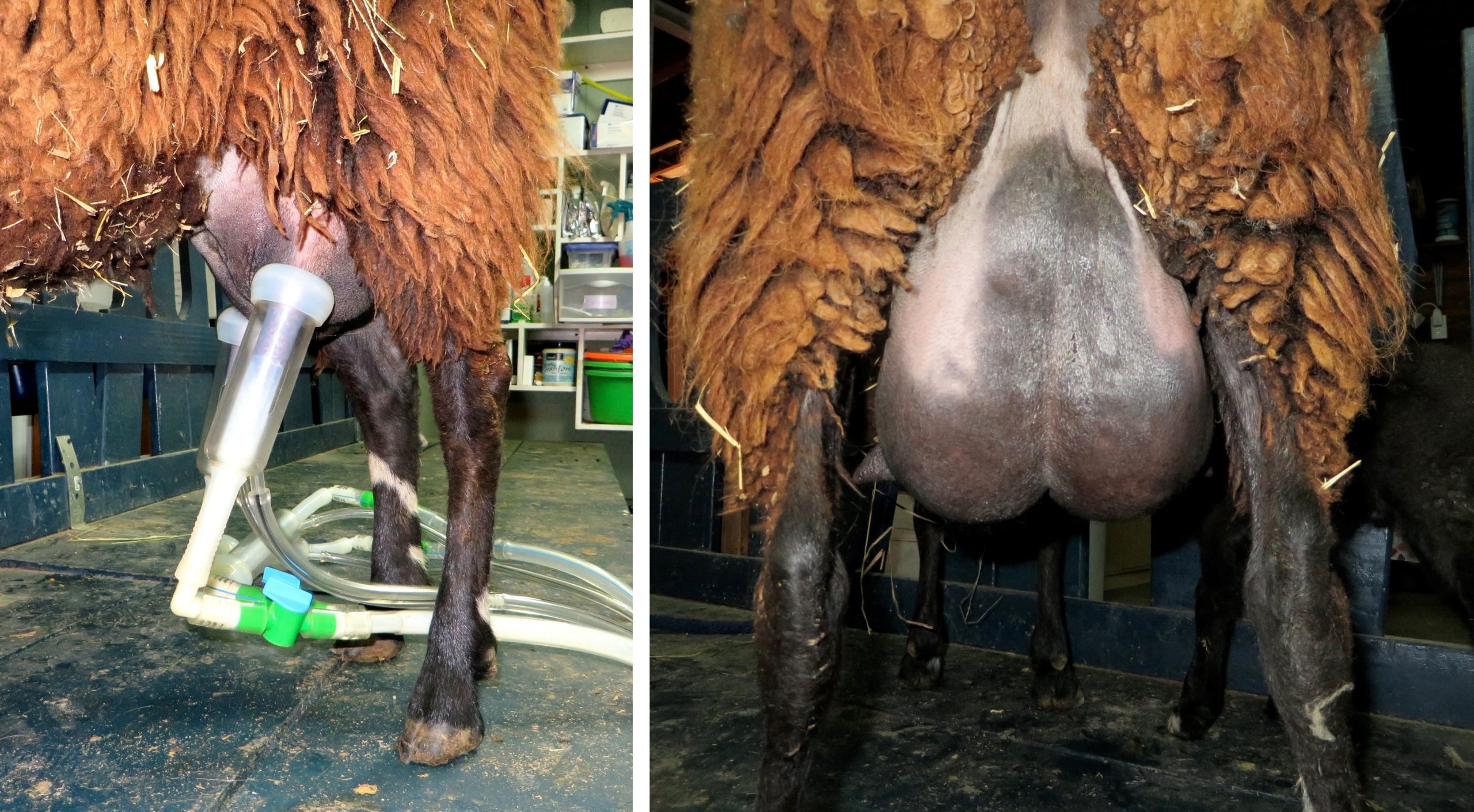
In mares and sows, each teat has two teat orifices because there are two independent glandular complexes per ‘mammary gland’. A gland complex is the single gland and associated duct system within a mammae. Depending on the species, each mammae can have from 1-14 gland complexes. On each mammae, the number of gland complexes coincides with the number of teat orifices:
- Ruminant 1
- Mare 2
- Sow 2
-
- Cow udder and teat. 7
-

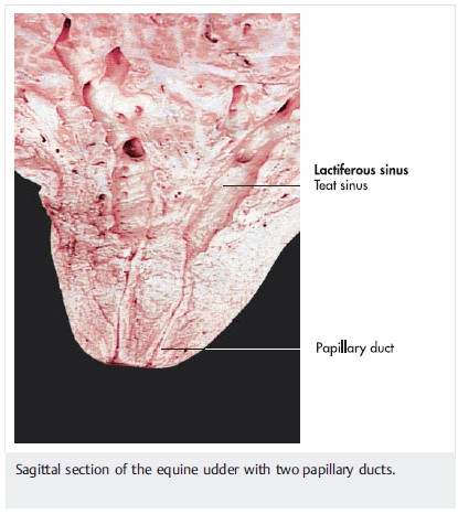
- Mare teat. 7
-

- Cow udder. 8
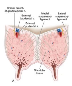
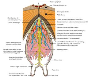
The udder is firmly attached and suspended from the ventral abdominal wall by 4 broad ligaments (laminae) that form the suspensory apparatus of the udder. The symphysial tendon gives rise to the left and right fibrous lateral laminae, which connect to the outer surfaces of the udder. The elastic abdominal tunic (remember? from the abdominal wall dissection!) gives rise to the pair of elastic medial laminae that attach to the medial surfaces of each half of the udder. Therefore, each half of the udder is supported by a separate sling, or basket of connective tissue formed by the medial and lateral laminae of that side and clinically this makes for an easier partial mastectomy when indicated e.g. severe chronic mastitis.
-
- A transverse section of the hind quarters of an adult dairy cow depicting the suspensory apparatus of the udder. 9
-
- Udder of cow, right lateral view. 2

-
- Udder of cow, transverse section through hind quarters 2
Instructor dissection/prosections: In the doe (suspensory apparatus is not as well developed in the mare so not readily dissected), carefully clean away the stumps of the left gracilis and adductor mm. to expose their origin on the symphysial tendon. Dissect ventrally over the left lateral aspect of the udder to identify the fibrous sheet of connective tissue, i.e. the lateral laminae on that side. Further in the doe, blunt dissection just caudodorsal to the mammary glands and moving cranially along the midline between left and right halves should expose a deeper plate of supporting tissue, the pair of medial laminae. Bluntly dissect on midline between the medial laminae to observe that the right and left halves of the udder can be cleanly divided.
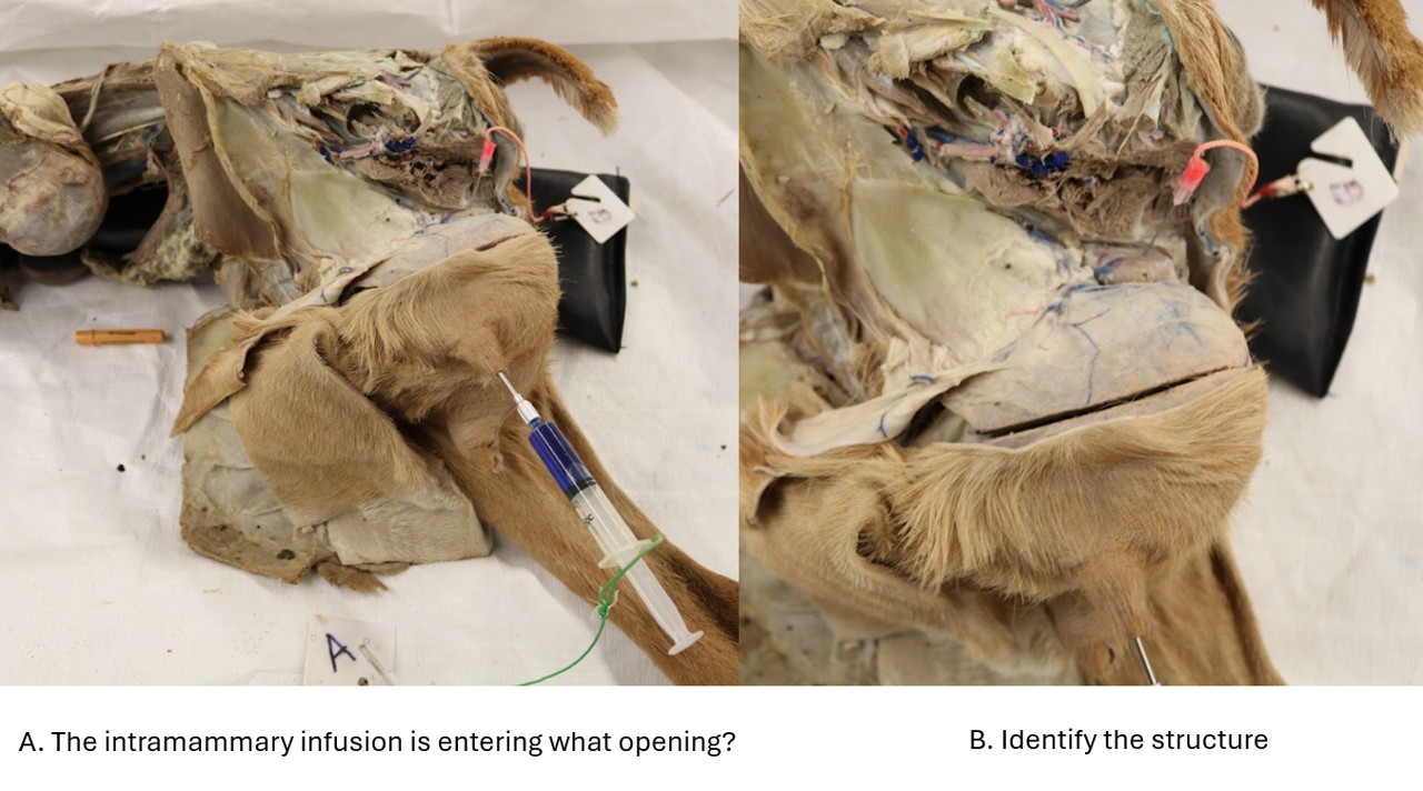
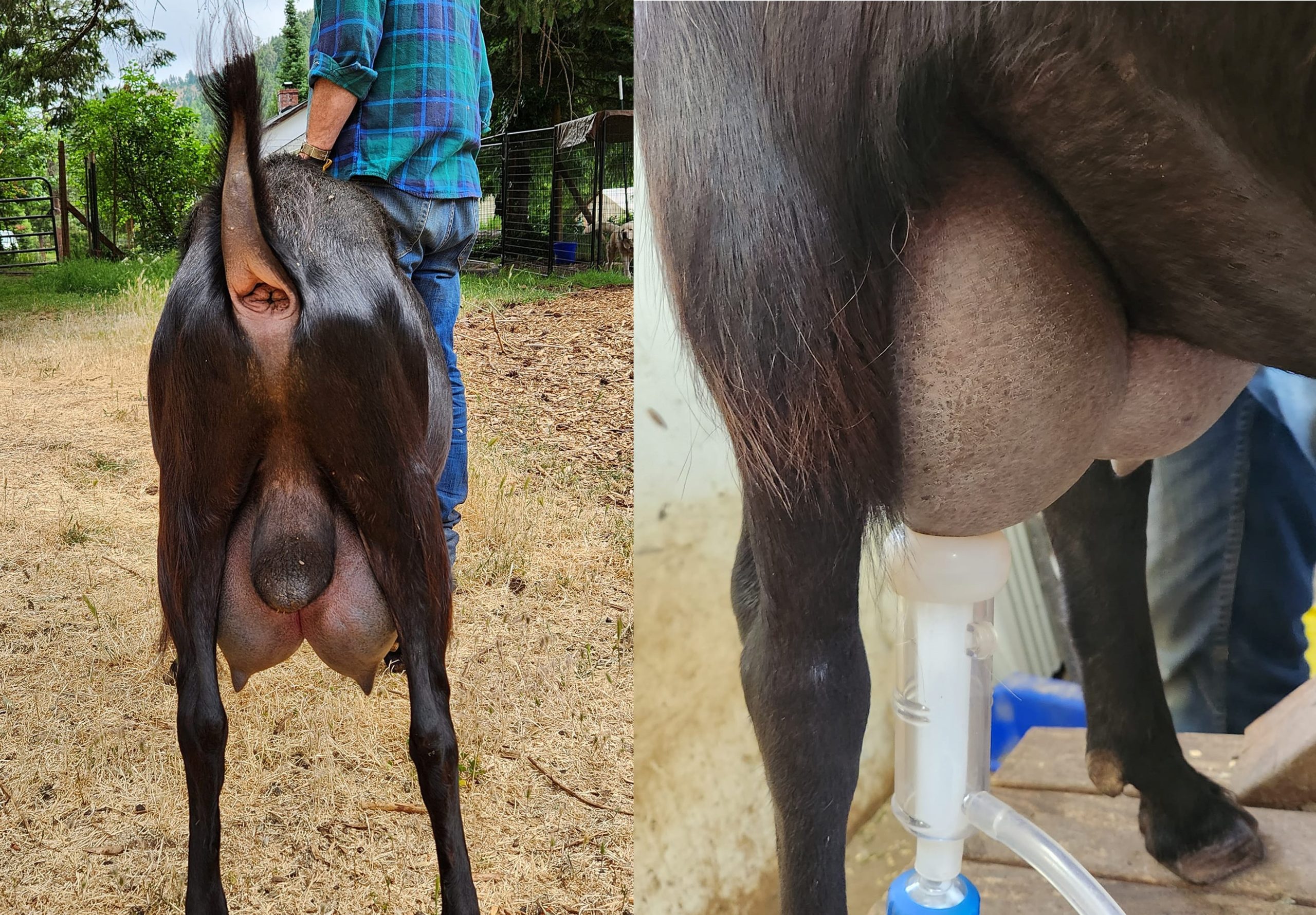
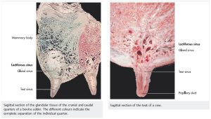
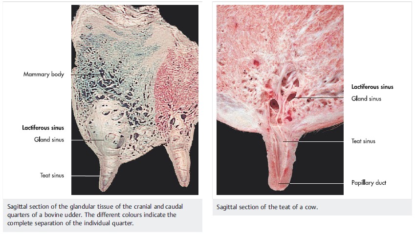
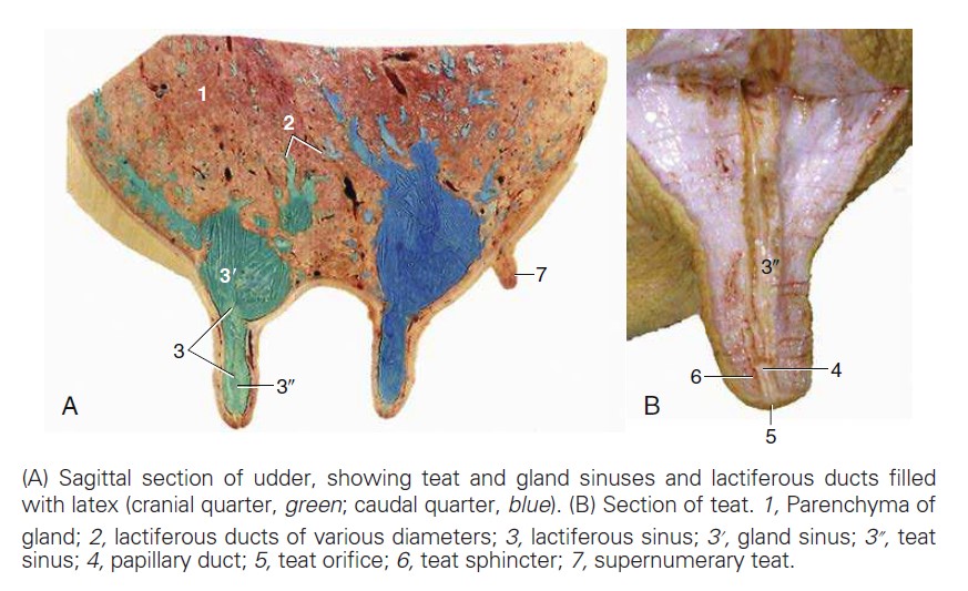
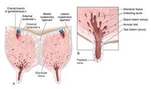
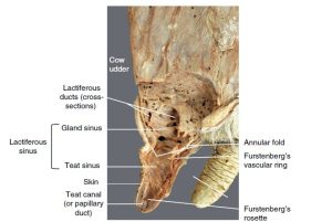
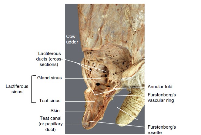
Incise a teat in half, longitudinally from tip to base, in a cranial to caudal direction to pass through the teat orifice(s). Then cut the lateral half of the teat away. Remove enough of the glandular tissue dorsal to the teat to open the cavity (gland sinus) that is continuous with the teat cavity (teat sinus). The teat sinus enters the teat canal, i.e. the short duct to the teat orifice and outside.
Observe: Identify the udder (cow, mare, doe, ewe), mammary glands (species variation in number), teats, and intermammary groove. In the ruminant, identify suspensory apparatus of the udder, i.e. the medial laminae and lateral laminae.
Teat anatomy
The combined teat sinus and gland sinus form a single reservoir, the lactiferous sinus. The teat sinus enters the teat canal, i.e. the short duct to the teat orifice and outside.
The mare has two separate lactiferous sinuses and teat canals (i.e. glandular duct systems) per side of udder, so there are two teat orifices per teat. The sow has 2-3 duct systems draining per teat.
In the cow, the teat canal (aka papillary duct or ‘streak canal’) is closed by longitudinal mucosal folds with the aid of a sphincter of smooth muscle and elastic tissue (known as Fürstenburg’s rosette). In the teat canal of the mare, sow, and ewe, there is no smooth muscle in the sphincter of the teat canal. Closure of this canal is accomplished by a dense network of elastic fibers.
In the mare, there are numerous sweat glands and sebaceous glands associated with the teat. The sebaceous glands are especially large near the teat canals, and it is the product of these large glands that is responsible for the phenomenon of “waxing” in the pregnant mare just prior to parturition.
-
-
(A) A transverse section of the hind quarters of an adult dairy cow depicting the suspensory apparatus of the udder.
(B) A longitudinal section of the teat and multiple glands depicting the milk collecting and entering system of a cow. 9
-
- Udder of cow, transverse section through hind quarters 2
-
-
Bovine (cow) teat and distal part of the udder:
longitudinal section. 12
Observe: In the ruminant, identify a teat sinus, gland sinus, lactiferous sinus, teat canal, and teat orifice(s). State how many of each of these structures exist in the mare, doe, and cow udder or one sow mammary gland.
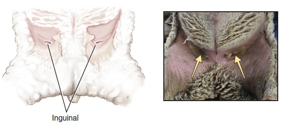
Unique to the ewe, located between the udder (or scrotum) and the medial surface of the thigh on each side, are the inguinal pouches, which emit an oily secretion thought to serve as a marker scent to help the ewe in identifying her own lamb(s).
-
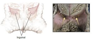
- The inguinal cutaneous pouch on sheep. 9
Mammary glands – vessels, lymphatics and innervation
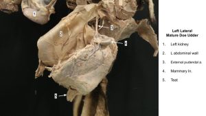
Instructor Dissection Only: In the doe, on the accessible left side, transect the lateral lamina in a horizontal manner dorsal to the udder. Reflect the lamina to reveal the mammary tissue of that side. Dissect dorsally towards the superficial inguinal ring area to identify vascular structures and lymph nodes.
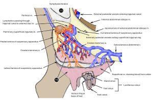
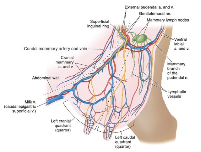
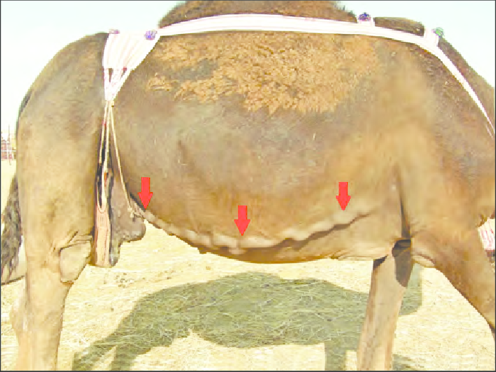
Observe: Dorsal to the mammary tissue identify the external pudendal artery and vein. The external pudendal a. has exited the inguinal canal at the superficial inguinal ring and is the primary arterial supply to each half of the udder (it divides into caudal and cranial mammary aa.; the cranial mammary a. = the caudal superficial epigastric a., the caudal mammary a. = the ventral labial a., in other species). The external pudendal vv. and the large ‘milk veins’, = anastomosed cranial and caudal superficial epigastric vv.) drain the udder (a third vein, the dorsal labial v. also provides caudal drainage). Most notable in the dairy cow, each subcutaneous abdominal vein follows a tortuous course along the abdomen until it penetrates the body wall at the so-called “milk well.” From here, the blood continues through the internal thoracic vein to the cranial vena cava. The external pudendal vv. provide the main venous drainage in the doe and ewe.
Numerous large, superficial lymphatics drain the mammary tissue to the mammary ln. (superficial inguinal ln.). Efferent lymphatics in the cow (and goat?) then continue through the inguinal canal, to the external iliac ln. (and in the mare to the proximal femoral ln.). The superficial lymphatics of the cow udder can be observed in the living animal, and their direction is primarily caudodorsal. These lymphatics can be distinguished from the superficial veins, which course in a more craniodorsal direction.
-

-
Blood supply lymphatics, and
innervation of the udder of a lactating cow. 9
-
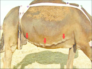
- Lactating camel showing the development of the milk vein (subcutaneously abdominal vein). M. Dioli
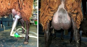
-
- Goat udder

Observe: Identify the mammary (superficial inguinal) lymph node. State the mammary lymph node efferent lymphatics in the cow and mare.
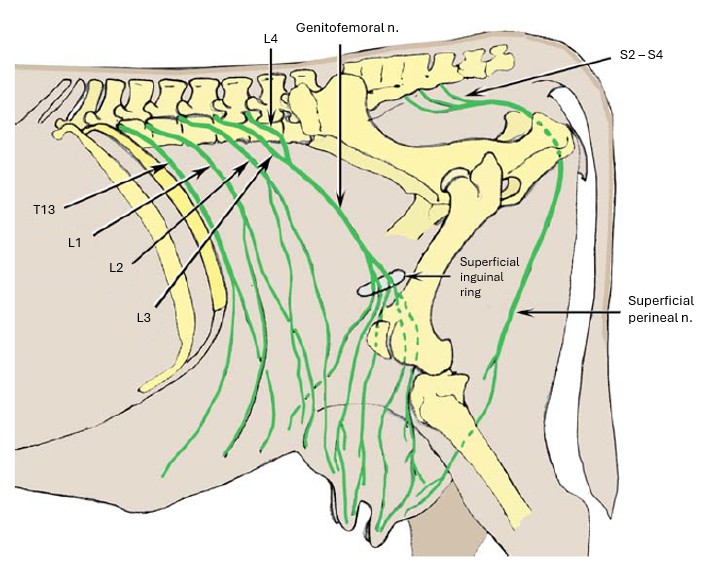
The udder of the cow is supplied by sensory and autonomic nerves. The sensory innervation is derived from the ventral cutaneous branches of L1 and L2, the genitofemoral nerve (L3 and L4), and the mammary (superficial perineal) branch of the pudendal nerve. Of these, the genitofemoral nerve is of major importance, because it innervates much of the skin, the teats, and most of the glandular tissue. The autonomic innervation is supplied by sympathetic fibers from the caudal mesenteric ganglion, which accompany the genitofemoral nerve through the inguinal canal.
Recall, to help with identification, that the genitofemoral n. travels closely related to the external pudendal vessels through the inguinal canal.
-
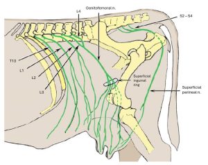
- Lumbar and superficial perineal nerves of ox2
Observe: In the ruminants and mare identify the external pudendal a/v and the genitofemoral n. What type of innervation does the genitofemoral nerve provide to the udder? What type of autonomic innervation supplies the udder (sympathetic or parasympathetic), and from which ganglion do these fibers originate?
Review Videos
Fetal Circulation – 14 min
Udder and placenta– 6 min
Placentation review – 10 min
Calf umbilical arteries – Watch until 3 min
Terms
| Placenta and associated structures | ||
| Term | Features | Species differences/comments |
| Chorioallantois | ||
| Chorion | ||
| Allantois | ||
| Amnion | ||
| Endometrium | ||
| Cotyledons | Ruminant only. | |
| Placenta | ||
| Endometrium | ||
| Chorionic villi | ||
| Epitheliochorial placenta | Mare, sow | |
| Synepitheliochorial placenta | Ruminants | |
| Endotheliochorial placenta | Carnivores | |
| Diffuse placenta | Mare, sow | |
| Cervical star | Mare | |
| Zonary placentas | Carnivore | |
| Cotyledonary placentas | Ruminant | |
| Caruncles | Ruminants only. Concave in small ruminants; convex in bovine | |
| Placentomes | Ruminant only | |
| Discoid placenta | Rodents and Primates (know conceptually) | |
| Hemochorial | Rodents and Primates (know conceptually) | |
| Fetal Circulation and Fetal Structures | ||
| Term | Features | Species differences/comments |
| Umbilical arteries | Identify in adult and fetal calf | |
| Umbilical cord | Do not ID contents, know conceptually | |
| Urachus | Becomes median ligament of bladder | ID in fetal calf |
| Round ligaments of the bladder | Remnants of umbilical arteries | |
| Lateral ligaments of the bladder | ||
| Umbilical vein | Conceptual | |
| Round ligament of the liver | Conceptual | |
| Allantoic cavity | ||
| Amniotic cavity | ||
| Median ligament of the urinary bladder | Urachus in fetus | |
Mammary Glands and Suspensory Apparatus |
||
| Term | Features | Species differences/comments |
| Mammary glands | ||
| Quarters | ||
| Udder | ||
| Intermammary groove | ||
| Teat | ||
| Teat sinus | ||
| Gland sinus | ||
| Lactiferous sinus | ||
| Teat canal | ||
| Teat orifices | ||
| Glandular complexes | ||
| Suspensory apparatus | ||
| Lateral laminae | ||
| Elastic medial laminae | ||
| Inguinal pouches | ||
Mammary Glands – Vessels, Lymphatics, and Innervation |
||
| Term | Features | Species differences/comments |
| External pudendal artery | ID only in ungulate | |
| External pudendal vein | ID only in ungulate | |
| Milk veins | Know conceptually | |
| Cranial superficial epigastric vv. | Know conceptually | |
| Caudal superficial epigastric vv. | Know conceptually | |
| Superficial inguinal ln. | Can be called Mammary ln. in ungulates | |
| External iliac ln. | Only in ruminant | |
| Proximal femoral ln. | Horse, analogous to external iliac, know conceptually | |
| Genitofemoral nerve | ||
| Caudal mesenteric ganglion | sympathetic fibers from this ganglion accompany the genitofemoral n. through the inguinal canal and supply sympathetic innervation to the udder. | |
Example Practical Exam Questions