
20 Animal Transport
At the end of this chapter you will be able to:
- Compare and contrast the transport of nutrients and gasses in animals with open and closed circulatory systems
- Describe how the human cardiovascular system works
- Compare and contrast the pulmonary and systemic circuits
- Describe how human veins and arteries work
- Describe the role the lymphatic system plays
Introduction
All living organisms need a way to move nutrients, gasses, water, and waste within their bodies. Some plants and animals are small enough and/or thin enough that they can rely on diffusion for this process; however, larger plants and animals need a more organized system to transport things from one part of their body to another. A common version is to have cells that act as “pipes” for such transport. In some cases, the vessels will be arranged in different sizes, from large to small. With this type of system, such as we see with human transport, you might have a large artery, leading to smaller arterioles and eventually to the smallest vessels – the capillaries. And it is here, at the smallest diameter, that you will see diffusion of materials and gasses. In other cases, such as we see with plants, the vessels are arranged end to end in a pipeline fashion without decreasing diameter, but diffusion is possible along the way with the small diameter vessels.
Animal Transport – Types of Circulatory Systems
Animals can either have no circulatory systems, relying instead on the diffusion of gasses and nutrients directly across their bodies or one of two types of circulatory systems: open or closed. The circulatory system varies from simple systems in invertebrates to more complex systems in vertebrates. The simplest animals, such as the sponges (Porifera) and rotifers (Rotifera), do not need a circulatory system because diffusion allows adequate exchange of water, nutrients, waste, and dissolved gases from an area of high concentration to an area of low concentration. Organisms that are more complex but still only have two layers of cells in their body plan, such as jellies (Cnidaria) and comb jellies (Ctenophora) also use diffusion through their epidermis and internally through the gastrovascular compartment ( gastrovascular cavity acts as both a mouth and an anus for the animal). Both their internal and external tissues are bathed in an aqueous environment and exchange fluids by diffusion on both sides. The exchange of fluids is assisted by the pulsing of the jellyfish body.
For more complex organisms, diffusion is not efficient for cycling gases, nutrients, and waste effectively through the body; therefore, more complex circulatory systems evolved. In an open system, an elongated beating heart pushes the hemolymph (analogous to blood but without respiratory pigments and gasses) through the body and muscle contractions help to move fluids. The larger more complex crustaceans, including lobsters, have developed arterial-like vessels to push blood through their bodies, and the most active mollusks, such as squids, have evolved a closed circulatory system and are able to move rapidly to catch prey. Closed circulatory systems are a characteristic of vertebrates; however, there are significant differences in the structure of the heart and the circulation of blood between the different vertebrate groups due to adaptation during evolution and associated differences in anatomy.
Use of Body Surface for Diffusion
Animals without circulatory systems tend to be small in size and/or flat in design. This is because diffusion, the movement of material from a high concentration to a low concentration without any energy investment, can only happen over short distances. Invertebrates, such as flatworms (in the phylum Platyleminthes) and cnidarinas have this type of transport.
For small multicellular organisms, diffusion across the outer membrane is sufficient to meet their oxygen needs. Gas exchange by direct diffusion across surface membranes is efficient for organisms less than 1 mm in diameter. In simple organisms, such as cnidarians and flatworms, every cell in the body is close to the external environment. Their cells are kept moist and gases diffuse quickly via direct diffusion. Flatworms are small, literally flat worms, which “breathe” through diffusion across the outer membrane. The flat shape of these organisms increases the surface area for diffusion, ensuring that each cell within the body is close to the outer membrane surface and has access to oxygen. If the flatworm had a cylindrical body, then the cells in the center would not be able to get oxygen.
Earthworms and amphibians use their skin (integument) as a respiratory organ. A dense network of capillaries lies just below the skin and facilitates gas exchange between the external environment and the circulatory system. The respiratory surface must be kept moist in order for the gases to dissolve and diffuse across cell membranes.
Open Circulatory System
Animals, such as insects, rely on a system called open. The term is in reference to the fact that after the heart pumps out “blood” referred to as hemolymph in short vessels, the vessels dump out directly into the main body of the animal, referred to as the coelom. The hemolymph is like blood, but lacks the respiratory pigments so it looks more green in color (as opposed to blood’s more red color) and transports nutrients, not oxygen as a result. Once the hemolymph’s nutrients are dumped into the open coelom, it is not contained in vessels (again it is an open system) and it can completely and quickly bathe all the cells and tissues in the animal. In this sense, diffusion is still in play and since diffusion only works over short distances, an animal with an open circulatory system is still not going to be large in size.
As described above, insect respiration is independent of its circulatory system and the hemolymph does not play a direct role in oxygen transport. Insects have a highly specialized type of respiratory system called the tracheal system, which consists of a network of small tubes that carries oxygen to the entire body. Because the circulatory system is not used primarily to move gasses, but instead the gas passes directly to the needed tissues, the tracheal system is the most direct and efficient respiratory system for getting oxygen to respiratory sites. The tubes in the tracheal system are made of a polymeric material called chitin.
Watch this short video on the open circulatory system of a grasshopper.
Insect bodies have openings, called spiracles, along the thorax and abdomen. These openings connect to the tubular network, allowing oxygen to pass into the body and regulating the diffusion of carbon dioxide and water vapor. Oxygen enters and leaves the tracheal system through the spiracles and directly bathes the cells of the animal.
Animals with open circulatory systems can move oxygen and gasses throughout their bodies quickly and efficiently, using less metabolic energy in doing so. Animals will have better body temperature control and since they have no blood pressure they are less vulnerable to external pressure. This means that animals living in the water can move to greater depths without being vulnerable to that pressure.
Animals with open circulatory system, however, they cannot grow to be large in size. Additionally, animals do not have strong immune systems with white blood cells that will attack and destroy foreign pathogens. Instead, these animals will block off the pathogen and try to contain it in one part of its body so it can’t cause problems elsewhere in the body.
In addition to insects, other arthropods have open systems. These include animals like lobsters, crabs, molluscs, oysters, snails and slugs.
Closed Circulatory System
More complex and larger animals rely on a closed circulatory system. This means that a heart pumps out blood through vessels that progressively get smaller in size, and eventually oxygen and nutrients can diffuse from one vessel (a capillary) into a cell. Diffusion from the vessel into the cell is occurring which means a vessel has to be very close to all cells in the body. Remember diffusion can only happen over short distances.
In a closed circulatory system, blood moves in one direction and animals can have more control over blood flow. The closed circuit also creates blood pressure, which allows blood to travel further distances, which in turn can allow for an animal to grow larger in size.
Animals such as earthworms, octopus, birds, amphibians, reptiles and mammals have closed systems. We will focus much more on the human system as an illustration of a closed system.
The Human Cardiovascular Circulatory System
The cardiovascular system, also called the circulatory system, is the organ system that transports materials to and from all the cells of the body. The materials carried by the cardiovascular system include oxygen from the lungs, nutrients from the digestive system, hormones from glands of the endocrine system, and waste materials from cells throughout the body. Transport of these and many other materials is necessary to maintain homeostasis of the body. The main components of the cardiovascular system are the heart, blood vessels, and blood (Fig 1).
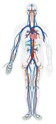
Figure 1: This simplified drawing of the cardiovascular system shows its main structures. The heart is shown in the chest in red. Blood vessels called arteries are also shown in red, and blood vessels called veins are shown in blue.
The heart is a muscular organ in the chest. It consists mainly of cardiac muscle tissue, and it pumps blood through blood vessels by repeated, rhythmic contractions. The heart has four inner chambers: a right atrium and ventricle, and a left atrium and ventricle (Fig 2). On each side of the heart, blood is pumped from the atrium to the ventricle below it, and from the ventricle out of the heart. The heart also contains several valves that allow blood to flow only in the proper direction through the heart.
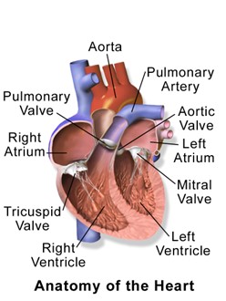
Figure 2: The right side of the heart includes the right atrium and right ventricle. The left side includes the left atrium and left ventricle.
The diagram labels the right side of the heart on the left side of the diagram, and vice versa. This is because it is assumed that in this diagram, the heart appears as if the patient was facing us – the patient’s left side is on our right side!
Unlike skeletal muscle, cardiac muscle routinely contracts without stimulation by the nervous system. Specialized cardiac muscle cells send out electrical impulses that stimulate the contractions. As a result, the atria and ventricles normally contract with just the right timing to keep blood pumping efficiently through the heart.
The blood vessels of the cardiovascular system are like a network of interconnected, one-way roads that range from superhighways to back alleys. Like a network of roads, the blood vessels are tasked with allowing the transport of materials from one place to another. There are three major types of blood vessels: arteries, veins, and capillaries (Fig 3).

Figure 3: This diagram represents the structure and functions of the different types of blood vessels int eh cardiovascular system.
Arteries are blood vessels that carry blood away from the heart (except for the arteries that actually supply blood to the heart muscle). Most arteries carry oxygen-rich blood, and one of their main functions is distributing oxygen to tissues throughout the body.
Blood flows through arteries largely because it is under pressure from the pumping action of the heart. It should be noted that coronary arteries, which supply heart muscle cells with blood, travel toward the heart, but not as part of the blood flow that travels through the chambers of the heart. Most arteries, including coronary arteries, carry oxygenated blood, but there are a few exceptions, most notably the pulmonary artery. This artery carries deoxygenated blood from the heart to the lungs, where it picks up oxygen and releases carbon dioxide. In virtually all other arteries, the hemoglobin in red blood cells is highly saturated with oxygen (95–100%). These arteries distribute oxygenated blood to tissues throughout the body.
The largest artery in the body is the aorta, which is connected to the heart and extends down into the abdomen. The aorta has high-pressure, oxygenated blood pumped directly into it from the left ventricle of the heart. The aorta has many branches, and the branches subdivide repeatedly, with the subdivisions growing smaller and smaller in diameter. The smallest arteries are called arterioles.
Veins are blood vessels that carry blood toward the heart. Most veins carry deoxygenated blood. Blood traveling through veins is not under pressure from the beating heart. It gets help moving along by the squeezing action of skeletal muscles, for example, when you walk or breathe. It is also prevented from flowing backward by valves in the larger veins. Veins are called capacitance blood vessels, because the majority of the body’s total volume of blood (about 60%) is contained within veins.
Most veins carry deoxygenated blood, but there are a few exceptions, including the four pulmonary veins. These veins carry oxygenated blood from the lungs to the heart, which then pumps the blood to the rest of the body. In virtually all other veins, hemoglobin is relatively unsaturated with oxygen (about 75%).
The two largest veins in the body are the superior vena cava — which carries blood from the upper body directly to the right atrium of the heart — and the inferior vena cava, which carries blood from the lower body directly to the right atrium (Fig 4). Like arteries, veins form a complex, branching system of larger and smaller vessels. The smallest veins are called venules. They receive blood from capillaries and transport it to larger veins. Each venule receives blood from multiple capillaries.
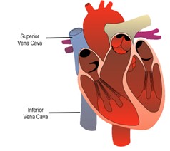
Figure 4: The Superior and Inferior Vena Cava are the largest veins in the body. They deliver deoxygenated blood directly to the right atrium.
Capillaries are the smallest blood vessels, and they connect arterioles and venules. As they pass through tissues, they exchange substances (including oxygen) with cells. This is where the important process of diffusion occurs. That is, gasses and nutrients move from an area of high concentration to an area of low concentration without the investment of ATP energy. Cells throughout the body need a constant supply of oxygen. They get oxygen from capillaries in the systemic circulation. The systemic circulation is just one of two interconnected circulations that make up the human cardiovascular system. The other circulation is the pulmonary system, which is where blood picks up oxygen to carry to cells. It takes blood about 20 seconds to make one complete transit through both circulations (Fig 5).

Figure 5: There are two main circuits through which blood flows in the cardiovascular system. In the pulmonary circuit, blood moves from the right side of the heart to the lungs and then back to the left side of the heart. In the systemic circuit, blood moves from the left side of the heart to the body tissues and then back to the right side of the heart.
The pulmonary circuit involves only the heart, the lungs, and the major blood vessels that connect them (Fig 6). Blood moves through the pulmonary circuit from the heart, to the lungs, and then back to the heart again, becoming oxygenated in the process. Specifically, the right ventricle of the heart pumps deoxygenated blood into the right and left pulmonary arteries. These arteries carry the blood to the right and left lungs, respectively. Oxygenated blood then returns from the right and left lungs through the two right and two left pulmonary veins. All four pulmonary veins enter the left atrium of the heart.

Figure 6: This diagram shows the heart, lungs, and major vessels that make up the pulmonary circulation. The colored arrows indicate the direction of blood flow — red for oxygenated blood and blue for relatively deoxygenated blood.
What happens to the blood while it is in the lungs? It passes through increasingly smaller arteries, and finally through capillary networks surrounding the alveoli (Fig 7). This is where gas exchange takes place. The deoxygenated blood in the capillaries picks up oxygen from the alveoli and gives up carbon dioxide to the alveoli. As a result, the blood returning to the heart in the pulmonary veins is almost completely saturated with oxygen.
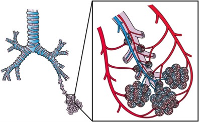
Figure 7: This diagram illustrates clusters of alveoli in the lungs, where gas exchange takes place with blood in capillaries as it passes through the pulmonary circulation.
The oxygenated blood that enters the left atrium of the heart in the pulmonary circulation then passes into the systemic circuit. This is the part of the cardiovascular system that transports blood to and from all of the tissues of the body to provide oxygen and nutrients, and to pick up wastes. It consists of the heart and blood vessels that supply the metabolic needs of all the cells in the body, including those of the heart and lungs.
In the systemic circulation, the left atrium pumps oxygenated blood to the left ventricle, which pumps the blood directly into the aorta, the body’s largest artery (Fig 8). Major arteries branching off the aorta carry the blood to the head and upper extremities. The aorta continues down through the abdomen and carries blood to the abdomen and lower extremities. The blood then returns to the heart through the network of increasingly larger veins of the systemic circulation. All of the returning blood eventually collects in the superior vena cava (upper body) and inferior vena cava (lower body), which empty directly into the right atrium of the heart.
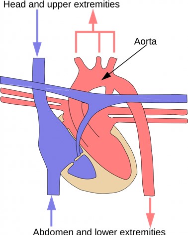
Figure 8: The systemic circulation includes the aorta (red), which carries oxygenated blood away from the heart to the rest of the body; and the inferior and superior venae cavae (blue), which return deoxygenated blood to the heart from the body. The coloured arrows in the diagram indicate the direction of blood flow — red for oxygenated and blue for deoxygenated.
Blood is a fluid connective tissue that circulates throughout the body in blood vessels by the pumping action of the heart. Blood carries oxygen and nutrients to all the body’s cells, and it carries carbon dioxide and other wastes away from the cells to be excreted. Blood also transports many other substances, defends the body against infection, repairs body tissues, and controls the body’s pH, among other functions.
The fluid part of blood is called plasma. It is a yellowish, watery liquid that contains many dissolved substances and blood cells. Types of blood cells in plasma include red blood cells, white blood cells, and platelets, all of which are illustrated in the photomicrograph (Fig 9) and described below.
Plasma makes up about 55% of blood by volume. It is about 92% water and contains many dissolved substances. Most of these substances are proteins, but plasma also contains trace amounts of glucose, mineral ions, hormones, carbon dioxide, and other substances. In addition, plasma contains blood cells. When the cells are removed from plasma, the remaining liquid is clear but yellow in color.
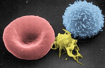
Figure 9: The three types of cells in blood are pictured here: red blood cell (left), platelet (center), and white blood cell (right).
Erythrocytes (red blood cells) have the main function of carrying oxygen in the blood. Red blood cells consist mostly of hemoglobin, a protein containing iron that binds with oxygen. This is the most numerous cells in blood. One microlitre of blood contains between 4.2 and 6.1 million red blood cells, and red blood cells make up about 25% of all the cells in the human body. The cytoplasm of a mature erythrocyte is almost completely filled with hemoglobin, the iron-containing protein that binds with oxygen and gives the cell its red color. In order to provide maximum space for hemoglobin, mature erythrocytes lack a cell nucleus and most organelles. They are little more than sacks of hemoglobin.
Leukocytes (white blood cells) are far fewer in number than red blood cells. They defend the body in various ways. White blood cells called phagocytes, for example, swallow and destroy pathogens, dead cells, and other debris in the blood. There are far fewer leukocytes than red blood cells in blood. There are normally only about 1,000 to 11,000 white blood cells per microlitre of blood. Unlike erythrocytes, leukocytes have a nucleus. White blood cells are part of the body’s immune system. They destroy and remove old or abnormal cells and cellular debris, as well as attack pathogens and foreign substances. There are five main types of white blood cells, which are differ in their specific immune functions:
| Type of Leukocyte | % of All Leukocytes | Main Function(s) |
| Neutrophil | 62% | Phagocytize (engulf & destroy) bacteria and fungi in blood |
| Eosinophil | 2% | Attack and kill large parasites; carry out allergic responses |
| Basophil | <1% | Release histamines in inflammatory responses |
| Lymphocyte | 30% | Attack and destroy virus-infected and tumor cells: create lasting immunity to specific pathogens |
| Monocyte | 5% | Phagocytize pathogens and debris in tissues |
Thrombocytes (platelets) are cell fragments involved in blood clotting. They stick to tears in blood vessels and to each other, forming a plug at the site of injury. They also release chemicals that are needed for clotting to occur.
Like erythrocytes, the thrombocytes lack a nucleus and are more numerous than white blood cells. There are about 150 thousand to 400 thousand thrombocytes per microliter of blood.
The main function of thrombocytes is blood clotting, or coagulation. This is the process by which blood changes from a liquid to a gel, forming a plug in a damaged blood vessel. If blood clotting is successful, it results in hemostasis, which is the cessation of blood loss from the damaged vessel. A blood clot consists of both platelets and proteins, especially the protein fibrin.
Coagulation begins almost instantly after an injury occurs to the endothelium of a blood vessel. Thrombocytes become activated and change their shape from spherical to star shaped. This helps them aggregate with one another (stick together) at the site of injury to start forming a plug in the vessel wall. Activated thrombocytes also release substances into the blood that activate additional thrombocytes and start a sequence of reactions leading to fibrin formation. Strands of fibrin crisscross the platelet plug and strengthen it, much as rebar strengthens concrete.
The average adult body contains between 4.7 and 5.7 liters of blood. More than half of that amount is fluid. Most of the rest of that amount consists of blood cells. In general, we can say that blood is a fluid connective tissue that circulates throughout the body through blood vessels of the cardiovascular system. What makes blood so special that it features in widespread myths? Although blood accounts for less than 10% of human body weight, it is quite literally the elixir of life. As blood travels through the vessels of the cardiovascular system, it delivers vital substances (such as nutrients and oxygen) to all of the cells and carries away their metabolic wastes. It is no exaggeration to say that without blood, cells could not survive. Indeed, without the oxygen carried in blood, cells of the brain start to die within a matter of minutes.
Blood performs many important functions in the body. Major functions of blood include:
- Supplying tissues with oxygen, which is needed by all cells for aerobic cellular respiration.
- Supplying cells with nutrients, including glucose, amino acids, and fatty acids.
- Removing metabolic wastes from cells, including carbon dioxide, urea, and lactic acid.
- Helping to defend the body from pathogens and other foreign substances.
- Forming clots to seal broken blood vessels and stop bleeding.
- Transporting hormones and other messenger molecules.
- Regulating the pH of the body, which must be kept within a narrow range (7.35 to 7.45).
- Helping regulate body temperature (through vasoconstriction and vasodilation).
Heart
The heart has a thick muscular wall that consists of several layers of tissue. Internally, the heart is divided into four chambers through which blood flows. Because of heart valves, blood flows in just one direction through the chambers.
The wall of the heart is made up of three layers, called the endocardium, myocardium, and pericardium (Fig 10).
- The endocardium is the innermost layer of the heart wall. It is made up primarily of simple epithelial cells. It covers the heart chambers and valves. A thin layer of connective tissue joins the endocardium to the myocardium.
- The myocardium is the middle and thickest layer of the heart wall. It consists of cardiac muscle surrounded by a framework of collagen. There are two types of cardiac muscle cells in the myocardium: cardiomyocytes — which have the ability to contract easily — and pacemaker cells, which conduct electrical impulses that cause the cardiomyocytes to contract. About 99 per cent of cardiac muscle cells are cardiomyocytes, and the remaining one per cent is pacemaker cells. The myocardium is supplied with blood vessels and nerve fibres via the pericardium.
- The pericardium is a protective sac that encloses and protects the heart. The pericardium consists of two membranes (visceral pericardium and parietal pericardium), between which there is a fluid-filled cavity. The fluid helps to cushion the heart, and also lubricates its outer surface.
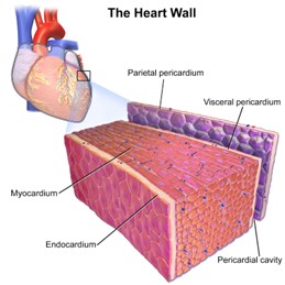
Figure 10: The wall of the heart is made up mainly of myocardium, which consists largely of cardiac muscle.
The four chambers of the heart include two upper chambers called atria (singular, atrium), and two lower chambers called ventricles (Fig 11). The atria are also referred to as receiving chambers, because blood coming into the heart first enters these two chambers. The right atrium receives deoxygenated blood from the upper and lower body through the superior vena cava and inferior vena cava, respectively. The left atrium receives oxygenated blood from the lungs through the pulmonary veins. The ventricles are also referred to as discharging chambers, because blood leaving the heart passes out through these two chambers. The right ventricle discharges blood to the lungs through the pulmonary artery, and the left ventricle discharges blood to the rest of the body through the aorta. The four chambers are separated from each other by dense connective tissue consisting mainly of collagen.
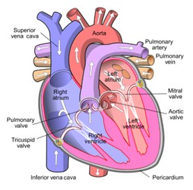
Figure 11: This cross-sectional diagram of the heart shows its four chambers and four valves. The white arrows indicate the direction of blood flow through the heart chambers.
The heart valves allow blood to flow from the atria to the ventricles, and from the ventricles to the pulmonary artery and aorta. The valves are constructed in such a way that blood can flow through them in only one direction, thus preventing the backflow of blood. The four valves are the:
- Tricuspid atrioventricular valve, (can be shortened to tricuspid AV valve) which allows blood to flow from the right atrium to the right ventricle.
- Bicuspid atrioventricular valve (also know as the mitral valve), which allows blood to flow from the left atrium to the left ventricle.
- Pulmonary semilunar valve, which allows blood to flow from the right ventricle to the pulmonary artery.
- Aortic semilunar valve, which allows blood to flow from the left ventricle to the aorta.
Blood circulates through the chambers of the heart and to the body as shown in Fig 12 below. The right atrium collects blood from two large veins, the superior vena cava (from the upper body) and the inferior vena cava (from the lower body). The blood that collects in the right atrium is pumped through the tricuspid valve into the right ventricle. From the right ventricle, the blood is pumped through the pulmonary valve into the pulmonary artery. The pulmonary artery carries the blood to the lungs, where it enters the pulmonary circulation, gives up carbon dioxide, and picks up oxygen. The oxygenated blood travels back from the lungs through the pulmonary veins (of which there are four), and enters the left atrium of the heart. From the left atrium, the blood is pumped through the mitral valve into the left ventricle. From the left ventricle, the blood is pumped through the aortic valve into the aorta, which subsequently branches into smaller arteries that carry the blood throughout the rest of the body. After passing through capillaries and exchanging substances with cells, the blood returns to the right atrium via the superior vena cava and inferior vena cava, and the process begins anew.
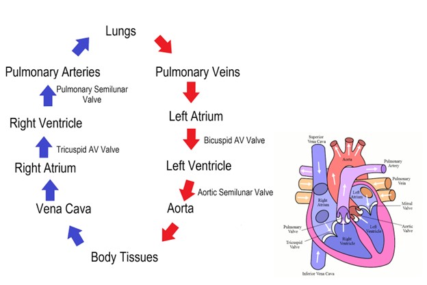
Figure 12: The flow chart in this diagram summarizes the pathway blood takes as it flows into, through, and out of the heart. Trace the path of blood flow in the diagram of the heart as you follow it through the flow chart.
The cardiac cycle refers to a single complete heartbeat, which includes one iteration of the lub and dub sounds heard through a stethoscope. During the cardiac cycle, the atria and ventricles work in a coordinated fashion so that blood is pumped efficiently through and out of the heart. The cardiac cycle includes two parts, called diastole and systole. During diastole, the atria contract and pump blood into the ventricles, while the ventricles relax and fill with blood from the atria. During systole, the atria relax and collect blood from the lungs and body, while the ventricles contract and pump blood out of the heart.
You can see this represented on an electrocardiogram (ECG or EKG).. Here we find wave called the PQRST (Fig 13) which can be broken down in the following way:
- Atrial Systole: During this brief phase, the atria contract, pushing blood into the ventricles. This is typically represented by the P-wave on an ECG.
- Ventricular Systole: Following atrial systole, the ventricles contract forcefully to eject blood into the pulmonary artery (right ventricle) and aorta (left ventricle). The QRS complex on an ECG corresponds to this phase, with the Q-wave representing the initial depolarization of the ventricles, the R-wave representing their main contraction, and the S-wave indicating their recovery or repolarization.
- Atrial Diastole: During this phase, both atria relax and begin to fill with blood from the venous circulation.
- Ventricular Diastole: After ventricular systole, the ventricles relax, creating a low-pressure environment that allows blood to flow into them from the atria. The T-wave on an ECG corresponds to the repolarization of the ventricles during this phase.
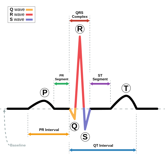
Figure 13: Schematic diagram of normal rhythm for a human heart as seen on ECG. Created by Agateller (Anthony Atkielski), converted to svg by atom., Public domain, via Wikimedia Commons
The PQRST waveforms on an ECG reflect the electrical activity of the heart as it goes through these phases of the cardiac cycle. Importantly, the sequence of events in the cardiac cycle ensures that blood is efficiently pumped through the heart and into the circulatory system, providing the body’s organs and tissues with the oxygen and nutrients they need.
The cardiac cycle considers how blood is pumped out of the heart and into the blood vessels. This requires pressure. Watch the following video on how blood pressure works.
Lymphatic System
Proteins and other large solutes cannot leave the capillaries. The loss of the watery plasma creates a hyperosmotic solution within the capillaries, especially near the venules. This causes about 85% of the plasma that leaves the capillaries to eventually diffuses back into the capillaries near the venules. The remaining 15% of blood plasma drains out from the interstitial fluid into nearby lymphatic vessels. The fluid in the lymph is similar in composition to the interstitial fluid. The lymph fluid passes through lymph nodes before it returns to the heart via the vena cava.
The lymphatic system consists of a complex network of vessels, tissues and organs (Fig 14). Most notably are the lymph nodes which are specialized organs that filter the lymph by percolation through a maze of connective tissue filled with white blood cells. The white blood cells remove infectious agents, such as bacteria and viruses, to “clean” the lymph before it returns to the bloodstream. After it is “cleaned,” the lymph returns to the heart by the action of smooth muscle pumping, skeletal muscle action, and one-way valves joining the returning blood near the junction of the venae cava entering the right atrium of the heart.
The lymphatic system helps maintain the balance of fluids in the body by collecting excess fluid and particulate matter from tissues and depositing them in the bloodstream.

Figure 14: The lymphatic system is composed of various vessels, tissues and organs. Attributed to Blausen.com staff (2014). “Medical gallery of Blausen Medical 2014”. WikiJournal of Medicine 1 (2). DOI:10.15347/wjm/2014.010. ISSN 2002-4436., CC, via Wikimedia Commons
Summary
Humans have a closed circulatory system with arteries and veins moving blood throughout the body. With two circuits, the pulmonary and the systemic, deoxygenated blood can be oxygenated (going from the heart to the lungs) at the same time that oxygenated blood is moving throughout the body. In addition to transporting oxygen, the blood also transports nutrients, hormones, and waste products throughout the body. This system is called “closed” because the blood circulates within a continuous set of vessels and does not come into direct contact with the body’s cells. Instead vessels decrease in size until the tiniest capillaries are within reach of all cells of the body and then diffusion of gases, nutrients, and metabolic waste can occur.
The heart is important to get the blood flowing. It is a muscular organ that acts as the central pump of the circulatory system. It consists of four chambers – two atria and two ventricles. The right atrium receives deoxygenated blood from the body, while the left atrium receives oxygenated blood from the lungs. The ventricles pump blood out of the heart to the lungs and the rest of the body.
Blood is composed of plasma, red blood cells, white blood cells, and platelets. Plasma is a liquid component that carries cells and dissolved substances. Red blood cells transport oxygen from the lungs to the body tissues and carry carbon dioxide back to the lungs for exhalation. White blood cells are involved in immune responses, while platelets aid in blood clotting.
In contrast, an open circulatory system is characterized by the absence of a closed network of blood vessels. Instead, hemolymph is pumped into the body cavity, where it directly bathes the tissues and organs. While this system is less efficient and controlled than a closed circulatory system, it serves the needs of simpler invertebrate organisms, like insects, and allows for the exchange of essential substances between the circulatory fluid and body cells very quickly.
Questions
Glossary
References
Kerr, SK, Weigel E, Spencer C, and D Garton. Types of Circulatory Systems. Organismal Biology. Georgia Institute of Technology.
Miller, C. Information on Human Circulatory System. Human Biology.
