
10 Prokaryotes
At the end of this chapter you will be able to:
- Describe the characteristics of prokaryotic organisms
- Compare and contrast Eubacteria (Bacteria) to Archaea
- Explain the importance and role that plasmids play for prokaryotic cells
- Describe how prokaryotes replicate themselves
- Compare and contrast the cell wall of Gram + and Gram – species of prokaryotes
- Describe the ways in which prokaryotes get energy and carbon for life processes
- Describe the roles that prokaryotes play in the environment
- Describe the benefits prokaryotes play with human health
Introduction
Prokaryotic cells are those that are without a nucleus or other membrane-bound organelles. Two kingdoms of life, Bacteria and Archaea, are prokaryotic. Prokaryotes were the first inhabitants on Earth, appearing 3.5 to 3.8 billion years ago. These organisms are abundant and ubiquitous; that is, they are present everywhere. In addition to inhabiting moderate environments, they are found in extreme conditions: from boiling springs to permanently frozen environments in Antarctica; from salty environments like the Dead Sea to environments under tremendous pressure, such as the depths of the ocean; and from areas without oxygen, such as a waste management plant, to radioactively contaminated regions, such as Chernobyl. Prokaryotes reside in the human digestive system and on the skin, are responsible for certain illnesses, and serve an important role in the preparation of many foods.
Prokaryotes are ubiquitous. They cover every imaginable surface where there is sufficient moisture, and they live on and inside of other living things. In the typical human body, prokaryotic cells outnumber human body cells by about ten to one. They comprise the majority of living things in all ecosystems. Some prokaryotes thrive in environments that are inhospitable for most living things. Prokaryotes recycle nutrients—essential substances (such as carbon and nitrogen)—and they drive the evolution of new ecosystems, some of which are natural and others man-made. Prokaryotes have been on Earth since long before multicellular life appeared.
Prokaryotes were the first forms of life on Earth, and they existed for billions of years before plants and animals appeared. The Earth and its moon are thought to be about 4.54 billion years old. This estimate is based on evidence from radiometric dating of meteorite material together with other substrate material from Earth and the moon. Early Earth had a very different atmosphere (contained less molecular oxygen) than it does today and was subjected to strong radiation; thus, the first organisms would have flourished where they were more protected, such as in ocean depths or beneath the surface of the Earth. At this time too, strong volcanic activity was common on Earth, so it is likely that these first organisms—the first prokaryotes—were adapted to very high temperatures. Early Earth was prone to geological upheaval and volcanic eruption, and was subject to bombardment by mutagenic radiation from the sun. The first organisms were prokaryotes that could withstand these harsh conditions.
Microbial Mats
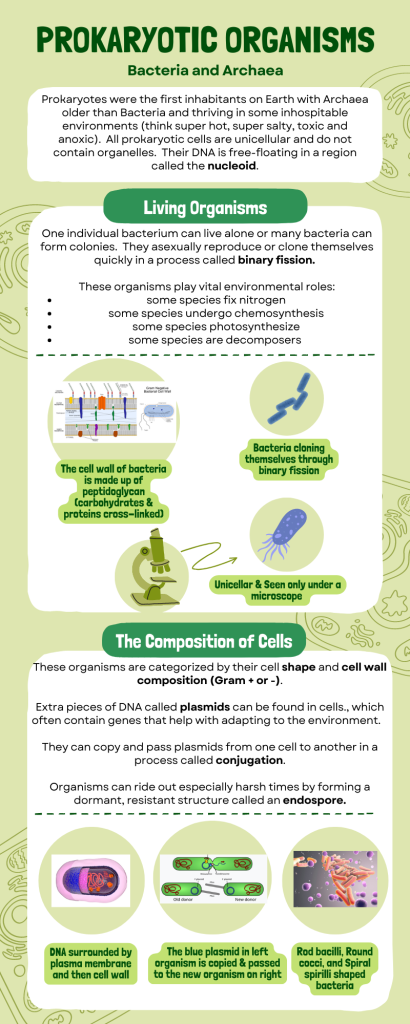
Microbial mats or large biofilms may represent the earliest forms of life on Earth; there is fossil evidence of their presence starting about 3.5 billion years ago. A microbial mat is a multi-layered sheet of prokaryotes (Fig 1) that includes mostly bacteria, but also archaea. Microbial mats are a few centimeters thick, and they typically grow where different types of materials interface, mostly on moist surfaces. The various types of prokaryotes that comprise them carry out different metabolic pathways, and that is the reason for their various colors. Prokaryotes in a microbial mat are held together by a glue-like sticky substance that they secrete called extracellular matrix.
The first microbial mats likely obtained their energy from chemicals found near hydrothermal vents. A hydrothermal vent is a breakage or fissure in the Earth’s surface that releases geothermally heated water. With the evolution of photosynthesis about 3 billion years ago, some prokaryotes in microbial mats came to use a more widely available energy source—sunlight—whereas others were still dependent on chemicals from hydrothermal vents for energy and food.

Figure 1: This (a) microbial mat, about one meter in diameter, grows over a hydrothermal vent in the Pacific Ocean in a region known as the “Pacific Ring of Fire.” The mat helps retain microbial nutrients. Chimneys such as the one indicated by the arrow allow gases to escape. (b) In this micrograph, bacteria are visualized using fluorescence microscopy. (credit a: modification of work by Dr. Bob Embley, NOAA PMEL, Chief Scientist; credit b: modification of work by Ricardo Murga, Rodney Donlan, CDC; scale-bar data from Matt Russell)
Stromatolites
Fossilized microbial mats represent the earliest record of life on Earth. A stromatolite is a sedimentary structure formed when minerals are precipitated out of water by prokaryotes in a microbial mat (Fig 2). Stromatolites form layered rocks made of carbonate or silicate. Although most stromatolites are artifacts from the past, there are places on Earth where stromatolites are still forming. For example, growing stromatolites have been found in the Anza-Borrego Desert State Park in San Diego County, California.
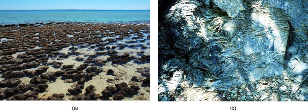
Figure 2: (a) These living stromatolites are located in Shark Bay, Australia. (b) These fossilized stromatolites, found in Glacier National Park, Montana, are nearly 1.5 billion years old. (credit a: Robert Young; credit b: P. Carrara, NPS)
Life in Moderate and Extreme Environments
Some organisms have developed strategies that allow them to survive harsh conditions. Prokaryotes thrive in a vast array of environments: some grow in conditions that would seem very normal to us, whereas others are able to thrive and grow under conditions that would kill a plant or animal. Almost all prokaryotes have a cell wall, a protective structure that allows them to survive in different and extreme osmotic conditions. Some soil bacteria can form endospores that resist heat and drought, thereby allowing the organism to survive until favorable conditions recur. These adaptations, along with others, allow bacteria to be the most abundant life form in all terrestrial and aquatic ecosystems.
Species of bacteria that are capable of making endospores have an advantage for “riding out” hard times. These species of bacteria produce endospores through a process called sporulation, which is often triggered by a lack of essential nutrients or other environmental stressors. The bacterial cell then undergoes an asymmetrical cell division. During this process, the original cell (known as the mother cell) divides into two unequal compartments: a larger mother cell and a smaller forespore, which will eventually become the endospore. The forespore, the smaller compartment, is surrounded by several protective layers. These layers include the inner membrane, peptidoglycan cell wall, and outer membrane. These layers provide structural support and protect the developing endospore from external threats. Next, a protein-rich spore coat is deposited around the forespore. The spore coat is a critical protective layer that helps shield the endospore from harmful environmental factors. Now that the endospore is complete, the mother cell undergoes lysis (breakdown), releasing the mature endospore into the environment. This endospore can remain dormant for extended periods. When conditions become favorable again, the endospore can germinate. This process involves activating metabolic processes, and shedding of the protective layers, along with the outgrowth of the vegetative cell. The vegetative cell can then resume normal bacterial growth and reproduction.
Other bacteria and archaea are adapted to grow under extreme conditions and are called extremophiles, meaning “lovers of extremes.” Extremophiles have been found in all kinds of environments: the depth of the oceans, hot springs, the Arctic, and the Antarctic, in very dry places, deep inside Earth, in harsh chemical environments, and high radiation environments, just to mention a few.
Other extremophiles, like radioresistant organisms, do not prefer an extreme environment (in this case, one with high levels of radiation), but have adapted to survive in it. For example, Deinococcus radiodurans is a prokaryote that can tolerate very high doses of ionizing radiation. It has developed DNA repair mechanisms that allow it to reconstruct its chromosome even if it has been broken into hundreds of pieces by radiation or heat.
These organisms give us a better understanding of prokaryotic diversity and open up the possibility of finding new prokaryotic species that may lead to the discovery of new therapeutic drugs or have industrial applications. Because they have specialized adaptations that allow them to live in extreme conditions, many extremophiles cannot survive in moderate environments.
There are many different groups of extremophiles: they are identified based on the conditions in which they grow best, and several habitats are extreme in multiple ways. For example, a soda lake is both salty and alkaline, so organisms that live in a soda lake must be both alkaliphiles and halophiles (Table 1).
Table 1: Extremophiles and Their Preferred Conditions
| Extremophile Type | Conditions for Optimal Growth |
| Acidophiles | pH 3 or below |
| Alkaliphiles | pH 9 or above |
| Thermophiles | Temperature 60–80 °C (140–176 °F) |
| Hyperthermophiles | Temperature 80–122 °C (176–250 °F) |
| Psychrophiles | Temperature of −15–10 °C (5–50 °F) or lower |
| Halophiles | Salt concentration of at least 0.2 M |
| Osmophiles | High sugar concentration |
One example of a very harsh environment is the Dead Sea, a hypersaline basin that is located between Jordan and Israel. Hypersaline environments are essentially concentrated seawater. In the Dead Sea, the sodium concentration is 10 times higher than that of seawater, and the water contains high levels of magnesium (about 40 times higher than in seawater) that would be toxic to most living things. Iron, calcium, and magnesium, elements that form divalent ions (Fe2+, Ca2+, and Mg2+), produce what is commonly referred to as “hard” water.
Taken together, the high concentration of divalent cations, the acidic pH (6.0), and the intense solar radiation flux make the Dead Sea a unique, and uniquely hostile, ecosystem (Bodaker et al. 2010; Fig 3).
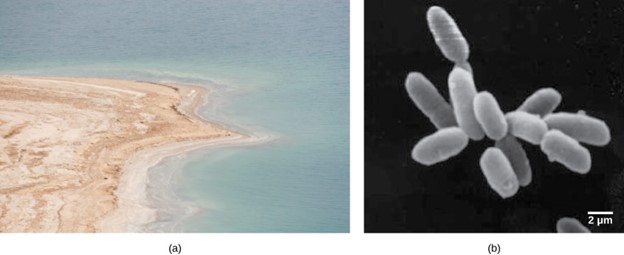
Figure 3: (a) The Dead Sea is hypersaline. Nevertheless, salt-tolerant bacteria thrive in this sea. (b) These halobacteria cells can form salt-tolerant bacterial mats. (credit a: Julien Menichini; credit b: NASA; scale-bar data from Matt Russell)
What sort of prokaryotes do we find in the Dead Sea? The extremely salt-tolerant bacterial mats include Halobacterium, Haloferax volcanii (which is found in other locations, not only the Dead Sea), Halorubrum sodomense, and Halobaculum gomorrense, and the Archaea Haloarcula marismortui, among others.
Prokaryotic Cell Structure
There are many differences between prokaryotic and eukaryotic cells. However, all cells have four common structures: the plasma membrane, which functions as a barrier for the cell and separates the cell from its environment; the cytoplasm, a jelly-like substance inside the cell; nucleic acids, the genetic material of the cell; and ribosomes, where protein synthesis takes place. Prokaryotes come in various shapes, but many fall into three categories: cocci (spherical), bacilli (rod-shaped), and spirilli (spiral-shaped) (Fig 4).

Figure 4: Prokaryotes fall into three basic categories based on their shape, visualized here using scanning electron microscopy: (a) cocci, or spherical (a pair is shown); (b) bacilli, or rod-shaped; and (c) spirilli, or spiral-shaped. (credit a: modification of work by Janice Haney Carr, Dr. Richard Facklam, CDC; credit c: modification of work by Dr. David Cox; scale-bar data from Matt Russell)
A prokaryotic cell is a simple, single-celled (unicellular) organism that lacks a nucleus, or any other membrane-bound organelle. Prokaryotic DNA is found in the central part of the cell: a darkened region called the nucleoid (Fig 5). Some prokaryotes have flagella, pili, or fimbriae. Flagella are used for locomotion, while most pili are used to exchange genetic material during a type of reproduction called conjugation. Many prokaryotes also have a cell wall and capsule. The cell wall acts as an extra layer of protection, helps the cell maintain its shape, and prevents dehydration. The capsule enables the cell to attach to surfaces in its environment.
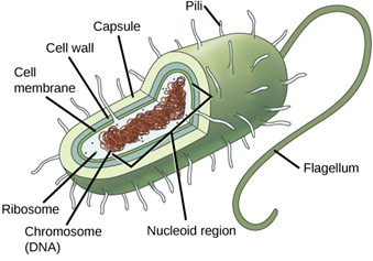
Figure 5: The features of a typical prokaryotic cell are shown.
Reproduction in prokaryotes is asexual and usually takes place by binary fission. The DNA of a prokaryote exists as a single, circular chromosome. Prokaryotes do not undergo mitosis. Rather the chromosome is replicated and the two resulting copies separate from one another due to the growth of the cell. The prokaryote, now enlarged, is pinched inward at its equator and the two resulting cells, which are clones, separate. Binary fission does not provide an opportunity for genetic recombination or genetic diversity, but prokaryotes can share genes by three other mechanisms. In transformation, the prokaryote takes in DNA found in its environment that is shed by other prokaryotes. If a nonpathogenic bacterium takes up DNA for a toxin gene from a pathogen and incorporates the new DNA into its own chromosome, it too may become pathogenic. In transduction, bacteriophages, the viruses that infect bacteria, sometimes also move short pieces of chromosomal DNA from one bacterium to another. Transduction results in a recombinant organism. Archaea are not affected by bacteriophages but instead have their own viruses that translocate genetic material from one individual to another. In conjugation, DNA is transferred from one prokaryote to another by means of a pilus, which brings the organisms into contact with one another. The DNA transferred can be in the form of a plasmid, a small circular piece of extrachromosomal DNA, or as a hybrid, containing both plasmid and chromosomal DNA. These three processes of DNA exchange are shown in Figure 6. Reproduction can be very rapid: a few minutes for some species. This short generation time coupled with mechanisms of genetic recombination and high rates of mutation result in the rapid evolution of prokaryotes, allowing them to respond to environmental changes (such as the introduction of an antibiotic) very quickly.
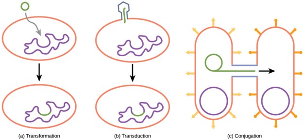
Figure 6: Besides binary fission, there are three other mechanisms by which prokaryotes can exchange DNA. In (a) transformation, the cell takes up prokaryotic DNA directly from the environment. The DNA may remain separate as plasmid DNA or be incorporated into the host genome. In (b) transduction, a bacteriophage injects DNA into the cell that contains a small fragment of DNA from a different prokaryote. In (c) conjugation, DNA is transferred from one cell to another via a mating bridge that connects the two cells after the sex pilus draws the two bacteria close enough to form the bridge.
Archaea and Bacteria: Two Kingdoms of Prokaryotes
The composition of the cell wall differs significantly between the Bacteria and Archaea. The composition of their cell walls also differs from the eukaryotic cell walls found in plants (cellulose) or fungi and insects (chitin). The cell wall functions as a protective layer, and it is responsible for the organism’s shape. Some bacteria have an outer capsule outside the cell wall. Other structures are present in some prokaryotic species, but not in others. For example, the capsule found in some species enables the organism to attach to surfaces, protects it from dehydration and attack by phagocytic cells, and makes pathogens more resistant to our immune responses. Some species also have flagella (singular, flagellum) used for locomotion, and pili (singular, pilus) used for attachment to surfaces. Plasmids, which consist of extra-chromosomal DNA, are also present in many species of bacteria and archaea.
The plasma membrane is a thin lipid bilayer (6 to 8 nanometers) that surrounds the cell and separates the inside from the outside. Its selectively permeable nature keeps ions, proteins, and other molecules within the cell and prevents them from diffusing into the extracellular environment, while other molecules may move through the membrane. The general structure of a cell membrane is a phospholipid bilayer composed of two layers of lipid molecules. In archaeal cell membranes, isoprene (phytanyl) chains linked to glycerol replace the fatty acids linked to glycerol in bacterial membranes. Some archaeal membranes are lipid monolayers instead of bilayers (Fig 7).
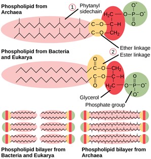
Figure 7: Archaeal phospholipids differ from those found in Bacteria and Eukarya in two ways. First, they have branched phytanyl sidechains instead of linear ones. Second, an ether bond instead of an ester bond connects the lipid to the glycerol.
The cytoplasm of prokaryotic cells has a high concentration of dissolved solutes. Therefore, the osmotic pressure within the cell is relatively high. The cell wall is a protective layer that surrounds some cells and gives them shape and rigidity. It is located outside the cell membrane and prevents osmotic lysis (bursting due to increasing volume). The chemical composition of the cell walls varies between archaea and bacteria, and also varies between bacterial species.
Bacterial cell walls contain peptidoglycan, composed of polysaccharide chains that are cross-linked by unusual peptides containing both L- and D-amino acids including D-glutamic acid and D-alanine. Proteins normally have only L-amino acids; as a consequence, many of our antibiotics work by mimicking D-amino acids and therefore have specific effects on bacterial cell wall development. There are more than 100 different forms of peptidoglycan. S-layer (surface layer) proteins are also present on the outside of cell walls of both archaea and bacteria.
Bacteria are divided into two major groups: Gram positive and Gram negative, based on their reaction to Gram staining. Note that all Gram-positive bacteria belong to one phylum; bacteria in the other phyla (Proteobacteria, Chlamydias, Spirochetes, Cyanobacteria, and others) are Gram-negative. The Gram staining method is named after its inventor, Danish scientist Hans Christian Gram (1853–1938). The different bacterial responses to the staining procedure are ultimately due to cell wall structure. Gram-positive organisms typically lack the outer membrane found in Gram-negative organisms (Fig 8). Up to 90 percent of the cell wall in Gram-positive bacteria is composed of peptidoglycan, and most of the rest is composed of acidic substances called teichoic acids. Teichoic acids may be covalently linked to lipids in the plasma membrane to form lipoteichoic acids. Lipoteichoic acids anchor the cell wall to the cell membrane. The cell wall of Gram + bacteria is thick. Gram-negative bacteria have a relatively thin cell wall composed of a few layers of peptidoglycan (only 10 percent of the total cell wall), surrounded by an outer envelope containing lipopolysaccharides (LPS) and lipoproteins. This outer envelope is sometimes referred to as a second lipid bilayer. The chemistry of this outer envelope is very different, however, from that of the typical lipid bilayer that forms plasma membranes.
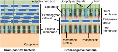
Figure 8: Gram-positive and -negative bacteria (credit: modification of work by “Franciscosp2″/Wikimedia Commons)
Underneath the microscope, Gram-positive bacteria will appear purple, while Gram-negative species will appear pink/red (Fig 9).
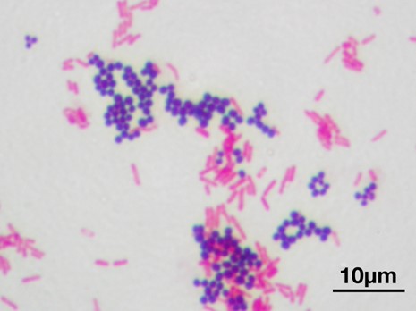
Figure 9: A microscopic image of a Gram stain of mixed Gram-positive cocci (Staphylococcus aureus ATCC 25923, purple) and Gram-negative bacilli (Escherichia coli ATCC 11775, pink). Y tambe, CC via Wikimedia Commons
Archaean cell walls do not have peptidoglycan. There are four different types of Archaean cell walls. One type is composed of pseudopeptidoglycan, which is similar to peptidoglycan in morphology but contains different sugars in the polysaccharide chain. The other three types of cell walls are composed of polysaccharides, glycoproteins, or pure protein.
Prokaryote Metabolism
Like all living things, prokaryotes need energy and carbon. They meet these needs in a variety of ways. Prokaryotes have just about every possible type of metabolism. They may get energy from light (photo) or chemical compounds (chemo). They may get carbon from carbon dioxide (autotroph) or other living things (heterotroph). Most prokaryotes are chemoheterotrophs. They depend on other organisms for both energy and carbon. Many break down organic wastes and the remains of dead organisms. They play vital roles as decomposers and help recycle carbon and nitrogen. Photoautotrophs are important producers. They are especially important in aquatic ecosystems.
Two major nutritional needs can be used to group prokaryotes. These are (1) carbon metabolism, their source of carbon for building organic molecules within the cells, and (2) energy metabolism, their source of energy used for growth.
In terms of carbon metabolism, prokaryotes are classified as either heterotrophic or autotrophic:
- Heterotrophic organisms use organic compounds, usually from other organisms, as carbon sources.
- Autotrophic organisms use carbon dioxide (CO2) as their only source or their main source of carbon. Many autotrophic bacteria are photosynthetic, and get their carbon from the carbon dioxide in the atmosphere. This process of capturing inorganic carbon and converting it to organic sugar molecules is known as carbon fixation.
Energy metabolism in prokaryotes is classified as one of the following:
- Phototrophic organisms capture light energy from the sun and convert it into chemical energy inside their cells.
- Chemotrophic organisms break down either organic or inorganic molecules to supply energy for the cell. Some chemotrophic organisms can also use their organic energy-supplying molecules as a carbon supply, which would make them chemoheterotrophs.
This is a nice summary video on Bacteria.
Role in the Environment
Prokaryotes play very important roles in the environment. As you read above, they can be producers, making food that drives food chains in communities. They can be decomposers, helping to cycle nutrients in the environment. Some species of bacteria can even convert unusable forms of an element into forms that can be used by other organisms. One primary example of this is the rare, but very important, species of bacteria that fix nitrogen. That is, species of prokaryotes can take nitrogen from the atmosphere that is not useable to living organisms and convert it into a useable form.
Nitrogen is a very important element for life because it is part of proteins and nucleic acids. It is a macronutrient, and in nature, it is recycled from organic compounds to ammonia, ammonium ions, nitrate, nitrite, and nitrogen gas by myriad processes, many of which are carried out only by prokaryotes. Prokaryotes are key to the nitrogen cycle (Fig 10). The largest pool of nitrogen available in the terrestrial ecosystem is gaseous nitrogen from the air, but this nitrogen is not usable by plants, which are primary producers. Gaseous nitrogen is transformed, or “fixed” into more readily available forms such as ammonia through the process of nitrogen fixation. Ammonia can be used by plants or converted to other forms.
Another source of ammonia is ammonification, the process by which ammonia is released during the decomposition of nitrogen-containing organic compounds. Ammonia released to the atmosphere, however, represents only 15 percent of the total nitrogen released; the rest is as N2 and N2O. Ammonia is catabolized anaerobically by some prokaryotes, yielding N2 as the final product. Nitrification is the conversion of ammonium to nitrite and nitrate. Nitrification in soils is carried out by bacteria belonging to the genera Nitrosomas, Nitrobacter, and Nitrospira. The bacteria perform the reverse process, the reduction of nitrate from the soils to gaseous compounds such as N2O, NO, and N2, a process called denitrification.
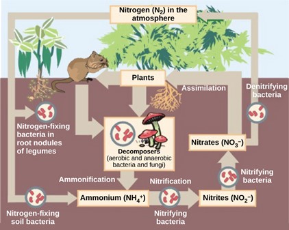
Figure 10: Prokaryotes play a key role in the nitrogen cycle. (credit: Environmental Protection Agency)
Humans also use bacteria in different ways, so their role is directly applicable depending on our needs. We can see the use of prokaryotes for food production as well as biotechnology. According to the United Nations Convention on Biological Diversity, biotechnology is “any technological application that uses biological systems, living organisms, or derivatives thereof, to make or modify products or processes for specific use.” The concept of “specific use” involves some sort of commercial application. Genetic engineering, artificial selection, antibiotic production, and cell culture are current topics of study in biotechnology. However, humans have used prokaryotes before the term biotechnology was even coined. In addition, some of the goods and services are as simple as cheese, bread, wine, beer, and yogurt, which employ both bacteria and other microbes, such as yeast, a fungus (Fig 11).
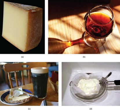
Figure 11: Some of the products derived from the use of prokaryotes in early biotechnology include (a) cheese, (b) wine, (c) beer and bread, and (d) yogurt. (credit bread: modification of work by F. Rodrigo/Wikimedia Commons; credit wine: modification of work by Jon Sullivan; credit beer and bread: modification of work by Kris Miller; credit yogurt: modification of work by Jon Sullivan)
Cheese production began around 4,000–7,000 years ago when humans began to breed animals and process their milk. Fermentation in this case preserves nutrients: milk will spoil relatively quickly, but when processed as cheese, it is more stable. As for beer, the oldest records of brewing are about 6,000 years old and refer to the Sumerians. Evidence indicates that the Sumerians discovered fermentation by chance. Wine has been produced for about 4,500 years, and evidence suggests that cultured milk products, like yogurt, have existed for at least 4,000 years.
Humans can also use prokaryotes to help clean up the environment. Microbial bioremediation is the use of prokaryotes (or microbial metabolism) to remove pollutants. Bioremediation has been used to remove agricultural chemicals (pesticides, fertilizers) that leach from soil into groundwater and the subsurface. Certain toxic metals and oxides, such as selenium and arsenic compounds, can also be removed from water by bioremediation. The reduction of SeO4−2 to SeO3−2 to Se0 (metallic selenium) is a method used to remove selenium ions from water. Mercury is an example of a toxic metal that can be removed from an environment by bioremediation. As an active ingredient of some pesticides, mercury is used in industry and is also a by-product of certain processes, such as battery production. Methyl mercury is usually present in very low concentrations in natural environments, but it is highly toxic because it accumulates in living tissues. Several species of bacteria can carry out the biotransformation of toxic mercury into nontoxic forms. These bacteria, such as Pseudomonas aeruginosa, can convert Hg+2 into Hg0, which is non-toxic to humans.
One of the most useful and interesting examples of the use of prokaryotes for bioremediation purposes is the cleanup of oil spills. The importance of prokaryotes to petroleum bioremediation has been demonstrated in several oil spills in recent years, such as the Exxon Valdez spill in Alaska (1989) (Fig 12), the Prestige oil spill in Spain (2002), the spill into the Mediterranean from a Lebanon power plant (2006), and more recently, the BP oil spill in the Gulf of Mexico (2010).
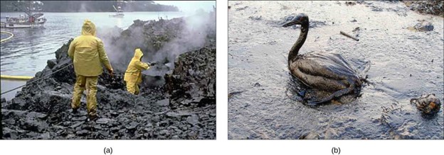
Figure 12: (a) Cleaning up oil after the Valdez spill in Alaska, workers hosed oil from beaches and then used a floating boom to corral the oil, which was finally skimmed from the water surface. Some species of bacteria are able to solubilize and degrade the oil. (b) One of the most catastrophic consequences of oil spills is the damage to fauna. (credit a: modification of work by NOAA; credit b: modification of work by GOLUBENKOV, NGO: Saving Taman)
To clean up these spills, bioremediation is promoted by the addition of inorganic nutrients that help bacteria to grow. Hydrocarbon-degrading bacteria feed on hydrocarbons in the oil droplet, breaking down the hydrocarbons. Some species, such as Alcanivorax borkumensis, produce surfactants that solubilize the oil, whereas other bacteria degrade the oil into carbon dioxide. In the case of oil spills in the ocean, ongoing, natural bioremediation tends to occur, since there are oil-consuming bacteria in the ocean before the spill. In addition to naturally occurring oil-degrading bacteria, humans select and engineer bacteria that possess the same capability with increased efficacy and spectrum of hydrocarbon compounds that can be processed. Under ideal conditions, it has been reported that up to 80 percent of the nonvolatile components in oil can be degraded within one year of the spill. Other oil fractions containing aromatic and highly branched hydrocarbon chains are more difficult to remove and remain in the environment for longer periods.
A final note centers on the direct use of microbes on or in our bodies. Human life is only possible due to the action of microbes, both those in the environment and those species that call us home. Internally, they help us digest our food, produce crucial nutrients for us, protect us from pathogenic microbes, and help train our immune systems to function correctly. Each individual has a normal microbial flora (also known as gut microbiota)—these terms simply refer to the collective of the bacteria living in each person’s stomach. When these bacteria counts change, it can cause digestive problems.
The bacteria that inhabit our skin and gastrointestinal tract do a host of good things for us. They protect us from pathogens, help us digest our food, and produce some of our vitamins and other nutrients. These activities have been known for a long time. More recently, scientists have gathered evidence that these bacteria may also help regulate our moods, influence our activity levels, and even help control weight by affecting our food choices and absorption patterns. The Human Microbiome Project has begun the process of cataloging our normal bacteria (and archaea) so we can better understand these functions.
A particularly fascinating example of our normal flora relates to our digestive systems. People who take high doses of antibiotics tend to lose many of their normal gut bacteria, allowing a naturally antibiotic-resistant species called Clostridium difficile to overgrow and cause severe gastric problems, especially chronic diarrhea. Trying to treat this problem with antibiotics only makes it worse. However, it has been successfully treated by giving the patients fecal transplants from healthy donors to reestablish the normal intestinal microbial community.
Scientists are also discovering that the absence of certain key microbes from our intestinal tract may set us up for a variety of problems. This seems to be particularly true regarding the appropriate functioning of the immune system. There are intriguing findings that suggest that the absence of these microbes is an important contributor to the development of allergies and some autoimmune disorders. Research is currently underway to test whether adding certain microbes to our internal ecosystem may help in the treatment of these problems as well as in treating some forms of autism.
Summary
Prokaryotes are a diverse and ancient group of microorganisms that lack a true nucleus and membrane-bound organelles. They constitute two distinct kingdoms of life: Bacteria and Archaea. Archaea were initially classified with bacteria, but they differ significantly in genetics, biochemistry, and cell membrane composition. Archaea often thrive in extreme environments and have unique molecular characteristics. Despite their simple cellular structure, both Archaea and Bacteria are fundamental to Earth’s ecosystems and have significant impacts on various aspects of life.
Key features of prokaryotes:
- Cellular Structure: Prokaryotic cells are characterized by their lack of a membrane-bound nucleus. Their genetic material, typically a single circular DNA molecule, is found in the nucleoid region. They lack membrane-bound organelles like mitochondria or chloroplasts.
- Size and Diversity: Prokaryotes come in a wide range of sizes and shapes, from spherical cocci to rod-shaped bacilli and spiral spirilla. They exhibit remarkable metabolic diversity and can thrive in diverse environments, including extreme conditions like hot springs, acidic environments, and deep-sea hydrothermal vents.
- Reproduction: Prokaryotes reproduce asexually through binary fission, where a single cell divides into two identical daughter cells. This rapid reproductive process contributes to their ability to adapt quickly to changing environments.
- Ecological Significance: Prokaryotes are vital to various ecological processes. They play essential roles in nutrient cycling, such as nitrogen fixation and decomposition. Many prokaryotes are symbiotic, forming partnerships with plants (e.g., nitrogen-fixing bacteria in root nodules) or animals (e.g., gut bacteria aiding digestion).
- Human Health and Industry: While some prokaryotes are pathogenic and cause diseases, others have positive impacts. Beneficial bacteria aid in digestion, vitamin synthesis, and immune system support. Prokaryotes are used in various industrial applications, including food production (fermentation), bioremediation (pollution cleanup), and pharmaceuticals (antibiotics).
Overall, prokaryotes are essential components of Earth’s biosphere, shaping ecosystems, influencing global processes, and contributing to the diversity of life. Their ability to adapt, evolve, and occupy diverse ecological niches underscores their importance in the grand tapestry of life on our planet.
Questions
Glossary
References
Bodaker, I, Itai, S, Suzuki, MT, Feingersch, R, Rosenberg, M, Maguire, ME, Shimshon, B, and others. 2010. Comparative community genomics in the Dead Sea: An increasingly extreme environment. The ISME Journal 4: 399–407, doi:10.1038/ismej.2009.141.
Kosal, E. 2023. Summary. NC State University.
Lumen Learning. Fundamentals of Biology I.
United Nations COnvention on Biological DIversity. Article 2: found at http://www.cbd.int/convention/articles/?a=cbd-02
