
19 Digestion
At the end of this chapter, you will be able to:
- Explain the processes of digestion and absorption
- Explain the specialized functions of the organs involved in processing food in the body
- Describe the ways in which organs work together to digest food and absorb nutrients
- Describe the essential nutrients required for cellular function that cannot be synthesized by the animal body
- Describe how excess carbohydrates and energy are stored in the body
Introduction
All living organisms need nutrients to survive. While plants can obtain nutrients from their roots and the energy molecules required for cellular function through the process of photosynthesis, animals obtain their nutrients through the consumption of other organisms. At the cellular level, the biological molecules necessary for animal function are amino acids, lipid molecules, nucleotides, and simple sugars. However, the food consumed consists of protein, fat, and complex carbohydrates. Animals must convert these macromolecules into the simple molecules required for maintaining cellular function. The conversion of the food consumed to the nutrients required is a multistep process involving digestion and absorption. During digestion, food particles are broken down into smaller components, which are later absorbed by the body. This happens by both physical means, such as chewing, and by chemical means.
One of the challenges in human nutrition is maintaining a balance between food intake, storage, and energy expenditure. Taking in more food energy than is used in activity leads to the storage of the excess in the form of fat deposits. The rise in obesity and the resulting diseases like type 2 diabetes makes understanding the role of diet and nutrition in maintaining good health all the more important.
Functions of the Digestive System
The digestive system has three main functions relating to food: digestion of food, absorption of nutrients from food, and elimination of solid food waste. Digestion is the process of breaking down food into components the body can absorb. It consists of two types of processes: mechanical digestion and chemical digestion. Mechanical digestion is the physical breakdown of chunks of food into smaller pieces, and it takes place mainly in the mouth and stomach. Chemical digestion is the chemical breakdown of large, complex food molecules into smaller, simpler nutrient molecules that can be absorbed by body fluids (blood or lymph). This type of digestion begins in the mouth and continues in the stomach, but occurs mainly in the small intestine.
After food is digested, the resulting nutrients are absorbed. Absorption is the process in which substances pass into the bloodstream or lymph system to circulate throughout the body. Absorption of nutrients occurs mainly in the small intestine. Any remaining matter from food that is not digested and absorbed passes out of the body through the anus in the process of elimination.
Types of Digestive Systems
Some animals do not have a digestive system, and other species have quite complex systems. For example, thin flatworms in the Platylhelminthes phylum engage in digestion using a relatively simple digestive system. Flatworms have a single opening to their digestive system, known as the “mouth,” which also serves as the “anus.” This type of digestive system is referred to as an incomplete digestive system. It means that the same opening is used for both taking in food and expelling waste, which can also be referred to as a gastrovascular cavity.
Flatworms are carnivorous or scavengers, depending on the species. They consume various types of food, such as small invertebrates, detritus, or decaying organic matter. When a flatworm encounters food, it uses a muscular tube called the pharynx to extend from its mouth and attach to the food item. The pharynx then releases digestive enzymes that start breaking down the food externally in a process called extracellular digestion, because it occurs outside the flatworm’s cells.
Once the food is partially digested externally, the flatworm uses its muscular body wall to engulf the softened food. The nutrients are absorbed through the body wall and directly enter the flatworm’s cells, where further intracellular digestion takes place. The digested nutrients are then distributed throughout the flatworm’s body, providing the necessary energy for cellular processes, growth, and reproduction. Whatever undigested or indigestible material remains after digestion is expelled through the same opening, the gastrovascular cavity (Fig 1).
The simplicity of the flatworm’s digestive system is reflective of the organism’s relatively simple body plan and limited size.
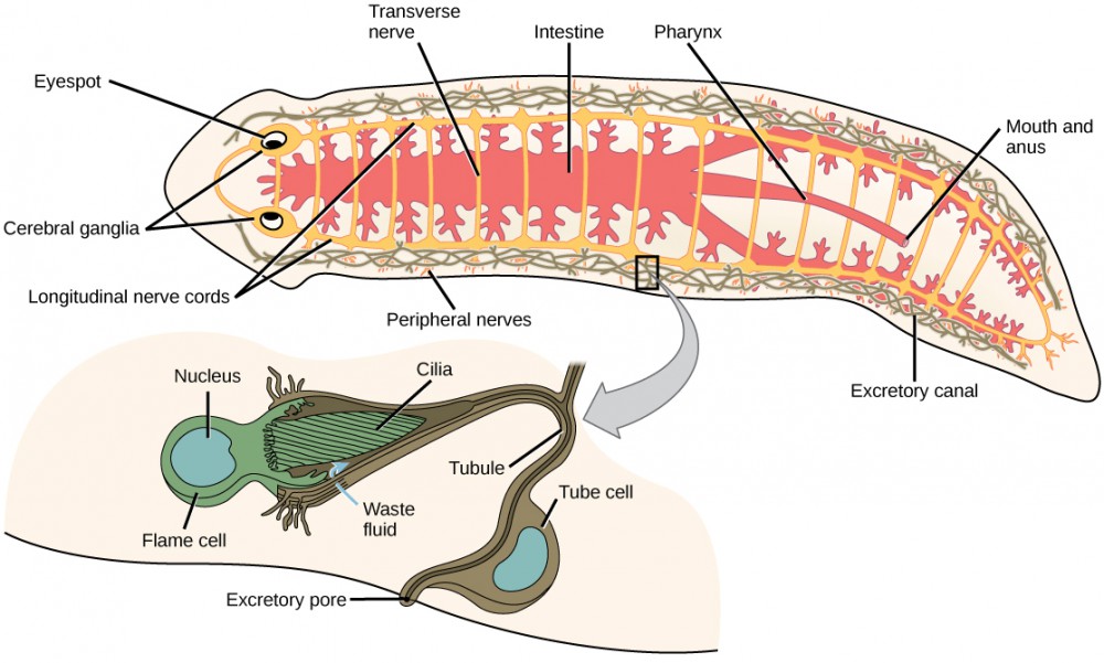
Figure 1: The digestive system (top) and excretory system (bottom) of a flatworm
In addition to the incomplete digestive system of flatworms, we can also see complete digestive systems in more complex animals. This complete digestive system is the most common in th animal kingdom and consists of a complete one-way digestive tract with separate openings for ingestion (mouth) and egestion (anus). The digestive system includes specialized organs such as the esophagus, stomach, small intestine, and large intestine. We will focus on this type of system as we delve deeper into the digestive system of humans.
Other types of digestive systems include:
- Crop-Gizzard Digestive System: Some animals, like birds, have a specialized digestive system with a crop and a gizzard. The crop stores and moistens the food before it enters the stomach, while the gizzard is a muscular structure that grinds and breaks down the food mechanically, often using swallowed stones or grit.
- Saccular Digestive System: Certain insects, like grasshoppers, have a saccular digestive system. It includes specialized regions such as the crop and the gastric caeca. The crop stores food temporarily, and the gastric caeca are blind sacs that secrete digestive enzymes to aid in further digestion.
- Rumen-Complex Digestive System: Ruminants, like cows and deer, possess a complex digestive system to break down cellulose-rich plant material. Their stomach has four compartments, including the rumen, reticulum, omasum, and abomasum. Microbes in the rumen help ferment cellulose, making it digestible.
- Filter-Feeding Digestive System: Filter-feeding animals, such as baleen whales and some marine invertebrates (e.g., clams, krill), have specialized structures to filter food particles from the water. They often possess feathery structures or baleen plates that trap food as water passes through.
- Detritivore Digestive System: Detritivores, such as earthworms and some insects, have digestive systems adapted to consuming decaying organic matter (detritus). Their digestive systems play a vital role in breaking down and recycling organic material in the ecosystem.
These examples illustrate the diversity of digestive systems in the animal kingdom, each of which has evolved to match the specific feeding habits, dietary needs, and environmental conditions of different species. The complexity of the digestive system often corresponds to the complexity of the organism’s body plan and its ecological role in the ecosystem.
Human Digestive System
Watch this video on how the digestive system works.
The process of digestion begins in the mouth with the intake of food (Fig 2). The teeth play an important role in masticating (chewing) or physically breaking food into smaller particles. The enzymes present in saliva also begin to chemically break down food. The food is then swallowed and enters the esophagus—a long tube that connects the mouth to the stomach. Using peristalsis, or wave-like smooth-muscle contractions, the muscles of the esophagus push the food toward the stomach. The stomach contents are extremely acidic, with a pH between 1.5 and 2.5. This acidity kills microorganisms, breaks down food tissues, and activates digestive enzymes. Further breakdown of food takes place in the small intestine where bile produced by the liver, and enzymes produced by the small intestine and the pancreas, continue the process of digestion. The smaller molecules are absorbed into the bloodstream through the epithelial cells lining the walls of the small intestine. The waste material travels onto the large intestine where water is absorbed and the drier waste material is compacted into feces; it is stored until it is excreted through the anus.
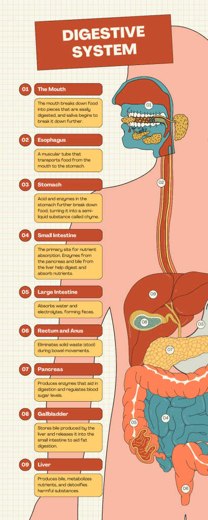
Figure 2: The components of the human digestive system
Both physical and chemical digestion begins in the mouth or oral cavity, which is the point of entry of food into the digestive system. The food is broken into smaller particles by mastication, the chewing action of the teeth. All mammals have teeth and can chew their food to begin the process of physically breaking it down into smaller particles.
The chemical process of digestion begins during chewing as food mixes with saliva, produced by the salivary glands (Fig 3). Saliva contains mucus that moistens food and buffers the pH of the food. Saliva also contains lysozyme, which has antibacterial action. It also contains an enzyme called salivary amylase that begins the process of converting starches in the food into a disaccharide called maltose. Another enzyme called lipase is produced by cells in the tongue to break down fats. The chewing and wetting action provided by the teeth and saliva prepare the food into a mass called the bolus for swallowing. The tongue helps in swallowing—moving the bolus from the mouth into the pharynx. The pharynx opens to two passageways: the esophagus and the trachea. The esophagus leads to the stomach and the trachea leads to the lungs. The epiglottis is a flap of tissue that covers the tracheal opening during swallowing to prevent food from entering the lungs.
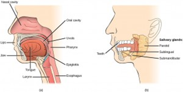
Figure 3: (a) Digestion of food begins in the mouth. (b) Food is masticated by teeth and moistened by saliva secreted from the salivary glands. Enzymes in the saliva begin to digest starches and fats. With the help of the tongue, the resulting bolus is moved into the esophagus by swallowing. (credit: modification of work by Mariana Ruiz Villareal)
The esophagus is a tubular organ that connects the mouth to the stomach. The chewed and softened food passes through the esophagus after being swallowed. The smooth muscles of the esophagus undergo peristalsis that pushes the food toward the stomach. The peristaltic wave is unidirectional—it moves food from the mouth to the stomach, and reverse movement is not possible, except in the case of the vomit reflex. The peristaltic movement of the esophagus is an involuntary reflex; it takes place in response to the act of swallowing.
Watch the video on by Mister Science to see peristalsis in action.
Ring-like muscles called sphincters form valves in the digestive system. The gastro-esophageal sphincter (or cardiac sphincter) is located at the stomach end of the esophagus. In response to swallowing and the pressure exerted by the bolus of food, this sphincter opens, and the bolus enters the stomach. When there is no swallowing action, this sphincter is shut and prevents the contents of the stomach from traveling up the esophagus. Acid reflux or “heartburn” occurs when the acidic digestive juices escape into the esophagus.
A large part of protein digestion occurs in the stomach (Fig 4). The stomach is a saclike organ that secretes gastric digestive juices.
Protein digestion is carried out by an enzyme called pepsin in the stomach chamber. The highly acidic environment kills many microorganisms in the food and, combined with the action of the enzyme pepsin, results in the catabolism of protein in the food. Chemical digestion is facilitated by the churning action of the stomach caused by contraction and relaxation of smooth muscles. The partially digested food and gastric juice mixture is called chyme. Gastric emptying occurs within two to six hours after a meal. Only a small amount of chyme is released into the small intestine at a time. The movement of chyme from the stomach into the small intestine is regulated by hormones, stomach distension and muscular reflexes that influence the pyloric sphincter.
The stomach lining is unaffected by pepsin and the acidity because pepsin is released in an inactive form and the stomach has a thick mucus lining that protects the underlying tissue.
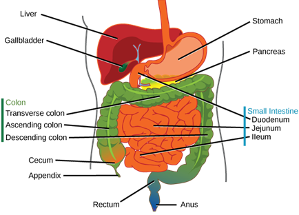
Figure 4: The stomach has an extremely acidic environment where most of the protein gets digested. (credit: modification of work by Mariana Ruiz Villareal)
Chyme moves from the stomach to the small intestine. The small intestine is the organ where the digestion of protein, fats, and carbohydrates is completed. The small intestine is a long tube-like organ with a highly folded surface containing finger-like projections called the villi. The top surface of each villus has many microscopic projections called microvilli. The epithelial cells of these structures absorb nutrients from the digested food and release them to the bloodstream on the other side. The villi and microvilli, with their many folds, increase the surface area of the small intestine and increase the absorption efficiency of the nutrients.
The human small intestine is over 6 m (19.6 ft) long and is divided into three parts: the duodenum, the jejunum and the ileum. The duodenum is separated from the stomach by the pyloric sphincter. The chyme is mixed with pancreatic juices, an alkaline solution rich in bicarbonate that neutralizes the acidity of chyme from the stomach. Pancreatic juices contain several digestive enzymes that break down starches, disaccharides, proteins, and fats. Bile is produced in the liver and stored and concentrated in the gallbladder; it enters the duodenum through the bile duct. Bile contains bile salts, which make lipids accessible to the water-soluble enzymes. The monosaccharides, amino acids, bile salts, vitamins, and other nutrients are absorbed by the cells of the intestinal lining.
The undigested food is sent to the colon from the ileum via peristaltic movements. The ileum ends and the large intestine begins at the ileocecal valve. The vermiform, “worm-like,” appendix is located at the ileocecal valve. The appendix of humans has a minor role in immunity.
The large intestine reabsorbs the water from indigestible food material and processes the waste material (Fig 4). The human large intestine is much smaller in length compared to the small intestine but larger in diameter. It has three parts: the cecum, the colon, and the rectum. The cecum joins the ileum to the colon and is the receiving pouch for the waste matter. The colon is home to many bacteria or “intestinal flora” that aid in the digestive processes. The colon has four regions, the ascending colon, the transverse colon, the descending colon and the sigmoid colon. The main functions of the colon are to extract the water and mineral salts from undigested food, and to store waste material (Fig 5).
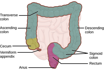
Figure 5: The large intestine reabsorbs water from undigested food and stores waste until it is eliminated. (credit: modification of work by Mariana Ruiz Villareal)
The rectum (Fig 5) stores feces until defecation. The feces are propelled using peristaltic movements during elimination. The anus is an opening at the far-end of the digestive tract and is the exit point for the waste material. Two sphincters regulate the exit of feces, the inner sphincter is involuntary and the outer sphincter is voluntary.
Accessory Organs & Gut Bacteria
The organs discussed above are the organs of the digestive tract through which food passes. Accessory organs add secretions and enzymes that break down food into nutrients. Accessory organs include the salivary glands, the liver, the pancreas, and the gall bladder. The secretions of the liver, pancreas, and gallbladder are regulated by hormones in response to food consumption.
The liver is the largest internal organ in humans and it plays an important role in the digestion of fats and detoxifying blood. The liver produces bile, a digestive juice that is required for the breakdown of fats in the duodenum. The liver also processes the absorbed vitamins and fatty acids and synthesizes many plasma proteins. The gallbladder is a small organ that aids the liver by storing bile and concentrating bile salts.
Besides its roles in digestion, the liver has many other vital functions:
- The liver synthesizes glycogen from glucose and stores the glycogen as required to help regulate blood sugar levels. It also breaks down the stored glycogen to glucose and releases it back into the blood as needed.
- The liver stores many substances in addition to glycogen, including vitamins A, D, B12, and K. It also stores the minerals iron and copper.
- The liver synthesizes numerous proteins and many of the amino acids needed to make them. These proteins have a wide range of functions. They include fibrinogen, which is needed for blood clotting; insulin-like growth factor (IGF-1), which is important for childhood growth; and albumen, which is the most abundant protein in blood serum and functions to transport fatty acids and steroid hormones in the blood.
- The liver synthesizes many important lipids, including cholesterol, triglycerides, and lipoproteins.
- The liver is responsible for the breakdown of many waste products and toxic substances. The wastes are excreted in bile or travel to the kidneys, which excrete them in urine.
The liver is clearly a vital organ that supports almost every other organ in the body. Because of its strategic location and diversity of functions, the liver is also prone to many diseases, some of which cause loss of liver function. There is currently no way to compensate for the absence of liver function in the long term, although liver dialysis techniques can be used in the short term. An artificial liver has not yet been developed, so liver transplantation may be the only option for people with liver failure.
The pancreas secretes bicarbonate that neutralizes the acidic chyme and a variety of enzymes for the digestion of protein, lipids, and carbohydrates (Fig 6).
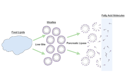 Figure 6: Bile from the liver and lipase from the pancreas help digest lipids in the small intestine
As an endocrine gland, the pancreas secretes several hormones, including insulin and glucagon, which circulate in the blood. The endocrine hormones are secreted by clusters of cells called pancreatic islets (or islets of Langerhans). As a digestive organ, the pancreas secretes many digestive enzymes and also bicarbonate, which helps neutralize acidic chyme after it enters the duodenum. The pancreas is stimulated to secrete its digestive substances when food in the stomach and duodenum triggers the release of endocrine hormones into the blood that reaches the pancreas via the bloodstream. The pancreatic digestive enzymes are secreted by clusters of cells called acini, and they travel through the pancreatic ducts to the duodenum. In the duodenum, they help to chemically break down carbohydrates, proteins, lipids, and nucleic acids in chyme. The pancreatic digestive enzymes include:
- Amylase, which helps digest starch and other carbohydrates.
- Trypsin and chymotrypsin, which help digest proteins.
- Lipase, which helps digest lipids.
- Deoxyribonucleases and ribonucleases, which help digest nucleic acids.
In addition to these bodily organs and structures, your large intestine houses bacteria that are very helpful in digestion. The large intestine is home to trillions of bacteria known as the “gut flora”. But don’t worry, most of these bacteria are helpful. Friendly bacteria live mostly in the large intestine and part of the small intestine. The acidic environment of the stomach does not allow bacterial growth.
Gut bacteria have several roles in the body. For example, intestinal bacteria:
- Produce vitamin B12 and vitamin K.
- Control the growth of harmful bacteria.
- Break down poisons in the large intestine.
- Break down some substances in food that cannot be digested, such as fibre and some starches and sugars. Bacteria produce enzymes that digest carbohydrates in plant cell walls. Most of the nutritional value of plant material would be wasted without these bacteria. These help us digest plant foods like spinach.
A wide range of friendly bacteria live in the gut. Bacteria begin to populate the human digestive system right after birth. Gut bacteria include Lactobacillus, the bacteria commonly used in probiotic foods such as yogurt, and E. coli bacteria. About a third of all bacteria in the gut are members of the Bacteroides species. Bacteroides are key in helping us digest plant food.
It is estimated that 100 trillion bacteria live in the gut. This is more than the human cells that make up you. It has also been estimated that there are more bacteria in your mouth than people on the planet — there are over 7 billion people on the planet!
The bacteria in your digestive system are from anywhere between 300 and 1,000 species. As these bacteria are helpful, your body does not attack them. They actually appear to the body’s immune system as cells of the digestive system, not foreign invaders. The bacteria actually cover themselves with sugar molecules removed from the actual cells of the digestive system. This disguises the bacteria and protects them from the immune system.
As the bacteria that live in the human gut are beneficial to us, and as the bacteria enjoy a safe environment to live, the relationship that we have with these tiny organisms is described as mutualism, a type of symbiotic relationship.
Lastly, keep in mind the small size of bacteria. Together, all the bacteria in your gut may weigh just about two pounds.
Getting Nutrients into Bloodstream
The goal of the digestive system is to break down food and get the nutrients into small enough molecules so that they can be transported throughout the body and used in the mitochondria of cells to make ATP energy. Large food eventually makes its way into a bolus, then chyme, and then building block materials of complex organic molecules such as glucose or amino acids.
It is the small building block that will make its way out of the small intestine, into the bloodstream, and into all the cells of the body for use in aerobic respiration.
The villi of the small intestine are therefore very important players in this process. As mentioned above, the villi are finger-like projections that are found throughout the small intestine (Fig 7). Each villus contains hundreds of microvilli and each villus contains a capillary network that will allow for diffusion of the nutrients out of the small intestine and into the capillaries. Remember that diffusion is the passive movement of materials from a high concentration to a low concentration. The small building blocks, like glucose, high in the small intestine will diffuse into the capillaries so they can move throughout the body and be distributed throughout the body.
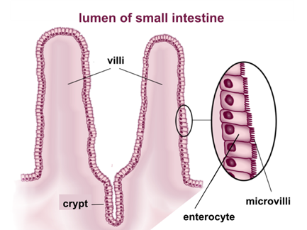
Figure 7: Drawing showing the relationship between villi and microvilli of the small intestine. The luminal surface of the enterocytes have microvilli (1 micrometer long) while the cell layer itself is folded to form villi (0.5-1.6 millimeters long) and crypts. Both serve to increase the total absorption surface of the intestine. BallenaBlanca, CC via Wikimedia Commons
Control of the Digestive Process
The process of digestion is controlled by both hormones and nerves. Hormonal control is mainly by endocrine hormones secreted by cells in the lining of the stomach and small intestine. These hormones stimulate the production of digestive enzymes, bicarbonate, and bile. The hormone secretin, for example, is produced by endocrine cells lining the duodenum of the small intestine. Acidic chyme entering the duodenum from the stomach triggers the release of secretin into the bloodstream. When the secretin returns via the circulation to the digestive system, it signals the release of bicarbonate from the pancreas. The bicarbonate neutralizes the acidic chyme. See Table 1 for a summary of the major hormones governing the process of chemical digestion.
| Hormone | Source Organ | Target Organ | Trigger | Result |
| Gastrin | Stomach walls | Stomach | High protein intake | HCL and pepsin release, stomach churning |
| Secretin | Duodenum | Pancreas; Gallbladder |
Acidic chyme entering the duodenum | Release sodium bicarbonate, release bile |
| Cholecystokinin (CCK) | Duodenum | Pancreas; Gallbladder |
Partially digested fat and protein in duodenum | Release lipase, trypsin, release bile |
Table 1: Major Hormones Governing Chemical Digestion
Nerves involved in digestion include those that connect digestive organs to the central nervous system, as well as nerves inside the walls of the digestive organs. Nerves connecting the digestive organs to the central nervous system cause smooth muscles in the walls of digestive organs to contract or relax as needed, depending on whether or not there is food to be digested. Nerves within digestive organs are stimulated when food enters the organs and stretches their walls. These nerves trigger the release of substances that speed up or slow down the movement of food through the GI tract and the secretion of digestive enzymes.
A final video to watch is a TED-Ed video on how your body knows when you are full.
Summary
Animal digestive systems are specialized structures and processes that allow organisms to obtain and process nutrients from their food. There are several types of digestive systems found in the animal kingdom, each adapted to the dietary and ecological needs of the species. Animals, like humans, can have a simple stomach system, while others have a more complex stomach, like ruminant. Here cows and sheep have a four-chambered stomach where food is first ingested and partially fermented in the first chamber (the rumen) before being regurgitated and chewed again (chewing cud). The food then passes through the other stomach chambers for further digestion and absorption. This system allows ruminants to extract nutrients from plant material that is difficult to digest. There are some invertebrates, like Hydra, which contain an incomplete digestive system where a single opening acts like a mouth and an anus. And some animals. like flatworms, might not even have a stomach at all and rely on diffusion of nutrients directly from the environment across their bodies into cells.
Focusing on complete digestive systems and humans, we find a series of both physical (like chewing with teeth and churning of the stomach) and chemical (like enzymes) factors working to break down large complex food into their simplest building blocks for absorption into the circulatory system. Once in the blood, sugars can be transported throughout the body and delivered to cells so the mitochondria can turn these simple sugars into ATP, which is the energy currency all cells use.
Questions
Glossary
References
Kosal, E. 2023. Getting Nutrients into Bloodstream and Types of Digestive Systems sections along with the Summary. NC State University.
Miller, C. 2020. Sections on Control of Digestive Process & Liver, Pancreas & Gut Bacteria. Human Biology.
Molnar, C. and J. Gair. Concepts of Biology. 1st Canadian Edition-GUnness
