
28.2 Base Pairing in DNA
Samples of DNA isolated from different tissues of the same species have the same proportions of heterocyclic bases, but samples from different species often have greatly differing proportions of bases. Human DNA, for example, contains about 30% each of adenine and thymine and about 20% each of guanine and cytosine. The bacterium Clostridium perfringens, however, contains about 37% each of adenine and thymine and only 13% each of guanine and cytosine. Note that in both examples the bases occur in pairs. Adenine and thymine are present in equal amounts, as are cytosine and guanine. Why?
In 1953, Rosalind Franklin, Maurice Wilkins, James Watson, and Francis Crick published scientific reports describing the secondary structure of DNA. According to their model, DNA under physiological conditions consists of two polynucleotide strands, running in opposite directions and coiled around each other in a double helix like the handrails on a spiral staircase. The two strands are complementary rather than identical and are held together by hydrogen bonds between specific pairs of bases, A with T and C with G. That is, whenever an A base occurs in one strand, a T base occurs opposite it in the other strand; when a C base occurs in one, a G occurs in the other (Figure 28.3). This complementary base-pairing thus explains why A and T are always found in equal amounts, as are G and C.
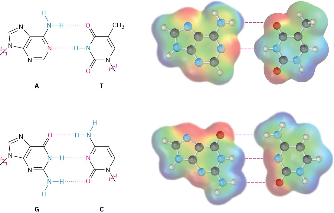
Figure 28.3 Hydrogen-bonding between base pairs in the DNA double helix. Electrostatic potential maps show that the faces of the bases are relatively neutral, while the edges have positive and negative regions. Pairing G with C and A with T brings together oppositely charged regions.
A full turn of a DNA double helix is shown in Figure 28.4. The helix is 20 Å wide, there are 10 base pairs per turn, and each turn is 34 Å in length. Notice in Figure 28.4 that the two strands of the double helix coil in such a way that two kinds of “grooves” result, a major groove 12 Å wide and a minor groove 6 Å wide. The major groove is slightly deeper than the minor groove, and both are lined by flat heterocyclic bases. As a result, a variety of other polycyclic aromatic molecules are able to slip sideways, or intercalate, between the stacked bases. Many cancer-causing and cancer-preventing agents function by interacting with DNA in this way.
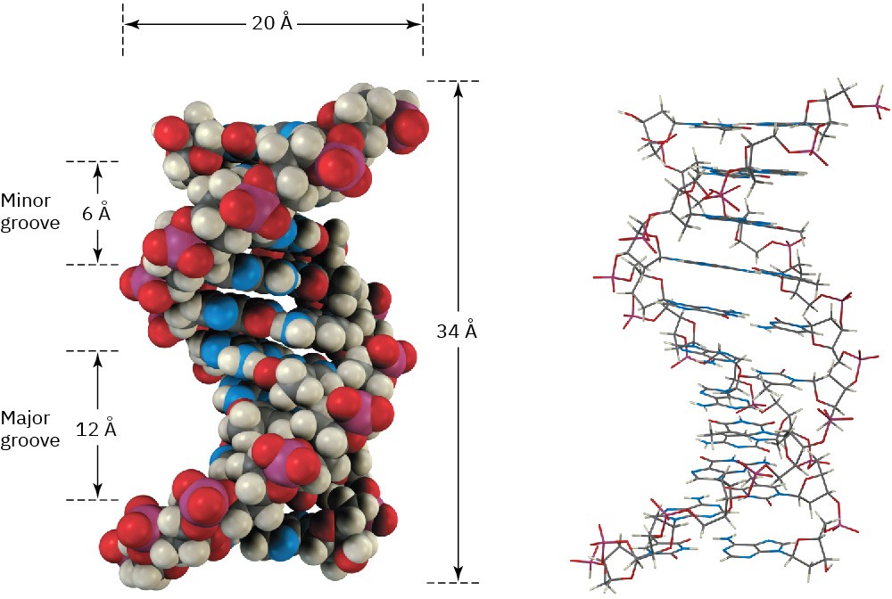
Figure 28.4 A turn of the DNA double helix in both space-filling and wire-frame formats. The sugar–phosphate backbone runs along the outside of the helix, and the amine bases hydrogen bond to one another on the inside. Both major and minor grooves are visible.
An organism’s genetic information is stored as a sequence of deoxyribonucleotides strung together in the DNA chain. For the information to be preserved and passed on to future generations, a mechanism must exist for copying DNA. For the information to be used, a mechanism must exist for decoding the DNA message and implementing the instructions it contains.
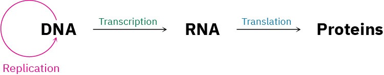
What Crick called the “central dogma of molecular genetics” says that the function of DNA is to store information and pass it on to RNA. The function of RNA, in turn, is to read, decode, and use the information received from DNA to make proteins. This view is greatly oversimplified but is nevertheless a good place to start. Three fundamental processes take place.
- Replication is the process by which identical copies of DNA are made so that information can be preserved and handed down to offspring.
- Transcription is the process by which genetic messages are read and carried out of the cell nucleus to ribosomes, where protein synthesis occurs.
- Translation is the process by which the genetic messages are decoded and used to synthesize proteins.
 Worked Example 28.1Predicting the Complementary Base Sequence in Double-Stranded DNAWhat sequence of bases on one strand of DNA is complementary to the sequence TATGCAT on another strand?StrategyRemember that A and G form complementary pairs with T and C, respectively, and then go through the sequence replacing A by T, G by C, T by A, and C by G. Remember also that the 5′ end is on the left and the 3′ end is on the right in the original strand.SolutionOriginal:(5′) TATGCAT (3′)Complement: (3′) ATACGTA (5′) or (5′) ATGCATA (3′)
Problem 28-3
What sequence of bases on one strand of DNA is complementary to the following sequence on another strand?
(5′) GGCTAATCCGT (3′)
